A cross-sectional observational study was performed in our department after obtaining prior approval from the Institute Ethics Committee. We initially reviewed CT thorax scans of 250 patients with RT-PCR-positive COVID-19 infection over one year as detailed in Fig. 1a. Lung nodules were present in 24 patients with COVID-19 infection of which four patients were eliminated from our study as their CT images were not suitable for radiomics analysis. Only patients in whom no other synchronous lung pathology was identified were included. The imaging features of COVID-19 lung nodules were studied in the remaining 20 patients and were assessed for size, type, margins, location, and the lobe and segment involved, Digital imaging and communications in medicine (DICOM) images of these 20 patients were thereafter subjected to segmentation analysis and radiomics post-processing.
Figure 1
(a) Flow diagram of the study of COVID-19-infected cases. (b) Flow diagram of the study of non-COVID-19-infected cases.
We exclusively examined patients with COVID pneumonia who exhibited solid pulmonary nodules only, totaling 20 cases, to ensure similarity with benign and malignant nodules. Our study involved individuals who underwent chest CT scans between the 3rd and 6th day after the onset of symptoms (1st week of the disease) and who tested positive for COVID-19 infection through RT-PCR. Along with these pulmonary nodules, the other common CT findings in these patients typically included ground glass opacities, crazy paving, and consolidation. However, our focus for radiomics analysis was solely on these pulmonary nodules.
We reviewed CT thorax of 1200 non-COVID-19 patients as shown in Fig. 1b. Lung nodules of compatible size were present in 133 cases. The final diagnosis was available in 97 patients, of whom 44 were benign and 53 were malignant nodules. The benign lesions were diagnosed based on histopathological diagnosis or correlation with clinical features and follow-up as per the Fleisher’s Society guidelines18. The final diagnosis in primary malignant lesions was arrived at based on histopathological diagnosis, and in metastasis based on the histopathological diagnosis of the lung nodule or primary tumor. The DICOM images of 40 benign nodules and 50 malignant nodules were subjected to segmentation analysis. Radiomics post-processing was done in 39 benign nodules and 49 malignant nodules. Radiomics analysis was not feasible in two patients. Subsequently, the radiomics texture analysis of COVID-19 lung nodules was compared separately with each radiomics analysis of benign non-COVID-19 benign lung nodules and malignant lung nodules.
The distribution of cases included in the final radiomics analysis included, n = 24 (22%) metastatic pulmonary nodules, n = 25 (23%) primary malignancies, n = 39 (36%) non-COVID benign nodules, and n = 20 (19%) COVID-related nodules (Fig. 2). The final diagnosis was available in all these nodules. n = 39 of the pulmonary nodules were found to be other benign, while n = 49 were malignant. The benign lesions were diagnosed based on histopathological diagnosis or correlation with clinical features and follow-up as per the Fleisher’s Society guidelines. The final diagnosis in primary malignant lesions was arrived at based on histopathological diagnosis, and metastasis was based on the histopathological diagnosis of the lung nodule or primary tumor. Only patients in whom no other synchronous lung pathology was identified were included to prevent overlap of pathologies. Patients with subsolid pulmonary nodules and Nodules with calcification were excluded from the study to compare purely solid pulmonary nodules. Of the other benign lesions (n = 39) analyzed using radiomics, 36% were septic emboli, 28%were benign lesions monitored long-term per Fleishner society guidelines, and the remainder comprised sarcoidosis, inflammatory conditions, pulmonary tuberculosis, benign carcinoid, hematoma, hydatid disease, Sjogren’s syndrome, and Wegner’s granulomatosis cases, as shown in Supplementary Information (Fig. S1). Among the malignant nodules (n = 49), there were 25 cases of primary lung malignancies and 24 cases of metastases. Primary lung malignancies consisted of adenocarcinoma (52%), squamous cell carcinoma (40%), and other types, as depicted in Supplementary Information (Fig. S2). The predominant source of metastases was breast carcinoma, with the remainder originating from various other primary organs, as depicted in Supplementary Information (Fig. S3).
Figure 2
Case distribution of the lung nodules included in the study.
CT acquisition
HRCT examinations were performed using one of the following multidetector computed tomography (MDCT) scanners: Phillips-brilliance 16 (Philips medical systems, Cleveland); GE EVO evolution 128 slices (GE healthcare, Princeton); and Siemens biograph horizon (Siemens AG, Munich). HR-CT images were obtained during breath-holding with the following parameters: 120 kV, 200 mA. The section thickness and reconstruction intervals were 0.65–0.80 mm. The CT images were sent to a picture archiving and communication system (PACS) to be interpreted at workstations.
Segmentation
The segmentation of the DICOM images of the pulmonary nodules, a critical initial step for accurate feature extraction, was performed manually by an expert radiologist using Insight Segmentation and Registration Toolkit (ITK-SNAP) software19 and was verified by three radiologists independently. The steps described above are shown in Figs. 3 and 4. By relying on the expert radiologist, we could delineate the nodules with a high degree of precision, particularly in terms of their shape and texture characteristics, which are crucial for subsequent radiomic analysis. Following the segmentation, we extracted radiomic features from the 3D representations of the nodules. The extraction process focused on a comprehensive set of features, including but not limited to, shape, size, intensity, texture, and wavelet features. The emphasis was on capturing a broad spectrum of information that reflects the underlying pathology and can be correlated with clinical outcomes. By combining expert radiological input with radiomic feature extraction techniques, we aimed to mitigate some of the challenges associated with parametric texture feature extraction.
Figure 3
A 57 year-old male patient with RT–PCR has proven COVID-19 pneumonia. (a,b) The axial section of the CT thorax in the lung window and soft tissue window shows a subpleural soft tissue nodule in the posterior segment of the left lower lobe. (c) Creation of ROI for segmentation. (d) 2D-segmented nodule. (e) 3D-volumetric rendering of the nodule. (f) Follow-up chest CT after 6 months revealed partial resolution of the nodule.
Figure 4
A 21 year-old lady with cough and hemoptysis. HPE: benign carcinoid tumor. (a,b) Axial section of CT thorax in lung window and soft tissue window showing a mass lesion in the posterior segment of the right lower lobe. (c) Creation of ROI for segmentation. (d) 2D segmented mass lesion. (e) 3D volumetric rendering of the mass lesion.
Radiomics analysis
Segmented lung nodules were used to extract different types of features. These features were classified into three categories: shape features (14), first-order features (18 features), and texture-based features (69 features). Texture-based features were of four types, namely gray level co-occurrence matrix (GLCM) features (24 features)20, gray-level run-length matrix (GLRLM) features (16 features)21, gray level size zone (GLSZM) features (16 features)15,22,23 and gray level dependence matrix (GLDM) features (13 features). Each radiomics feature was given a feature rank based on a random forest classifier. Out of 101, the top 10 features were selected for classification algorithms according to Anand et al.24. Figures 5 and 6 show the top 10 selected radiomics features with rank, and Tables 1 and 2 summarize their feature importance values. Several classification algorithms, such as SUPPORT VECTOR MACHine (SVM)25, multi-layer perceptron (MLP), naive Bayes, discriminant analysis, and decision tree26, were applied to selected feature matrices to classify benign and malignant nodules. SVM with the linear kernel (L-SVM) and radial basis function kernel (RBF–SVM) were used as SVM variants. Linear discriminant analysis (LDA) and quadratic discriminant analysis (QDA) were used in the category of discriminant analysis. We have also experimented with MLP classifiers for different hyperparameters which include activation, layers/number of neurons, and learning rate. To evaluate the performance of classifiers, confusion matrices were drawn on the test set. Accuracy, sensitivity, specificity, precision, and F1-measure were calculated for each classifier.
Figure 5
Figure 6
(a) Ten important features used for the classification of COVID-19 and non-COVID-19 benign lung nodules. (b) Comparison plot of the most prominent feature.
Table 1 COVID-19 vs non-COVID-19 Benign lung nodules-different performance metrics for different classifiers obtained on the test data set.
Table 2 Experiment 2: COVID-19 vs malignant lung nodules-different performance metrics for different classifiers obtained on the test data set.
While many state-of-the-art approaches in medical image analysis today do use deep learning methods, in our experiments they showed poor performance with an accuracy of at most 55%. We evaluated models such as ResNet, DenseNet, and Vision Transformer for the same but due to the limited data available, the models showed poor performance27. The radiomic features provide a more robust basis for training on limited data as compared to the deep learning approaches.
Ethical clearance
The study was performed after obtaining prior approval from the Institutional Research Ethics Committee—Sri Ramachandra Institute of Higher Education and Research (CSP–MED/19/SEP/56/122) and all methods were performed by relevant guidelines and regulations.
Informed consent
Informed consent was obtained from all subjects and/or their legal guardians involved in the study.
"My name is George Irungu and I am 37 years old. I am a pharmaceutical technician at North Kinangop Catholic Hospital in Nyandarua County.
I have battled tuberculosis three times. I was first diagnosed with pulmonary tuberculosis in 2005 when I was in secondary school. I started treatment in August 2005 and finished after six months. I was tested and found to be free of TB. However, due to remnants of previous infections, I contracted the disease again in 2022.
My symptoms were a persistent cough that lasted for a month, night sweats, fever, general weakness and loss of appetite.
At first, I was in doubt. I was tested again and the diagnosis was confirmed. I was given the standard six-month course of TB medication. After finishing the medication, I was tested to confirm that I was free of TB. Six months later the symptoms came back. The TB test was negative, but the cough would not go away.
I was so scared. I remember sweating profusely on my bed. I would wake up every morning to sheets soaked with sweat. I lost weight every day. I remember that I weighed 64 kilos before I got sick, but when TB recurred, I dropped to 50 kilos.
There was that fear of infecting my children and my colleagues at work. My contacts were screened and tested and they went for X-rays and they were found to be negative for TB infections.
A Computed Tomography (CT) Scan was conducted and showed that my lungs had cavitation, small holes in the lungs. Further tests were conducted and I underwent a lobectomy, a surgery to remove one of the lobes of the lungs.
The infected lobe was removed to prevent the infections from spreading to the other lobes. There were still remnants of TB bacteria in my lungs and I had to restart the treatment again because it was not clear whether the remaining parts of my lungs were clear of the TB. Now this was the third time.
The lungs have sections called lobes. The right lung has 3 lobes. The left lung has 2 lobes. A lobectomy may be done when a problem is found in just part of a lung.
After undergoing lobectomy, my journey towards recovery was a difficult one. After a piece of my lungs was removed, breathing became difficult for several months. I used to exercise a lot to improve my general health.
So far, I have experienced full recovery. I can run, swim and I can properly function post TB treatment.
When I contracted TB in 2005, I was in denial, which I attribute partly to the stigma associated with the disease.
My family got worried when they learnt I had TB. They thought it was a bad disease which was untreatable. Some people associate the condition with other diseases like HIV/Aids or sexually transmitted infections. Others insinuated that I was cursed.
My aunt told me, "oooh you have TB? That's a very bad disease. Ooh noo."
I urge members of the public to go for TB screening in the nearest health facilities and for those on TB treatment to complete medication so as to get cured.
I would also like to urge my fellow health workers to go for routine TB screening as well as actively screen patients when they visit the facility.”
Tuberculosis is caused by a bacterium called Mycobacterium tuberculosis. Kenya is among 30 high TB burden countries in the world.
Pulmonary tuberculosis affecting the lungs, is the commonest form of TB causing a distinctive cough. Apart from Pulmonary tuberculosis, the bacteria can also cause TB of the bladder, spine, skin, brain and even the heart. But there are types of TB in which individuals don’t experience such symptoms that is; latent TB. In this case, one’s immune system fights the bacteria and the disease is not transmissible.
On January 21, the Ministry of Health National TB Programme in collaboration with Centre for Health Solutions-Kenya through its USAID-funded Tuberculosis Accelerated Response and Care II (TB ARC II) and other partners launched a plan to combat tuberculosis in Kenya.
The plan, dubbed National Strategic Plan for Tuberculosis, Leprosy and Lung Health is a five-year (2024-28) strategic plan that aims to ensure quality care for TB, leprosy and lung diseases for all Kenyans.
Public Health and Professional Standards Principal Secretary Mary Muthoni raised concern about what she termed as the scourge that is TB. The PS said that the ministry is committed to further reducing the cases of TB reported in a year by 2030.
In 2022, Kenya reported a total of 90,841 TB cases, an increase compared to the 77,854 cases reported in 2021. In the same year, 17,000 people died of TB.
Kenya recorded a 32 percent decline in TB incidence and a 42 percent mortality reduction between 2015-2020.
“Alarming as this figure is, it represents only 68 percent of the estimated 133,000 TB cases that were likely to emerge that year, leaving 32 percent undiagnosed and untreated,” the PS said.
However, Muthoni regretted the emergence of drug-resistant TB cases, totaling 756 cases last year, which highlights the urgent need for a comprehensive and coordinated response.
“The strategic plan does not only acknowledge the existing challenges in TB control but also proposes practical interventions to address the root causes of these barriers,” she said, identifying partnership and accountability as the key pillars towards a united front in this battle.
An estimated 10.6 million people fell ill with TB across the globe, including 5.8 million men, 3.5 million women and 1.3 million children according to the World Health Organisation.
Tuberculosis is coming back to Germany – here is the x-ray of a lung
Source: dpa
You can listen to our WELT podcasts here
In order to display embedded content, your revocable consent to the transmission and processing of personal data is necessary, as the providers of the embedded content require this consent as third party providers [In diesem Zusammenhang können auch Nutzungsprofile (u.a. auf Basis von Cookie-IDs) gebildet und angereichert werden, auch außerhalb des EWR]. By setting the switch to “on”, you agree to this (revocable at any time). This also includes your consent to the transfer of certain personal data to third countries, including the USA, in accordance with Art. 49 (1) (a) GDPR. You can find more information about this. You can revoke your consent at any time using the switch and privacy at the bottom of the page.
The number of cases of tuberculosis in Germany has been declining for years. The war in Ukraine is also causing more cases to be registered again. Experts describe the risks – and what you should avoid because of possible infection.
After years of decline in reported tuberculosis cases in Germany, the number of cases rose again last year from a low level. Around 4,480 cases were registered in 2023, as the Robert Koch Institute announced. In the previous year there were around 4,080 cases, in 2021 around 3,930.
“The background for these latest developments is currently the immigration of people seeking protection from Ukraine,” it said. Tuberculosis is significantly more common in the country. According to the RKI, three quarters of the people who receive a tuberculosis diagnosis in this country were born outside Germany.
Last year, according to estimates by the WorldHealth Organization (WHO), 10.6 million people worldwide became ill with tuberculosis and 1.3 million died from it. According to the RKI, countries in Southeast Asia, such as the Philippines, Indonesia and India, as well as countries in southern Africa, such as Lesotho and Zimbabwe, are particularly affected. In Europe the focus is on Eastern Europe.
also read
Tuberculosis is transmitted through the respiratory tract through aerosols containing bacteria and can be cured with medication in the vast majority of cases. In the case of a latent infection, illness can be prevented. A latent infection occurs when you are infected with tuberculosis bacteria without becoming ill. The treatment lasts at least six months.
also read
According to the RKI, the majority of those affected (70 percent) suffer from pulmonary tuberculosis. An infection is manifested by a cough, which in rare cases can be bloody. Occasionally, chest pain and shortness of breath occur. “If the cough persists for a long time, tuberculosis should also be considered and further examined using an X-ray examination of the lungs in accordance with existing recommendations,” recommended RKI President Lars Schaade.
Here you will find content from third parties
In order to display embedded content, your revocable consent to the transmission and processing of personal data is necessary, as the providers of the embedded content require this consent as third party providers [In diesem Zusammenhang können auch Nutzungsprofile (u.a. auf Basis von Cookie-IDs) gebildet und angereichert werden, auch außerhalb des EWR]. By setting the switch to “on”, you agree to this (revocable at any time). This also includes your consent to the transfer of certain personal data to third countries, including the USA, in accordance with Art. 49 (1) (a) GDPR. You can find more information about this. You can revoke your consent at any time using the switch and privacy at the bottom of the page.
According to the RKI, people who have had close and long-term contact with people who have infectious pulmonary tuberculosis, as well as people with inadequately treated previous tuberculosis, are particularly at risk. “HIV, smoking, alcohol and drug addiction, malnutrition, diabetes mellitus and living conditions such as homelessness, previous imprisonment and poverty are also risk factors.”
Before the pandemic, according to the experts, there were many positive developments to end the global tuberculosis epidemic by 2035 in accordance with a WHO strategy. “The COVID-19 pandemic then led to painful setbacks in many countries that have to be laboriously recovered.”
Carbapenem-resistant Klebsiella pneumoniae (CRKP) is a notorious pathogen worldwide that can cause pneumonia, bloodstream infections, urinary tract infections or wound infections, which are often difficult or even impossible to treat.1 Carbapenems often serve as the final effective line of defense against infections caused by multidrug-resistant K. pneumoniae, and multiple mechanisms contribute to the development of drug-resistant strains.2 In China, the prevalence of CRKP has shown an alarming upward trend and has become a serious threat to public health due to high drug resistance, hypervirulence, and high fatality rates.3,4 Worryingly, this trend has been observed globally.5
Chronic obstructive pulmonary disease (COPD) is a complex and heterogeneous lung disease characterized by persistent, progressive airflow obstruction. Acute exacerbation of chronic obstructive pulmonary disease (AECOPD) is a state in which respiratory symptoms of COPD are rapidly exacerbated, with bacterial infection being one of the main causes.6 When AECOPD is complicated by bronchial infection or pneumonia, in-hospital mortality significantly increases, particularly in elderly patients.7 In 2015, 99.9 million Chinese adults suffered from COPD, making it the third most common chronic disease after hypertension and diabetes in China.8 Acute exacerbation increases the frequency of hospitalization in COPD patients, seriously affects the quality of life, and commonly accompanies a heavy economic burden.9
Previous studies have reported the association of Haemophilus influenzae or Streptococcus pneumoniae with AECOPD.10 However, pathogen types in COPD patients’ sputum appear to vary by population and geographic location. In China, some studies have identified K. pneumoniae as one of the most common pathogens (Pseudomonas aeruginosa, Acinetobacter baumannii and Staphylococcus aureus in addition) in the sputum of AECOPD patients.11,12 In recent years, there has been a rapid escalation in the drug resistance of CRKP, concurrent with a persistent surge in reported mortality rates associated with this formidable pathogen.13 At present, CRKP is mainly reported in medical institutions, and evidence indicating its proliferation beyond the confines of hospital settings has been found.14 COPD patients often visit healthcare institutions, potentially increasing the risk of future CRKP infection. In fact, CRKP is a pathogen closely associated with respiratory infection, and its impact tends to be more severe in elderly patients.15 Simultaneously, studies have shown a substantial increase in the risk of mortality following CRKP infection in individuals diagnosed with COPD.16,17
However, there is a paucity of current studies on CRKP-induced pneumonia in AECOPD patients. Here, we reported the prevalence of CRKP-induced pneumonia in AECOPD patients, and we retrospectively analyzed the clinical characteristics and prognostic factors of COPD patients with acute exacerbation due to CRKP-induced pneumonia. The results will help the diagnosis and treatment of CRKP-induced pneumonia in AECOPD patients.
Methods
Study Design
We conducted a retrospective case-control study in Hunan Provincial People’s Hospital (the First Affiliated Hospital of Hunan Normal University). The study period spanned from January 1, 2016 to December 31, 2022, and the subjects were AECOPD patients hospitalized for K. pneumoniae-induced pneumonia during this period. The diagnosis of AECOPD was made by the pulmonologist according to the Global Initiative for Chronic Obstructive Lung Disease (GOLD) guidelines, the patient’s COPD history, and the clinical presentation of acute changes in symptoms.18 The diagnosis of K. pneumoniae-induced pneumonia was based on the patient’s clinical manifestations (characterized by intensified cough, increased sputum volume or purulent sputum, fever, shortness of breath, or a combination of these symptoms), findings from lung imaging (infiltrates detected on chest X-ray or CT scans), elevated peripheral white blood cell counts, and deep sputum or bronchoalveolar lavage fluid cultures positive for K. pneumoniae. All cases presented with either AECOPD as the primary diagnosis or a primary diagnosis of pulmonary infection, with AECOPD identified as a secondary diagnosis. Patients received at least one antibiotic with in vitro antimicrobial activity within 5 days of the pneumonia diagnosis. Patients with negative sputum or bronchoalveolar lavage cultures, non-K. pneumoniae cultured, respiratory tract colonization, incomplete hospitalization records, active pulmonary tuberculosis, or co-infection (defined as bacterial pathogens other than K. pneumoniae cultured within 48 hours),19 as well as those without AECOPD or pulmonary infection as the main diagnosis, were excluded. In cases where a patient had multiple hospitalizations for K. pneumoniae-induced pneumonia, only the initial hospitalization was documented for the study.
Data Collection
The patient’s hospitalization information was searched through medical records, including demographic data, smoking habits, admission routes, comorbidities, previous acute exacerbation (defined as at least one documented history of institutional visit due to acute exacerbation of respiratory symptoms in the past year), and prior use of carbapenems. Data collection also encompassed the results of blood cell tests, C-reactive protein tests, arterial blood gas tests, liver function tests, kidney function tests, serum ion tests, and coagulation tests conducted at the time of infection. Neutrophil-lymphocyte ratio (NLR) and platelet-lymphocyte ratio (PLR) were calculated from blood cell analysis results. Stable-phase pulmonary function results were also recorded. Using the extracted data, the Acute Physiology and Chronic Health Evaluation II (APACHE II) score for each patient was calculated based on the worst variable obtained at the time of infection to assess disease severity.20 Simultaneously, the treatment process and outcome of each patient were documented.
Microbiological Analysis
Deep sputum samples or bronchoalveolar lavage fluid samples were cultured in the clinical microbiology laboratory for identification and antibacterial susceptibility testing by VITEK MS (bioMérieux, Marcy-l’Étoile, France) or VITEK-2 compact system (bioMérieux, Marcy-l’Étoile, France). The interpretive criteria for tigecycline were based on the Food and Drug Administration (FDA) guidelines, while those for colistin were based on the European Committee on Antimicrobial Susceptibility Testing (EUCAST) guidelines. The other drugs were interpreted according to the M100 performance standards for antimicrobial susceptibility testing of the Clinical Laboratory Standards Institute (CLSI) 2015–2021 editions. CRKP was defined by K. pneumoniae with resistance to imipenem or meropenem. Multidrug-resistant (MDR) strains were defined according to the previous study.21 Patients were divided into the CRKP-induced pneumonic acute exacerbation (CRKPpAE) group and the non-CRKP-induced pneumonic acute exacerbation (non-CRKPpAE) group.
Statistical Analysis
Enumeration data were expressed as frequency (%), and the chi-squared test or Fisher’s exact probability method was used for comparison between groups. Normality of the distribution of numerical variables was checked by the Kolmogorov–Smirnov test. Normally distributed variables were described using mean ± standard deviation (SD) and compared using independent samples t-test. Non-normally distributed variables were described using median with 1st and 3rd quartiles and compared using the Mann–Whitney U-test. After checking for collinearity, variables associated with CRKP-induced pneumonia (p value <0.10) were allowed into a forward conditional binary logistic regression model to identify independently associated variables. The fitness of the multivariate model was performed using the Hosmer-Lemeshow goodness-of-fit test. All statistical analyses were conducted using IBM SPSS Statistics software, version 21.0 (SPSS Inc, Chicago, IL, USA). A two-tailed p value <0.05 was considered statistically significant.
Results
Patient Information and Clinical Characteristics
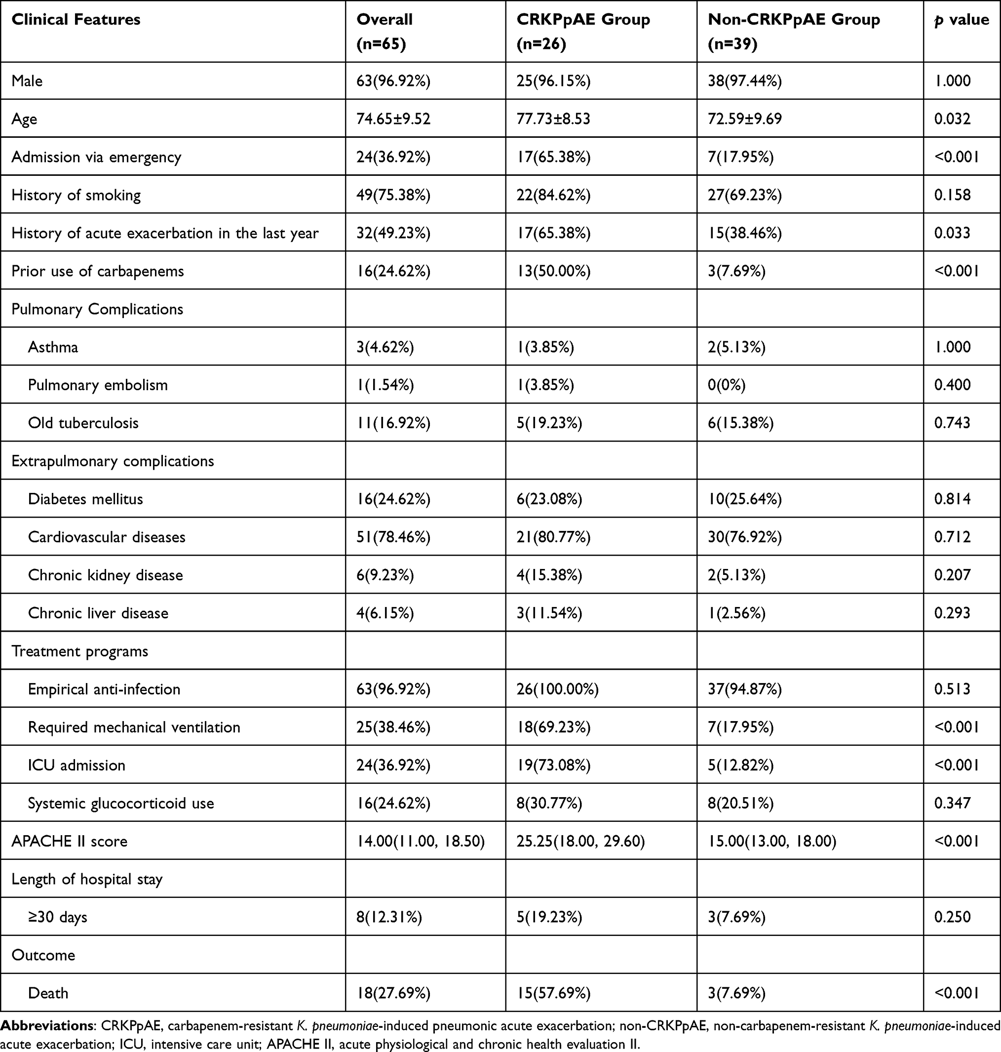
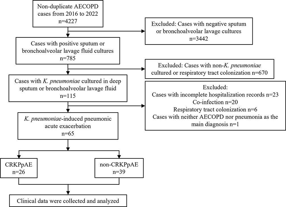
4227 non-duplicated AECOPD cases were investigated, and ultimately 65 cases hospitalized for acute exacerbation due to K. pneumoniae-induced pneumonia were analyzed from January 1, 2016 to December 31, 2022 (Figure 1). Most patients were male (63/65, 96.9%), the minimum age was 54 years old, and the average age was 74.65±9.52 years old. The CRKPpAE group comprised 26 patients, while the non-CRKPpAE group consisted of 39 patients. Patients in the CRKPpAE group were older than those in the non-CRKPpAE group (77.73±8.53 and 72.59±9.69 years, respectively, p=0.032). Patients in the CRKPpAE group were admitted more often through the emergency department (17/26, 65.38% and 7/39, 17.95%, respectively, p<0.001). A history of acute exacerbation in the past year (17/26, 65.38% and 15/39, 38.46%, respectively, p=0.033) and prior use of carbapenems (13/26, 50.00% and 3/39, 7.69%, respectively, p<0.001) were significantly associated with CRKPpAE. Patients in the CRKPpAE group generally required mechanical ventilation (18/26, 69.23% and 7/39, 17.95%, respectively, p<0.001) and ICU admission (19/26, 73.08% and 5/39, 12.82%, respectively, p<0.001), and showed more severe disease severity [The median of APACHE II scores with interquartile range were 25.25 (18.00, 29.60) and 15.00 (13.00, 18.00), respectively, p<0.001]. In addition, the risk of in-hospital death was significantly higher in the CRKPpAE group compared to the the non-CRKPpAE group (all-cause mortality 15/26, 57.69% and 3/39, 7.69%, respectively, p<0.001) (Table 1).
Table 1 Clinical Features of CRKP-Induced Pneumonia in AECOPD Patients
Figure 1 Flow chart of the study population.
Abbreviations: AECOPD, acute exacerbation of chronic obstructive pulmonary disease; CRKPpAE, carbapenem-resistant K. pneumoniae-induced pneumonic acute exacerbation; non-CRKPpAE, non-carbapenem-resistant K. pneumoniae-induced pneumonic acute exacerbation.
Univariate Analysis of Laboratory Test Results
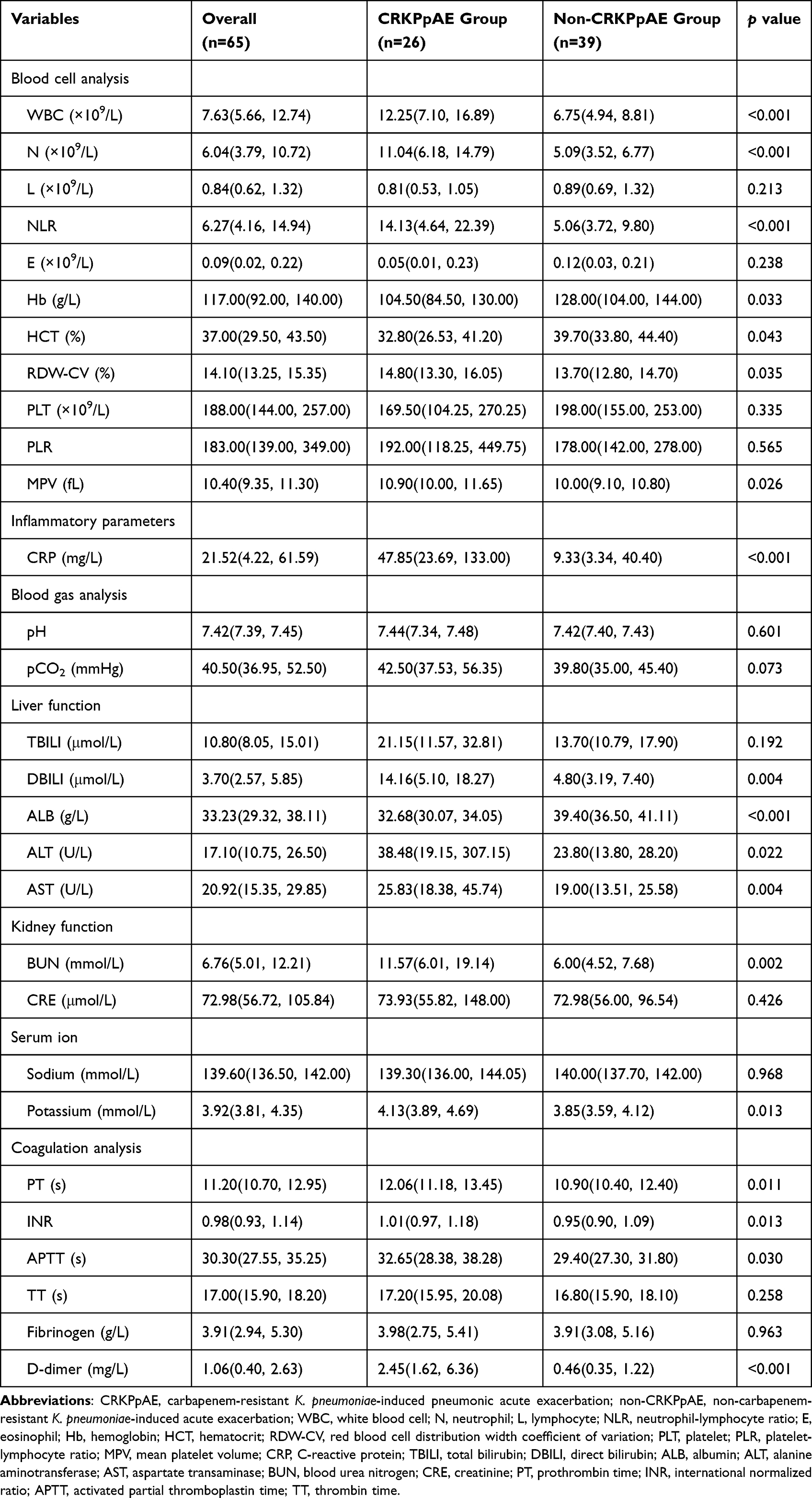
By comparing the results of laboratory tests, the white blood cell counts [12.25(7.10, 16.89) and 6.75(4.94, 8.81) ×109/L, respectively, p<0.001], neutrophil counts [11.04(6.18, 14.79) and 5.09(3.52, 6.77) ×109/L, respectively, p<0.001], NLR levels [14.13(4.64, 22.39) and 5.06(3.72, 9.80), respectively, p<0.001], red blood cell distribution width coefficient of variation values [14.80(13.30, 16.05) and 13.70(12.80, 14.70) %, respectively, p=0.035] and mean platelet volume values [10.90(10.00, 11.65) and 10.00(9.10, 10.80) fL, respectively, p=0.026] of patients in the CRKPpAE group were significantly higher than those in the non-CRKPpAE group. However, hemoglobin values [104.50(84.50, 130.00) and 128.00(104.00, 144.00) g/L, respectively, p=0.033] and hematocrit values [32.80(26.53, 41.20) and 39.70(33.80, 44.40) %, respectively, p=0.043] in the CRKPpAE group were lower than those in the non-CRKPpAE group. C-Reactive protein values [47.85 (23.69, 133.00) and 9.33 (3.34, 40.40) mg/L, respectively, p<0.001] in the CRKPpAE group were significantly higher than those in the non-CRKPpAE group. For the CRKPpAE patients, there was a more serious liver function damage [direct bilirubin values: 14.16(5.10, 18.27) and 4.80(3.19, 7.40) μmol/L, respectively, p=0.004, alanine aminotransferase values: 38.48(19.15, 307.15) and 23.80(13.80, 28.20) U/L, respectively, p=0.022, aspartate transaminase values: 25.83(18.38, 45.74) and 19.00(13.51, 25.58) U/L, respectively, p=0.004], and a higher risk of hypoalbuminemia [albumin values: 32.68(30.07, 34.05) and 39.40(36.50, 41.11) g/L, respectively, p<0.001]. Blood urea nitrogen values [11.57(6.01, 19.14) and 6.00(4.52, 7.68) mmol/L, respectively, p=0.002] of patients in the CRKPpAE group were higher than those in the non-CRKPpAE group, and there was a significant difference in the serum potassium levels between the two groups [4.13(3.89, 4.69) and 3.85(3.59, 4.12) mmol/L, respectively, p=0.013]. Through coagulation analysis, the CRKPpAE group exhibited higher values in prothrombin time [12.06(11.18, 13.45) and 10.90(10.40, 12.40) s, respectively, p=0.011], international normalized ratio [1.01(0.97, 1.18) and 0.95(0.90, 1.09), p=0.013], activated partial thromboplastin time [32.65(28.38, 38.28) and 29.40(27.30, 31.80) s, respectively, p=0.030] and D-dimer [2.45(1.62, 6.36) and 0.46(0.35, 1.22) mg/L, respectively, p<0.001] compared to the non-CRKPpAE group (Table 2). Stable-phase pulmonary function results were accessible for a subset of just 13 patients, all of whom were categorized in the non-CRKPpAE group. Due to the limited sample size, a comparative analysis of stable-phase pulmonary function results was not conducted.
Table 2 Comparison of Laboratory Test Results Between the CRKPpAE Group and the Non-CRKPpAE Group
Multivariate Analysis of Risk Factors
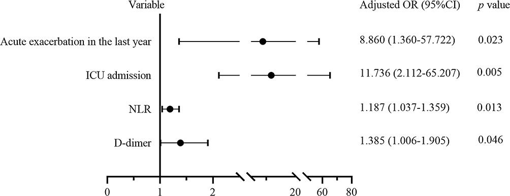
The factors that significant at p <0.10 in univariate analysis were allowed into the logistic regression model to identify independent risk factors. We found that a history of acute exacerbation in the past year (OR=8.860, 95% CI: 1.360–57.722, p=0.023), ICU admission (OR=11.736, 95% CI: 2.112–65.207, p=0.005), higher NLR levels (OR=1.187, 95% CI: 1.037–1.359, p=0.013) and higher D-dimer levels (OR=1.385, 95% CI: 1.006–1.905, p=0.046) were independently associated with CRKPpAE (Figure 2).
Figure 2 Multivariate analysis of clinical characteristics of patients with CRKPpAE.
Abbreviations: ICU, intensive care unit; NLR, neutrophil-lymphocyte ratio.
Comparison of Drug Resistance Between CRKP and Non-CRKP
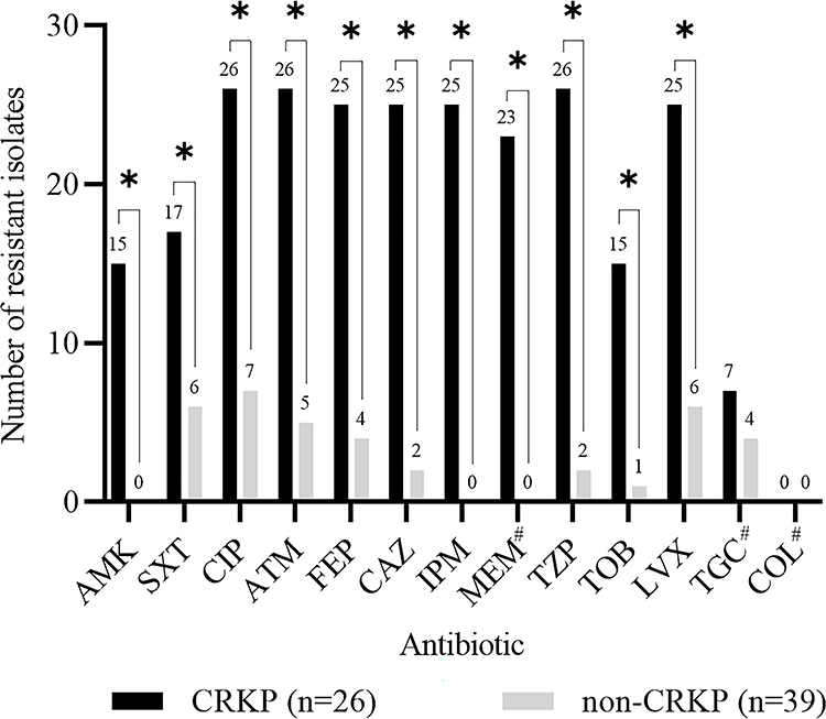
CRKP isolates exhibited significantly higher resistance to the majority of tested antimicrobials [including amikacin (57.69% and 0% respectively), trimethoprim/sulfamethoxazole (65.38% and 15.38% respectively), ciprofloxacin (100.00% and 17.95% respectively), aztreonam (100.00% and 12.82% respectively), cefepime (96.15% and 10.26% respectively), ceftazidime (96.15% and 5.13% respectively), imipenem (96.15% and 0% respectively), meropenem (95.83% and 0% respectively), piperacillin/tazobactam (100.00% and 5.13% respectively), tobramycin (57.69% and 2.56% respectively), and levofloxacin (96.15% and 15.38% respectively), all p<0.001] compared to non-CRKP isolates (Figure 3). Strains resistant to tigecycline were found (CRKP, 7/24, 29.17% and non-CRKP, 4/33, 12.12%, respectively), while all strains exhibited sensitivity to colistin. All CRKP isolates were MDR strains (26/26, 100.00%), while 5 non-CRKP isolates were MDR strains (5/39, 12.82%).
Figure 3 Comparison of antimicrobial resistance between CRKP and non-CRKP. *The difference was statistically significant, p<0.001. #Only 24 CRKP and 33 non-CRKP participated in the antimicrobial susceptibility test.
CRKP infections are constantly being reported, and CRKP-induced pneumonia in AECOPD patients deserves more attention. Here, we conducted a retrospective study of acute exacerbation of COPD caused by CRKP-induced pneumonia in a tertiary hospital in China, reported the clinical features and mortality of CRKP-induced pneumonia, and analyzed factors related to prognosis.
Our results showed that the mortality rate of acute exacerbation of COPD caused by CRKP-induced pneumonia reached a staggering 57.69%. It is higher than the 42.1% in-hospital mortality rate reported in another study on CRKP infection in respiratory intensive care unit.22 Of the patients with CRKPpAE, 65.38% had at least one documented medical visit for acute exacerbation in the past year, a frequency higher than that observed in the non-CRKPpAE group. The CRKP-induced pneumonia could be attributed to healthcare-associated infections. Current research data on CRKPpAE are very limited. Previous studies have found exacerbation in the past year to be an independent risk factor for exacerbation readmission in patients with COPD.23 We speculate that a history of acute exacerbation led to a decline in lung health, providing an opportunity for CRKP infection. Studies have shown that CRKP carries more drug resistance and virulence determinants than non-CRKP, which make it more invasive.24 Together with the increased chances of contact with CRKP during frequent medical visit, CRKPpAE occurred. The existence of drug resistance and virulence determinants may be the reasons why patients with CRKPpAE were admitted to ICU. On the other hand, impairment of tracheal clearance in patients and wide distribution of CRKP in the ICU may also contribute to the above association.25,26 It is noteworthy that many studies have shown that prior use of carbapenems was an independent risk factor for CRKP infection.25,27 However, in our study, this association was not statistically significant through multivariate analysis, possibly owing to the relatively modest sample size in our study.
NLR and PLR are emerging markers closely associated with poor prognosis in AECOPD patients.28 In our study, higher NLR levels were independently associated with CRKPpAE by multivariate analysis, suggesting that CRKPpAE resulted in more severe inflammatory responses. Coagulation abnormalities often occur in AECOPD patients, with infection being one of the significant contributing factors.29 We found that higher D-dimer levels were independently associated with CRKPpAE, indicating that coagulation disorders were more severe when CRKPpAE occurred, and vigilance for thrombosis should be maintained.
In our study, CRKP not only resistant to carbapenems, but also had high resistance to many other types of antibacterial drugs, and all of them were MDR strains. Although isolates resistant to tigecycline were identified, they all remained sensitive to colistin. The emergence of drug resistance in these strains posed significant challenges to clinical treatment. Colistin and tigecycline could be regarded as potential treatment choices, and there was also the option to consider combination therapy.30 Notably, MDR strains were also found in non-carbapenem-resistant isolates. Proactively controlling the spread of resistant strains will aid in the successful recovery of patients undergoing treatment.
This was a single-center retrospective study and was limited by a relatively small sample size. Larger-scale research data will help increase the credibility of the conclusions. The incidence of CRKP-induced pneumonia in patients with AECOPD may be underestimated due to the limitations of sputum culture and co-infection. Unfortunately, we were unable to collect stable-phase pulmonary function data for all patients. Furthermore, we were not able to study the impact of early CRKP respiratory colonization on infection and outcomes. Given that patients with COPD often seek medical help, and CRKP is widespread in the hospital environment, analyzing the status of CRKP colonization in the respiratory tract of these patients would be of great value.
As a result, our report showed that CRKP-induced pneumonia significantly contributed to the acute exacerbation of COPD. The frequent exacerbations and medical visits might have contributed to the occurrence of CRKPpAE events. The salient clinical features of these patients were mainly manifested by more severe inflammatory response and coagulation disturbance, and the risk of ICU admission was significantly increased. CRKP showed high resistance to a variety of antibacterial drugs, which brought challenges to treatment. Our findings will contribute to clinical decision-making in CRKPpAE.
Conclusion
Our study reveals a significantly higher mortality rate in AECOPD with CRKP-induced pneumonia compared to non-CRKP-induced pneumonia. Additionally, a history of acute exacerbation in the last year, ICU admission, higher NLR levels, and elevated D-dimer levels are independently associated with AECOPD due to CRKP-induced pneumonia.
Data Sharing Statement
The data that support the findings of this study are available from the corresponding author upon reasonable request.
Ethics Approval and Informed Consent
This study was approved by the Ethics Committee of Hunan Provincial People’s Hospital (the First Affiliated Hospital of Hunan Normal University) (approval number [2023]-160). Informed consent was waived due to the retrospective study and no intervention in patient treatment. This research was conducted in compliance with the tenets of the Helsinki Declaration.
Author Contributions
All authors made a significant contribution to the work reported, whether that is in the conception, study design, execution, acquisition of data, analysis and interpretation, or in all these areas; took part in drafting, revising or critically reviewing the article; gave final approval of the version to be published; have agreed on the journal to which the article has been submitted; and agree to be accountable for all aspects of the work.
Disclosure
The authors report no conflicts of interest in this work.
References
1. Wang M, Earley M, Chen L, et al. Clinical outcomes and bacterial characteristics of carbapenem-resistant Klebsiella pneumoniae complex among patients from different global regions (CRACKLE-2): a prospective, multicentre, cohort study. Lancet Infect Dis. 2022;22(3):401–412. doi:10.1016/S1473-3099(21)00399-6
2. Minerdi D, Loqui D, Sabbatini P. Monooxygenases and antibiotic resistance: a focus on carbapenems. Biology. 2023;12(10):1316. doi:10.3390/biology12101316
3. Hu F, Zhu D, Wang F, et al. Current status and trends of antibacterial resistance in China. Clin Infect Dis. 2018;67:S128–S134. doi:10.1093/cid/ciy657
4. Chen J, Ma H, Huang X, et al. Risk factors and mortality of carbapenem-resistant Klebsiella pneumoniae bloodstream infection in a tertiary-care hospital in China: an eight-year retrospective study. Antimicrob Resist Infect Control. 2022;11:161. doi:10.1186/s13756-022-01204-w
5. Lan P, Jiang Y, Zhou J, et al. A global perspective on the convergence of hypervirulence and carbapenem resistance in Klebsiella pneumoniae. J Glob Antimicrob Resist. 2021;25:26–34. doi:10.1016/j.jgar.2021.02.020
6. Agustí A, Celli BR, Criner GJ, et al. Global initiative for chronic obstructive lung disease 2023 report: gold executive summary. Eur Respir J. 2023;61:2300239. doi:10.1183/13993003.00239-2023
7. Song Y, Chen R, Zhan Q, et al. The optimum timing to wean invasive ventilation for patients with AECOPD or COPD with pulmonary infection. Int J Chron Obstruct Pulmon Dis. 2016;11:535–542. doi:10.2147/COPD.S96541
8. Wang C, Xu J, Yang L, et al. Prevalence and risk factors of chronic obstructive pulmonary disease in China (the China Pulmonary Health [CPH] study): a national cross-sectional study. Lancet. 2018;391:1706–1717. doi:10.1016/S0140-6736(18)30841-9
9. Kunadharaju R, Sethi S. Treatment of acute exacerbations in chronic obstructive pulmonary disease. Clin Chest Med. 2020;41:439–451. doi:10.1016/j.ccm.2020.06.008
10. Decramer M, Janssens W, Miravitlles M. Chronic obstructive pulmonary disease. Lancet. 2012;379(9823):1341–1351. doi:10.1016/S0140-6736(11)60968-9
11. Lin SH, Kuo PH, Hsueh PR, et al. Sputum bacteriology in hospitalized patients with acute exacerbation of chronic obstructive pulmonary disease in Taiwan with an emphasis on Klebsiella pneumoniae and Pseudomonas aeruginosa. Respirology. 2007;12(1):81–87. doi:10.1111/j.1440-1843.2006.00999.x
12. Ma X, Cui J, Wang J, et al. Multicentre investigation of pathogenic bacteria and antibiotic resistance genes in Chinese patients with acute exacerbation of chronic obstructive pulmonary disease. J Int Med Res. 2015;43(5):699–710. doi:10.1177/0300060515587577
13. Rodrigo-Troyano A, Sibila O. The respiratory threat posed by multidrug resistant Gram-negative bacteria. Respirology. 2017;22(7):1288–1299. doi:10.1111/resp.13115
14. Esposito F, Cardoso B, Sellera FP, et al. Expansion of healthcare-associated hypervirulent KPC-2-producing Klebsiella pneumoniae ST11/KL64 beyond hospital settings. One Health. 2023;17:100594. doi:10.1016/j.onehlt.2023.100594
15. Zhang Y, Wang Q, Yin Y, et al. Epidemiology of carbapenem-resistant Enterobacteriaceae infections: report from the China CRE network. Antimicrob Agents Chemother. 2018;62(2):e01882–17. doi:10.1128/AAC.01882-17
16. Qian Y, Bi Y, Liu S, et al. Predictors of mortality in patients with carbapenem-resistant Klebsiella pneumoniae infection: a meta-analysis and a systematic review. Ann Palliat Med. 2021;10(7):7340–7350. doi:10.21037/apm-21-338
17. Gonçalves Barbosa LC, Silva ESJA, Bordoni GP, et al. Elevated mortality risk from CRKp associated with comorbidities: systematic review and meta-analysis. Antibiotics. 2022;11(7):874. doi:10.3390/antibiotics11070874
18. Vestbo J, Hurd SS, Agustí AG, et al. Global strategy for the diagnosis, management, and prevention of chronic obstructive pulmonary disease: GOLD executive summary. Am J Respir Crit Care Med. 2013;187(4):347–365. doi:10.1164/rccm.201204-0596PP
19. Chen IR, Huang PH, Wu PF, et al. Clinical characteristics and outcomes of 56 patients with pneumonia caused by carbapenem-resistant Klebsiella pneumoniae. J Glob Antimicrob Resist. 2021;25:326–330. doi:10.1016/j.jgar.2021.03.028
20. Knaus WA, Draper EA, Wagner DP, et al. APACHE II: a severity of disease classification system. Crit Care Med. 1985;13(10):818–829. doi:10.1097/00003246-198510000-00009
21. Magiorakos AP, Srinivasan A, Carey RB, et al. Multidrug-resistant, extensively drug-resistant and pandrug-resistant bacteria: an international expert proposal for interim standard definitions for acquired resistance. Clin Microbiol Infect. 2012;18(3):268–281. doi:10.1111/j.1469-0691.2011.03570.x
22. Zhang H, Wang J, Zhou W, et al. Risk factors and prognosis of carbapenem-resistant Klebsiella pneumoniae infections in respiratory intensive care unit: a retrospective study. Infect Drug Resist. 2021;14:3297–3305. doi:10.2147/IDR.S317233
23. Wei X, Ma Z, Yu N, et al. Risk factors predict frequent hospitalization in patients with acute exacerbation of COPD. Int J Chron Obstruct Pulmon Dis. 2018;13:121–129. doi:10.2147/COPD.S152826
24. Yang X, Dong N, Chan EW, et al. Carbapenem resistance-encoding and virulence-encoding conjugative plasmids in Klebsiella pneumoniae. Trends Microbiol. 2021;29(1):65–83. doi:10.1016/j.tim.2020.04.012
25. Lou T, Du X, Zhang P, et al. Risk factors for infection and mortality caused by carbapenem-resistant Klebsiella pneumoniae: a large multicentre case-control and cohort study. J Infect. 2022;84(5):637–647. doi:10.1016/j.jinf.2022.03.010
26. Zhang Y, Yu S, Chen C, et al. Comprehensive surveillance and sampling reveal carbapenem-resistant organism spreading in tertiary hospitals in China. Infect Drug Resist. 2022;15:4563–4573. doi:10.2147/IDR.S367398
27. Li J, Li Y, Song N, Chen Y. Risk factors for carbapenem-resistant Klebsiella pneumoniae infection: a meta-analysis. J Glob Antimicrob Resist. 2020;21:306–313. doi:10.1016/j.jgar.2019.09.006
28. Zinellu A, Zinellu E, Mangoni AA, et al. Clinical significance of the neutrophil-to-lymphocyte ratio and platelet-to-lymphocyte ratio in acute exacerbations of COPD: present and future. Eur Respir Rev. 2022;31:220095. doi:10.1183/16000617.0095-2022
29. Liu M, Hu R, Jiang X, et al. Coagulation dysfunction in patients with AECOPD and its relation to infection and hypercapnia. J Clin Lab Anal. 2021;35:e23733. doi:10.1002/jcla.23733
30. Agyeman AA, Bergen PJ, Rao GG, Nation RL, Landersdorfer CB. A systematic review and meta-analysis of treatment outcomes following antibiotic therapy among patients with carbapenem-resistant Klebsiella pneumoniae infections. Int J Antimicrob Agents. 2020;55(1):105833. doi:10.1016/j.ijantimicag.2019.10.014
Tuberculosis, commonly referred to as TB, is an infectious disease caused by bacteria. The symptoms of TB can be problematic and can prove to be fatal if left unattended. Therefore, in this blog, we will unravel the enigma surrounding this infectious disease that has left an indelible mark on human history. Stay with this blog as we discuss the causes of tuberculosis, how it happens, what its subtle symptoms are, and unveil the array of treatments that modern medicine has devised to combat this condition.
What is Tuberculosis?
Tuberculosis is a bacterial infection that primarily affects your lungs but is capable of spreading to other organs of your body.
When you inhale air containing the TB bacteria, these microscopic invaders find a home in your lungs and start multiplying, marking the initiation of the infection. TB usually affects in 3 stages. As the bacteria settle, they form tubercles, clusters that induce inflammation within your lung tissue, marking the primary stage, often characterised by a persistent cough and, at times, chest pain.
Despite the symptoms, the infection can enter a dormant stage for years (latent TB), akin to a hibernation within your body. There is a possibility that this dormant stage will turn into an active TB stage later. Moreover, if your immune system weakens, the bacteria can resurface, leading to the active stage of TBdisease.
How Common is Tuberculosis?
You may be surprised to learn that TB remains a global health concern, affecting millions each year.
The World Health Organization estimates that over 10 million people annually contract TB, with around 1.5 million succumbing to the disease. Despite medical advancements, TB persists, particularly in regions with limited healthcare access. Therefore, this is just a reminder that, even in the 21st century, TB continues to impact communities worldwide, emphasising the importance of public health initiatives and global collaboration to address and reduce its incidence.
Are There Different Types of Tuberculosis?
Certainly, there are different types oftuberculosis, each presenting unique challenges for diagnosis and treatment.
One distinct type is pulmonary tuberculosis, which affects your lungs and is often characterised by a persistent cough, chest pain, and respiratory issues.
Extra-pulmonary TB involves infections in organs beyond your lungs, such as the kidneys, bones, liver, brain or lymph nodes, posing diagnostic complexities.
Multidrug-resistant tuberculosis (MDR-TB) and extensively drug-resistant tuberculosis (XDR-TB) have emerged as formidable variants. MDR-TB resists the two most potent TB drugs, isoniazid and rifampicin, while XDR-TB additionally withstands second-line medications. These drug-resistant forms heighten the difficulty of managing and treating the disease, necessitating specialised approaches.
Latent TB infection where you may carry the bacteria without exhibiting symptoms.
What Causes Tuberculosis?
Tuberculosis is caused by the bacterium Mycobacterium tuberculosis. While tuberculosis is contagious, it typically requires prolonged exposure to an infected individual for transmission to occur easily. This means that casual contact, such as sitting next to someone on a bus or sharing utensils, will not cause the infection. However, factors like weakened immune systems can increase your vulnerability to TB disease.
How is Tuberculosis Spread?
Tuberculosis spreads through the air when an infected person with active TB disease expels respiratory droplets into the environment through coughing, sneezing, or talking. These tiny droplets, containing the bacterium Mycobacterium tuberculosis, can be unknowingly inhaled by individuals in close proximity, leading to new infections.
The transmission of TB is more likely to occur in enclosed spaces with poor ventilation, making crowded areas such as public transportation or prisons a higher risk for infection. However, it is important to note that not everyone exposed to the bacteria becomes infected. Factors like the duration and area of exposure, as well as the infectiousness of the source case, play an important role in determining the transmission rates. Moreover, if you are suffering from conditions like HIV/AIDS or malnutrition, you can be more sensitive to contracting and developing active TB after exposure.
What are the Signs and Symptoms of Tuberculosis?
TB symptoms include a persistent cough, often with sputum or blood. Thrdr TB symptoms can be subtle initially, potentially leading to delayed diagnosis.
Chest pain, fatigue, weight loss, and night sweats are common indicators of TB infection.
Loss of appetite is another common TB symptom.
Difficulty breathing and fever may accompany the above TB symptoms.
Latent TB infection may not present noticeable signs or symptoms.
What Kinds of Tests are Used to Diagnose Tuberculosis?
Several tests are employed to diagnose tuberculosis, each serving a specific purpose in the identification of the infection:
Tuberculin Skin Test: The Tuberculin Skin Test (TST) injects a small amount of tuberculin under your skin to assess your immune system's reaction.
IGRA: Interferon-Gamma Release Assays (IGRAs) measure specific immune system substances released in your body's response to TB antigens.
Imaging analysis: Chest X-rays or CT scans can detect TB-related abnormalities in your lungs.
Sputum test: This test examines your respiratory secretions for Mycobacterium tuberculosis, confirming active TB.
PCR: Molecular tests, like the Polymerase Chain Reaction (PCR), rapidly identify TB DNA for quicker results.
Culture techniques: Culturing techniques involve growing bacteria from your sample to confirm TB presence and determine drug susceptibility.
How Do I Know if I Should Get Tested for Tuberculosis?
Determining whether you should get tested for tuberculosis involves considering various factors related to your health and potential exposure risks:
Symptoms: If you are experiencing symptoms of TB, such as a persistent cough, chest pain, fatigue, weight loss, or night sweats, it is advisable to seek medical attention. These symptoms may indicate an active TB infection.
Contact with TB Patients: If you have been in close contact with someone diagnosed with active TB, you may be at risk of infection. Close and prolonged exposure increases the likelihood of transmission, warranting testing.
High-Risk Groups: Individuals in high-risk groups, such as those with weakened immune systems (due to conditions like HIV/AIDS or certain medications), healthcare workers, and individuals residing in or travelling to areas with high TB prevalence, should consider testing.
Health Screenings: If you are undergoing routine health screenings, discuss your risk factors with your doctor. Some healthcare settings may include TB testing as part of routine check-ups.
Immunosuppressive Conditions: If you are suffering from conditions that compromise the immune system, such as diabetes, cancer, kidney disease or certain medical treatments, you should discuss TB testing with your healthcare provider.
How is Tuberculosis Treated?
Tuberculosis treatment typically involves a combination of antimicrobial medications to eradicate the bacteria, prevent recurrence, and minimise the risk of drug resistance.
The standard tuberculosis treatment regimen consists of a multi-drug approach, often incorporating isoniazid, rifampicin, ethambutol, rifapentine and pyrazinamide. You should adhere to the prescribed medication regimen for successful tuberculosis treatment and to prevent the development of drug-resistant strains.
Monitoring and periodic assessments are essential throughout tuberculosis treatment to gauge your response and ensure your well-being. With advancements in healthcare, tuberculosis treatment has become more streamlined, offering improved outcomes.
Complications/Side Effects of Tuberculosis Treatment
Tuberculosis treatment, while essential, may pose complications and side effects in your body. Common issues that you may experience include gastrointestinal disturbances, liver toxicity, and skin reactions. Rifampicin, a key medication, can cause your bodily fluids to turn orange, and some drugs may interact negatively with other medications. Severe complications, though rare, may include drug-induced hepatitis.
How Soon After Starting Treatment for Active TB Will I Feel Better?
Improvement in symptoms after starting tuberculosis treatment for active TB varies. While some individuals experience relief within weeks, others may take months. Prompt medical attention, coupled with completing the full course of treatment, increases the likelihood of a quicker response.
Can Tuberculosis be Cured?
Yes, you can completely cure tuberculosis with appropriate and timely medical treatment.
What Can You Do to Prevent the Spreading of Tuberculosis?
For tuberculosis prevention, you can take several proactive measures. For instance:
If diagnosed with active TB, make sure you take all your medications and follow your doctor's advice correctly.
You should attend scheduled follow-up appointments for monitoring to track your progress and adjust treatment as needed.
If in close contact with a TB patient, undergo testing and consider preventive treatment to minimise the risk of infection.
Practice good respiratory hygiene by covering your mouth and nose when coughing or sneezing to prevent the release of infectious droplets. Regularly wash your hands.
Maintain proper ventilation in living and working spaces, as TB spreads more easily in enclosed areas.
Promote awareness in your community about the importance of early detection, treatment, and following preventive measures.
Is There a Vaccine to Prevent Tuberculosis?
Yes, a tuberculosis vaccine called Bacillus Calmette-Guérin (BCG) exists. Administered in infancy, the BCG tuberculosis vaccine is widely used in countries with high TB prevalence. While it provides partial protection against severe forms of TB in children, its efficacy against adult pulmonary TB varies.
Research into improving the tuberculosis vaccine and developing new ones continues, highlighting the ongoing efforts to tackle this disease.
What is the Outlook (Prognosis) for Someone With Tuberculosis?
With prompt and adequate treatment, the prognosis for tuberculosis is generally favourable. You can experience improvement within weeks to a few months. Regular medical monitoring ensures effective management and reduces the risk of complications, contributing to a positive outlook for your recovery.
When Should I See My Healthcare Provider?
If you experience symptoms like a bad cough (more than two weeks), chest discomfort, weakness, weight loss, and fever, consult a doctor promptly. Additionally, seek medical attention if you have been in close contact with someone diagnosed with TB. If you are in a high-risk group, discuss this with your doctor.
Conclusion
Tuberculosis remains a global health challenge, and we need more awareness and proactive measures. Early diagnosis, following the doctor's prescription religiously, and preventive strategies are very important. With ongoing research and collective efforts, there is hope for improved diagnostics, treatments, and, eventually, a world where the impact of tuberculosis is significantly reduced. If you or your loved ones suspect TB infection and want to get tested precisely and swiftly, Metropolis Healthcare is the number 1 choice! Offering a diverse range of TB tests such as Tuberculin test, IGRA test, sputum test and other blood tests. So, book your test today!
Acute exacerbations of chronic obstructive pulmonary disease (AECOPD) is a leading cause of disease-associated morbidity and mortality among patients with chronic obstructive pulmonary disease (COPD).1 AECOPD also accounts for impaired lung function and poor quality of life and is the largest component of the socioeconomic burden of COPD.2–4 Early assessment of the severity of AECOPD may facilitate risk-stratified clinical management, including outpatient treatment or early supported discharge for patients with mild AECOPD and timely escalation or appropriate palliation for patients with severe AECOPD.5 Several prognostic scores that stratify patients hospitalized for AECOPD according to their risk of short-term mortality have been published, the most notable being BAP-65 and DECAF.6,7 Most of the scores performed well in the derivation cohort, but the results of subsequent validation studies were controversial.8,9 Thus, current international guidelines do not recommend the use of a prognostic score for predicting the risk of adverse outcomes among patients with AECOPD admitted to the hospital. The Global Initiative on Obstructive Lung Disease (GOLD) has been recommending a classification of AECOPD severity based on post facto medication use and hospitalizations,4 which cannot provide practical information for making clinical decisions.
In light of this, a group of international COPD experts recently proposed a new severity classification of AECOPD, called the Rome proposal, through a Delphi process based on a thorough literature review and discussion.10 In this new classification, six objectively measured variables are used to mark the event severity: dyspnea (assessed by a visual analog scale (VAS), which is on a scale of 0–10, arterial oxygen saturation (SaO2), respiratory rate (RR), heart rate (HR), serum C-reactive protein (CRP) and, in selected cases, arterial blood gases (ABG). Based on these variables, AECOPD is subsequently classified as mild, moderate or severe. However, since the severity classification of AECOPD by the Rome proposal is based on the Delphi methodology, its predictive performance needs to be validated in real-world settings. To date, a few studies applied this new severity classification in small and single-center cohorts with AECOPD,11–13 but to our knowledge, it has not been validated in large multicenter cohorts or in Chinese populations with AECOPD.
The aim of this study is to assess the validity of the Rome severity classification in distinguishing the severity of AECOPD based on short-term mortality and other adverse outcomes, including ICU admission, MV and IMV, et al, through a large, real-world and multicenter cohort of patients hospitalized for AECOPD in China.
Materials and Methods
Ethical Considerations
Our study complies with the Declaration of Helsinki. And it was approved by the Ethics Committee on Biomedical Research, West China Hospital of Sichuan University, and the Ethics Committee of the other nine academic medical centers that participated. Written informed consent was obtained from all participants.
Study Design and Participants
We performed a secondary analysis based on the data collected from the prospective, multicenter and noninterventional cohort study, MAGNET AECOPD (MAnaGement aNd advErse ouTcomes in inpatients with acute exacerbation of COPD) Registry study (ChiCTR2100044625) in China. In the MAGNET study, adult inpatients diagnosed with AECOPD were consecutively enrolled between September 2017 and July 2021 in ten hospitals and followed-up by telephone, outpatient visits, or rehospitalization when necessary. The admission, arrangement of auxiliary examinations and treatment of patients were at the discretion of the attending physicians, and no additional direct intervention was performed. The inclusion criteria, as well as the diagnosis criteria of AECOPD include: (1) a history of COPD defined according to 3 items: 1) exposure to risk factors (eg, tobacco smoking, specific environmental exposure); 2) long-term dyspnea (progressive, on exertion or persistent), chronic cough, or sputum production; 3) post-bronchodilator spirometry testing performed (forced expiratory volume in 1-second/forced vital capacity ratio (FEV1/FVC) <70%); and (2) an acute worsening of respiratory symptoms resulting in additional therapy. Patients were excluded from the analysis if they met any of the following criteria: (1) age less than 40 years; (2) no available information on parameters to assess the severity according to the Rome classification, including HR, RR, CRP, SaO2 or the evidence of VAS scores.
Data Collection and Severity Classification
A standardized case report form including baseline demographics, comorbidities, symptoms, vital signs, laboratory tests, radiological findings, treatments and adverse outcomes was completed for every patient enrolled in the MAGNET AECOPD Registry study. RR, HR and other vital signs were taken and recorded within 2 hours after admission. Almost all blood test results were obtained within 24 hours of admission.
Based on the information gathered at admission, all AECOPD patients were categorized as mild, moderate, or severe (Table S1). First, severe AECOPD events are defined by arterial blood gas values indicating hypercapnia (PaCO2>45 mm Hg) and acidosis (pH< 7.35). If one’s arterial blood gas values could be obtained and show hypoxemia (PaO2<60 mmHg) and/or hypercapnia (PaCO2 >45 mmHg) but no acidosis (pH >7.35), the patient would be identified as moderate. Patients were also classified as moderate when they met at least 3 of the 5 variables: dyspnea VAS≥5, RR≥24 breaths/min, HR≥95 bpm, CRP≥10 mg/L or resting SaO2<92% when breathing ambient air or usual oxygen prescription. The other patients were directly categorized as mild. As VAS scores were not routinely obtained in clinical practice, we retrospectively determined VAS scores based on the medical records and nurse’s description of the severity of dyspnea on admission.
Study Outcomes
The primary outcome was defined as 60-day all-cause mortality after admission. Secondary outcomes involved in-hospital all-cause mortality, ICU admission, MV, IMV and length of stay (LOS). The usage of glucocorticoids and antibiotics was also included in the analysis, as they can reflect disease severity.
Statistical Analysis
Quantitative variables with normal distribution were denoted as the mean values with standard deviation (SD) and compared using ANOVA tests. Quantitative variables with skewed distribution were depicted as medians with interquartile ranges (IQRs) and were compared using the Mann–Whitney U-test. The Kolmogorov–Smirnov test was used to assess the normality of distributions. Qualitative variables (categorical variables) were displayed as absolute frequencies with percentages, and Pearson’s chi-squared test (Fisher’s exact test for frequencies <5) was used for group comparisons. Dunn’s test with a Bonferroni correction for multiple comparisons was applied. Univariate regression analysis reporting odds ratios (ORs) with 95% confidence intervals (95% CIs) was conducted to determine whether there was a relationship between the Rome severity classification and adverse outcomes. Time-to-event analyses were performed with Kaplan‒Meier curves to evaluate the cumulative risks of 60-day mortality among the mild, moderate, and severe groups. All statistical analyses were conducted using SPSS version 22.0 (IBM, New York, United States). All P values were two-tailed, and a P value <0.05 indicated a statistically significant difference.
Results
Study Population
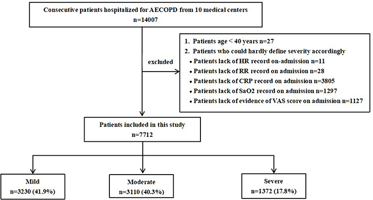
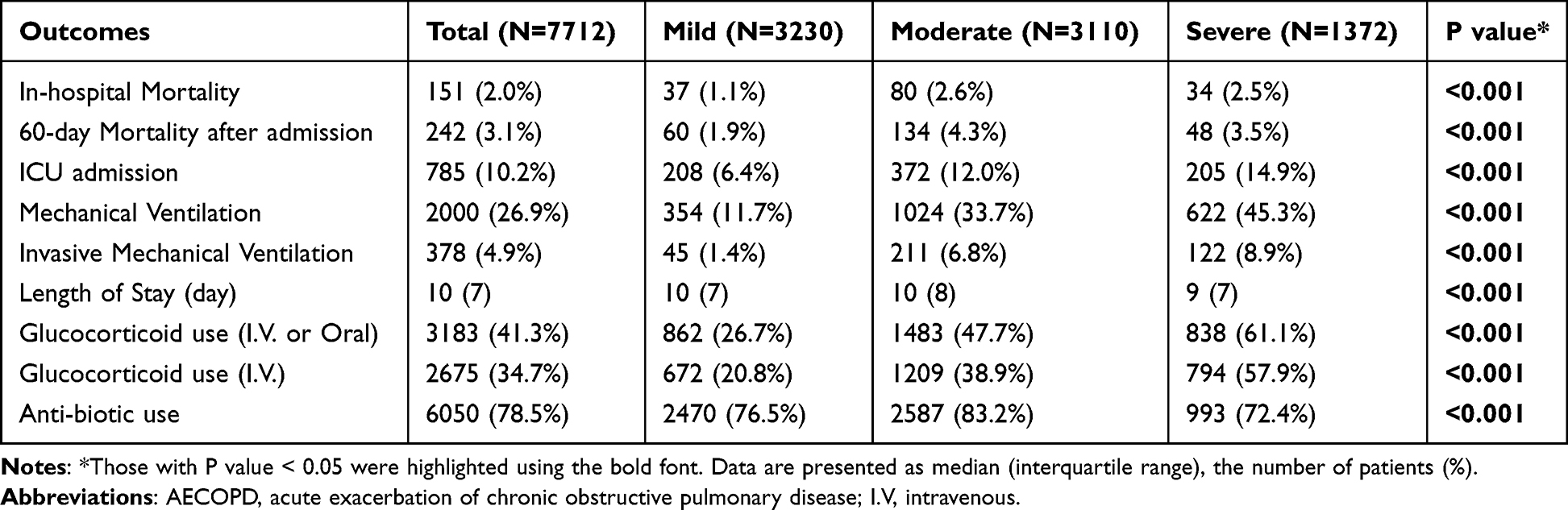
A total of 14,007 patients were consecutively enrolled in the MAGNET AECOPD Registry study. Among them, 7712 were included in this analysis. The main reasons for exclusion were as follows: (1) age less than 40 years (n=27); (2) lacking HR record on admission (n=11); (3) lacking RR record on admission (n=28); (4) lacking CRP record (n=3805); (5) lacking SaO2 record on admission (n=1297); and (6) lacking evidence of VAS on admission (n=1127). The mean age of the population was 72.68±10.61 years, and 77.6% were male. Approximately 18.9% were active smokers. According to the Rome severity classification, 3230 (41.88%) AECOPD inpatients were categorized as mild, 3110 (40.33%) were categorized as moderate and 1372 (17.79%) were categorized as severe. The 60-day mortality and in-hospital mortality rates were 3.1% and 2%, respectively. A total of 785 (10.2%) patients were admitted to the ICU during the hospital stay, 2000 (26.9%) patients received MV, and 378 (4.9%) patients received IMV during their hospital stay. The flow chart of the study is shown in Figure 1.
Figure 1 Flow chart of the study.
Abbreviations: AECOPD, acute exacerbation of chronic obstructive pulmonary disease; HR, heart rate; bpm, beats per minute; RR, respiration rate; CRP, C-reactive protein; SaO2, Arterial Oxygen Saturation; VAS, visual analog scale.
Baseline and Clinical Characteristics of Included AECOPD Patients
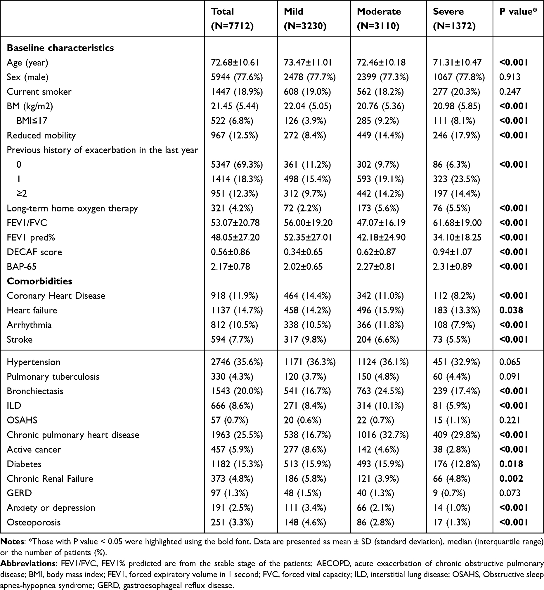
The baseline characteristics are summarized according to the severity of AECOPD based on the Rome classification in Table 1. Gender and current smoker distribution were comparable among groups. It is surprising that the patients who experienced a mild AECOPD event were older, while those who experienced a severe AECOPD event were slightly younger. Patients were more likely to experience reduced mobility, a history of exacerbation in the last year, or accept long-term home oxygen therapy if their severity gradings were classified more severe. A negative association was observed between body mass index (BMI) and severity of AECOPD, as the proportion of BMI≤17 kg/m2 in the mild group were less than those in the moderate or severe group. Additionally, the FEV1 pred% of the mild group was significantly higher compared to the moderate or severe group. Higher DECAF scores and BAP-65 scores were related to more severe severity according to the Rome classification. The incidence of most comorbidities, including coronary heart disease, heart failure, arrhythmia, stroke, bronchiectasis, interstitial lung disease (ILD), chronic pulmonary heart disease, active cancer, diabetes, chronic renal failure, anxiety or depression and osteoporosis, was higher in the mild or moderate group than in the severe group. The prevalence of hypertension, pulmonary tuberculosis, obstructive sleep apnea-hypopnea syndrome (OSAHS) and gastroesophageal reflux disease (GRED) was generally comparable among the groups.
Table 1 Baseline Characteristics of Patients According to AECOPD Severity Based on the Rome Severity Classification
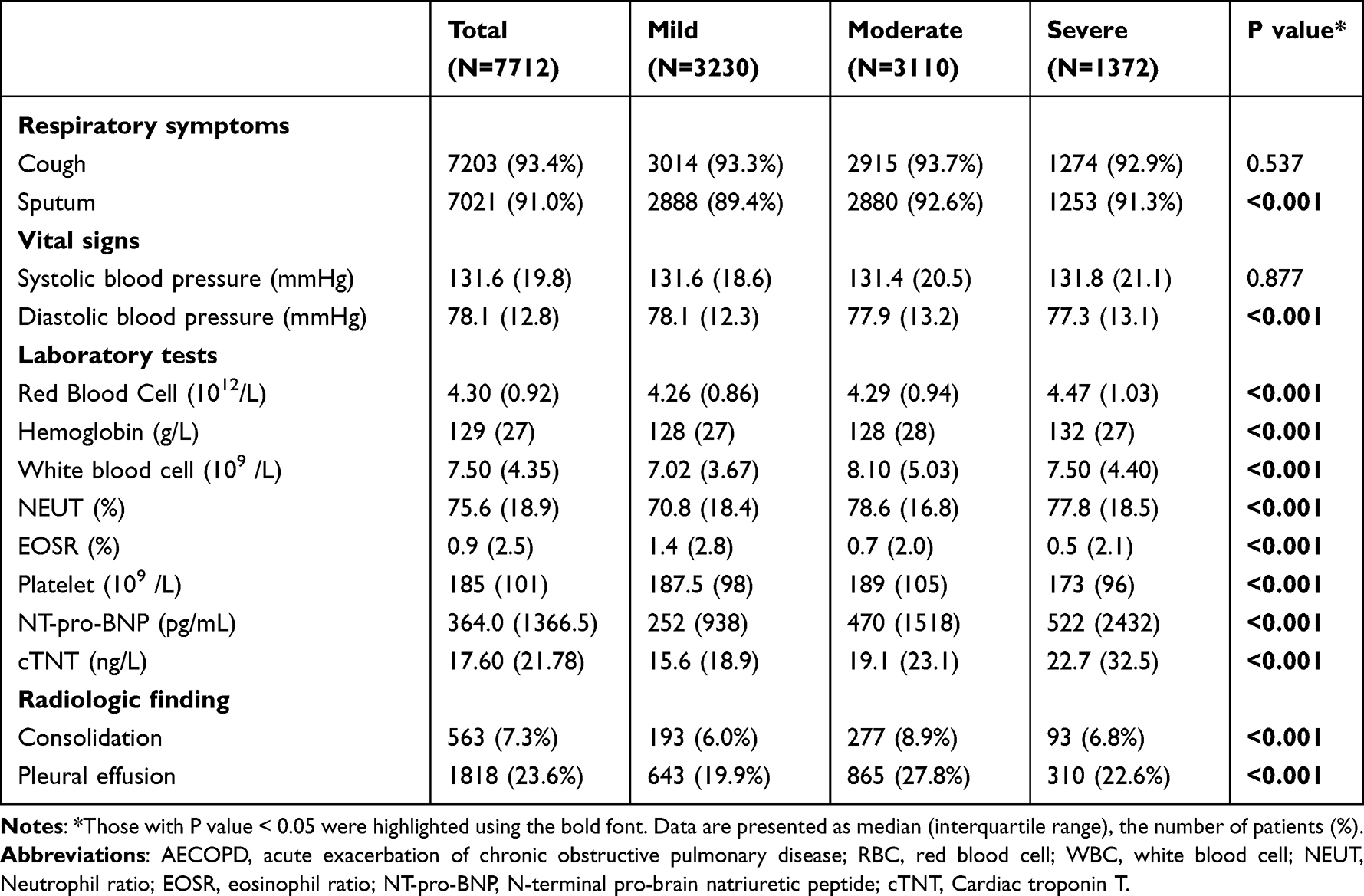
Table 2 shows symptoms, vital signs and laboratory parameters for patients stratified according to the Rome classification. The moderate and severe groups were more likely to have sputum and lower diastolic blood pressure than the mild group. The complete blood counts demonstrated higher red blood cell (RBC), white blood cell (WBC) and neutrophil percentages in the moderate and severe groups compared with the mild group; in contrast, the moderate and severe groups had lower eosinophil percentages than the mild group. The levels of N-terminal pro-B-type natriuretic peptide (NT-pro-BNP) and troponin T (cTNT) were higher in the moderate and severe groups than in the mild group. Radiologic abnormalities, including consolidation and pleural effusion, were more commonly seen in patients classified as moderate.
Table 2 Clinical Features of Patients According to AECOPD Severity Based on the Rome Severity Classification
Variables Included in the Rome Severity Classification
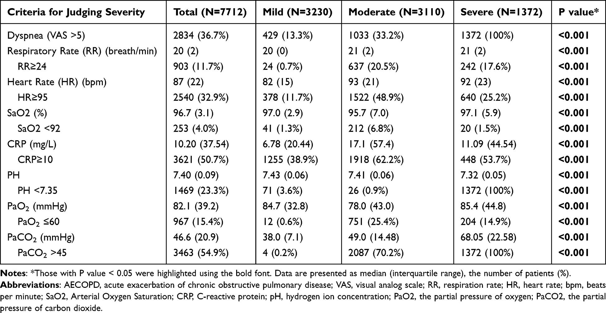
As shown in Tables 3 and S2, patients in the moderate group had significantly faster RR (21 vs 20 breaths/min), HR (93 vs 82 bpm), higher CRP (17.1 vs 6.78 mg/L), PaCO2 (49.0 vs 38.0 mmHg), and proportion of dyspnea with VAS score >5 (33.2% vs 13.3%) as well as lower levels of SaO2 (95.7 vs 97.0%) and PaO2 (78.0 vs 84.7 mmHg) than those in the mild group (all P < 0.001). The value of PaCO2 and proportion of VAS score >5 indicated a significantly worse breathing condition in patients classified as severe compared with those classified as moderate (68.05 vs 49.0 mmHg, 100% vs 33.2%, respectively). However, the value of CRP (11.09 vs 17.1 mg/L) was unexpectedly lower and the level of PaO2 (85.4 vs 78.0 mmHg) was unexpectedly higher in the severe group than in the moderate group.
Table 3 Criteria for Determining AECOPD Severity According to the Rome Classification
Clinical Outcomes According to the Rome Severity Classification
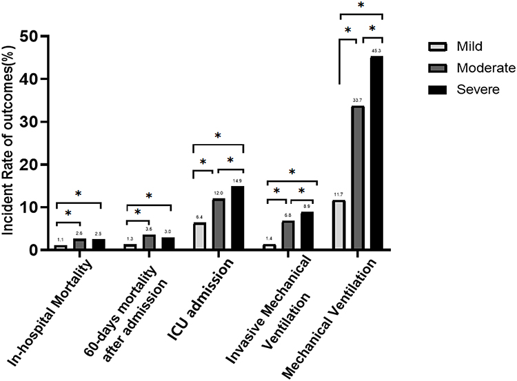
The comparison of outcomes is shown in Table 4, and the multiple comparison is shown in Figure 2. The incidence of ICU admission (6.4% vs 12.0% vs 14.9%, P <0.001), mechanical ventilation (11.7% vs 33.7% vs 45.3%, P <0.001) and invasive mechanical ventilation (1.4% vs 6.8% vs 8.9%, P <0.001) increased with the severity of AECOPD from the mild group to the severe group according to the Rome severity classification. Although the moderate and severe groups had higher 60-day mortality than the mild group (3.5% vs 1.9%, 4.3% vs 1.9%, P <0.05, respectively), mortality in the severe group was very close to that in the moderate group (3.5% vs 4.3%, P >0.05). The results for in-hospital mortality showed the same trend, difference of in-hospital mortality between the moderate and severe groups was not significant (2.5% vs 2.6%, P >0.05) despite the moderate group and severe group had higher mortality than the mild group respectively (2.6% vs 1.1%, 2.5% vs 1.1%, P <0.05). The administration of systemic glucocorticoids increased with the severity of AECOPD from the mild group to the severe group (P<0.001), while the moderate group received antibiotics more often than the mild or severe group.
Table 4 Outcomes According to AECOPD Severity Based on the Rome Classification
Figure 2 Multiple comparison of adverse outcomes in inpatients with varied AECOPD severity according to the Rome classification (*P value<0.05).
Abbreviation: ICU, intensive care unit.
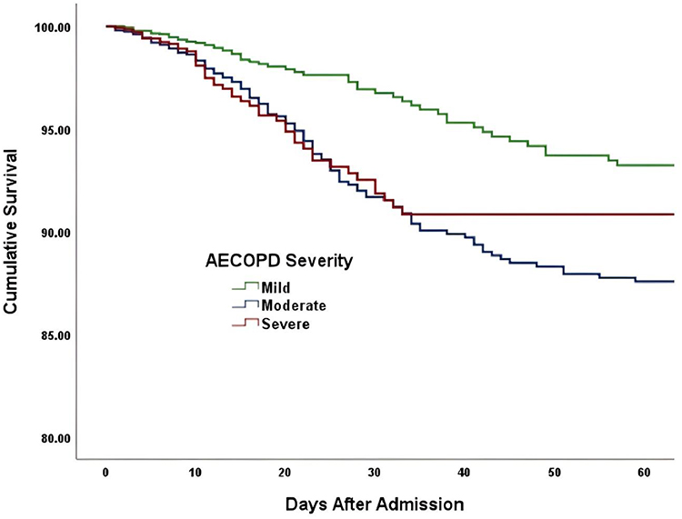
The results of univariate logistic analysis on adverse outcomes in inpatients with AECOPD are shown in Table 5 and Table 6. Similarly, the risk of death at 60 days after admission was significantly higher in the moderate and severe groups than in the mild group (ORs: 2.38 vs 1, 1.92 vs 1.00, respectively, P=0.001), while there was no significant difference in the risk of 60-day mortality between the severe group and the moderate group (ORs: 0.81 vs 1.00, P=0.206). The increase in the Rome severity classification was significantly associated with an increased risk of ICU admission (mild vs moderate vs severe ORs: 1.00 vs 1.97 vs 2.55, P <0.001) and IMV (mild vs moderate vs severe ORs: 1.00 vs 5.15 vs 6.91, P <0.001). The Kaplan–Meier curves also demonstrated similar results (Figure 3). That is, the severe and moderate groups had significantly worse 60-day survival than the mild group (both P <0.05), but the survival was not significantly different between the severe group and the moderate group (P >0.05).
Table 5 Univariate Logistic Analysis on 60-Days Mortality in Inpatients with AECOPD
Table 6 Univariate Logistic Analysis on ICU Admission and Invasive Ventilation Use in Inpatients with AECOPD
Figure 3 Kaplan–Meier estimates in-hospital survival in patients with varied AECOPD severity.
Abbreviation: AECOPD, acute exacerbation of chronic obstructive pulmonary disease.
Discussion
Through a large multicenter cohort of AECOPD patients, we revealed that the Rome severity classification could excellently distinguish the risk of ICU admission, MV and IMV. However, more studies are needed to determine whether it can reliably discriminate the risk of short-term mortality.
Several studies have been published, aiming to validate the Rome severity classification in AECOPD patients. A retrospective study conducted in 200 Spanish inpatients with AECOPD revealed that the Rome classification lacked the capacity to classify the severity of AECOPD compared with the Spanish COPD Guidelines (GesEPOC) classification.11 Carmen et al applied the Rome classification to a cohort of 364 hospitalized patients with AECOPD in the Netherlands and found that the Rome classification can differentiate between exacerbation events with different short-term mortality rates.12 The study conducted by Lee et al in Korea found excellent performance of the Rome classification for predicting ICU admission and the need for noninvasive mechanical ventilation (NIV) or IMV and acceptable performance for predicting in-hospital mortality by comparing the Rome classification with the DECAF score and GesEPOC 2021 criteria.13 Our findings concerning the association between the Rome severity classification and the need for ICU admission and MV were highly consistent with Lee’s findings. However, the Rome classification worked poorly in our cohort compared to Lees and Carmen’s studies in identifying the difference in mortality rates between the moderate and severe groups. The inconsistent results might be attributed to differences in the characteristics of patient cohorts and the geographical setting of different studies. The global variability in the available resources to treat patients with AECOPD and local customs may affect the criteria for hospital visits and admissions and thus may also contribute to the differences in study results. Notably, the three studies mentioned above are all single-center, small-sample studies, which inevitably induces selection bias and weakens the power of these studies. In the present study, we consecutively included unselected inpatients with AECOPD from 10 tertiary general hospitals in China, which should represent real-world situations.
The physiologic parameters (VAS, HR, RR, SaO2) and inflammatory biomarkers (CRP) included in the Rome severity classification are all easy to obtain and have been tested as prognostic factors of AECOPD in some previous studies. The VAS offers the benefit of quantitatively representing ventilatory demand on a scale from 0 to 10,14 and the scale has been validated against respiratory loads in patients with COPD.15 The proportion of VAS scores >5 increased with disease severity in our cohort. Several studies showed that an elevated HR or RR was associated with exacerbation,16,17 readmission9,18 and correspondingly early mortality in COPD patients.19,20 We also found faster HR and RR in the moderate and severe groups than in the mild group in the study. Furthermore, the two variables could be measured easily and noninvasively in the clinic.21 It is not difficult to foresee that the two variables could facilitate the application of the Rome classification in the management of COPD, as smartphones and wearable device technology are gradually used to facilitate early detection and treatment of AECOPD.22,23 CRP, an acute-phase protein that can be measured accurately within minutes at the point of care, is a biomarker for assessing AECOPD since elevated CRP is associated with the need for antibiotics and higher mortality.24,25 Although using CRP as a marker of airway inflammation may lack specificity, CRP is widely recognized as a useful and sensitive marker of infections and AECOPD.26 SaO2 is a reflection of gas exchange in AECOPD patients. It is more practical and widely available to measure pulse oximetry in all clinical settings, although SaO2 is less reliable than arterial blood gas analysis. Some studies showed that a reduction in oxygen saturation was associated with AECOPD risk;23,27 unfortunately, the change in SaO2 from baseline was not used to distinguish mild versus moderate events because of its unavailability in this post-hoc analysis. Therefore, future studies are needed to validate the reduction in SaO2 in assessing the severity of AECOPD. Acute respiratory failure with hypoxemia and/or hypercapnia and acidosis is the first and possibly most important clinical feature observed in patients with severe AECOPD,28 and the mortality risk is higher at lower pH values.29 Based on this fact, arterial blood gas analysis was the only criterion that was used to determine the severe group in the Rome classification. Theoretically, the incorporation of objective, easy and ready-to-measure variables in this proposed classification may assist in a better delineation of clinically different AECOPD.
However, the Rome classification should not be considered as a complete version. First, the criteria included in the severity classification are single-minded based on a review of the literature and discussion. ABG alone may not be sufficient to determine severe AECOPD events. It is unquestionable that COPD is a heterogeneous illness underpinned by diverse clinical characteristics and pathophysiological mechanisms.30 Although there are several markers that potentially indicate AECOPD severity, none have received widespread acceptance. As stated by Ramakrishnan et al, the different endotypes of AECOPD severity cannot be assessed by solely focusing on inflammatory or pathophysiological parameters.31 Our research team previously discovered that blood urea nitrogen (BUN), a component of BAP-65, was linked to an increased risk of in-hospital death and other adverse outcomes in AECOPD inpatients.6,32 Moreover, the baseline parameters, especially dyspnea in the stable stage, may have an impact on how this exacerbation may turn out.33,34 Comorbidities like heart failure, arrhythmia, coronary heart disease, and diabetes, which are highly prevalent in COPD patients, also contribute to event severity in the real-life clinical setting. Therefore, baseline parameters and the existence of comorbidities may also need to be considered to incorporate into the severity assessment for AECOPD. Second, the threshold setting seems to be arbitrary. Taking CRP as an example, in our research, serum CRP levels were interestingly the highest in moderate events, with 62.2% of patients having a CRP above 10 mg/L. Similar results were also found by the two studies mentioned earlier and conducted in the Netherlands and Korea, respectively.12,13 This revealed that a cutoff value of 10 mg/L may lack specificity to distinguish a moderate event from a severe one, and the mechanism linking CRP to the exacerbations of COPD may be complex and requires further researches. Probably due to the two flaws mentioned above, the Rome classification is valid in discriminating between mild and more severe AECOPD but fails to distinguish between moderate and severe AECOPD. Last, it is currently difficult to suggest optimal medical treatments according to AECOPD severity based on the Rome classification. In this cohort study, patients admitted with mild Rome AECOPD were slightly older and suffered from more comorbidities, suggesting that these factors, rather than the severity of acute respiratory events, contributed to the indication for hospitalization. Therefore, whether the Rome classification could be used as an indicator for hospitalization for AECOPD is debated. In addition, patients in the severe group needed more ICU admissions and mechanical ventilation than those in the moderate group, while the mortality did not increase accordingly. A possible explanation is that the strengthened treatments received by severe AECOPD patients may contribute to an improve in short-term survival; a more likely explanation is that this classification does lack a distinction between moderate and high mortality risks because of the flaws mentioned above. Consequently, the Rome classification needs to be optimized in the selection of variables and corresponding thresholds based on prospectively studies.
To our knowledge, this is the first large-scale multicenter cohort study to validate the Rome classification. Moreover, the consecutive inclusion of unselected inpatients with AECOPD and comprehensive collection of information in our study ensured high data quality and true associations between the Rome classification and risk of adverse outcomes in the real-world setting. Importantly, this study provided important initial insights into the distinctive value of the proposed classification in the Chinese population, and the Rome severity classification is the first step to differentiate clinically different AECOPD. Nevertheless, our study has several limitations. First, the VAS score was retrospectively evaluated based on the medical records and nurse’s description of the severity of dyspnea on admission, which inevitably leads to a bias. Fortunately, not all patients require a VAS score to be classified, and only those who already meet two out of the 5 moderate criteria need a VAS score to determine whether they are moderate. In addition, an exact VAS score was not necessary; alternatively, we only needed to evaluate whether the patient had significant dyspnea (VAS≥5 vs <5), which facilitates the accuracy of this retrospective evaluation based on detailed medical records on admission. Second, our study only allows the applicability of the Rome classification to be assessed in the hospital setting and not in the primary care setting, and how the Rome classification works in outpatient clinics or communities is still unclear. Finally, the exclusion of patients because of missing data may result in selection biases and affect the external validity of the results. But the original data is from an noninterventional cohort study, and the arrangement of auxiliary examinations and treatment of patients were at the discretion of the attending physicians, which should reflect the real-world application circumstance of Roma classification in China. Additionally, we found that the excluded patients tend to have milder conditions, with lower mortality rates (data not shown) and a greater likelihood of being classified to the mild group. So even if these patients were included, we believe the results would hardly change. Thus, optimization and prospective validation of the classification and a deeper exploration of its prognostic applicability in making therapeutic decisions remain pending.
Conclusion
It is essential for an AECOPD severity assessment tool to accurately identify patients at high risk for adverse outcomes. In this large cohort study, the Rome severity classification demonstrated excellent performance in predicting ICU admission and the need for MV or IMV but failed to distinguish the risk of short-time mortality between the moderate and severe groups. Studies are warranted to validate whether it could accurately evaluate the risk of mortality, and further optimization may still be needed before clinical application.
Abbreviations
AECOPD, Acute exacerbations of chronic obstructive pulmonary disease; COPD, Chronic obstructive pulmonary disease; GOLD, Global Initiative for Chronic Obstructive Lung Disease; MAGNET AECOPD, MAnaGement aNd advErse ouTcomes in inpatients with acute exacerbation of COPD) Registry; FEV1/FVC, forced expiratory volume in 1-second/forced vital capacity ratio; HR, heart rate; bpm, beats per minute; RR, respiration rate; CRP, C-reactive protein; VAS, visual analog scale; PaCO2, arterial carbon dioxide tension; pH, hydrogen ion concentration; PO2, arterial oxygen tension; SaO2, Arterial Oxygen Saturation; ICU, intensive care unit; MV, mechanical ventilation; IMV, invasive mechanical ventilation; LOS, length of stay; SD, standard deviation; ANOVA, one-way analysis of variance; IQR, interquartile range; ORs, odds ratios; 95% Cis, 95% confidence intervals; SPSS, Statistic Package for Social Science; BMI, body mass index; Kg/m2, kilogram per square meter; ILD, interstitial lung disease; LTOT, Long-term home oxygen therapy; RBC, red blood cell; WBC, White blood cell; NEUT, neutrophil percentage; EOSR, Percentage of eosinophils; SBP, Systolic blood pressure; DBP, diastolic blood pressure; NT-pro-BNP, N-terminal pro-brain natriuretic peptide; cTNT, Cardiac troponin T; NIV, noninvasive mechanical ventilation; GesEPOC, Spanish COPD Guidelines; BUN, serum urea nitrogen.
Data Sharing Statement
The data will be shared on reasonable request to the corresponding author.
Ethics Approval and Informed Consent
This study was approved by the Ethics Committee of the ten academic medical centers that participated. Written informed consent was obtained from all the participants. This study complies with the Declaration of Helsinki.
Funding
This study was supported by the National Natural Science Foundation of China (82370021), the Sichuan Science and Technology Program (2022YFS0262), the Suzhou Collaborative Medical Health Foundation (Y117) and National Key Research Program of China (2016YFC1304202).
Disclosure
The authors report no conflicts of interest in this work.
References
1. Adeloye D, Song P, Zhu Y, et al. Global, regional, and national prevalence of, and risk factors for, chronic obstructive pulmonary disease (COPD) in 2019: a systematic review and modelling analysis. Lancet Respir Med. 2022;10(5):447–458.
2. Halpin DMG, Decramer M, Celli BR, Mueller A, Metzdorf N, Tashkin DP. Effect of a single exacerbation on decline in lung function in COPD. Respir Med. 2017;128:85–91. doi:10.1016/j.rmed.2017.04.013
3. Hurst JR, Skolnik N, Hansen GJ, et al. Understanding the impact of chronic obstructive pulmonary disease exacerbations on patient health and quality of life. Eur J Intern Med. 2020;73:1–6. doi:10.1016/j.ejim.2019.12.014
4. A global strategy for prevention, diagnosis and management of COPD: 2022 report. Available from: goldcopd.org/2022-gold-reports-2/. Accessed January9, 2024.
5. Kim V, Aaron SD. What is a COPD exacerbation? Current definitions, pitfalls, challenges and opportunities for improvement. Europ Resp J. 2018;52(5):1801261. doi:10.1183/13993003.01261-2018
6. Tabak YP, Sun X, Johannes RS, Gupta V, Shorr AF. Mortality and need for mechanical ventilation in acute exacerbations of chronic obstructive pulmonary disease: development and validation of a simple risk score. Arch Intern Med. 2009;169(17):1595–1602. doi:10.1001/archinternmed.2009.270
7. Echevarria C, Steer J, Heslop-Marshall K, et al. Validation of the DECAF score to predict hospital mortality in acute exacerbations of COPD. Thorax. 2016;71(2):133–140. doi:10.1136/thoraxjnl-2015-207775
8. Huang Q, He C, Xiong H, et al. DECAF score as a mortality predictor for acute exacerbation of chronic obstructive pulmonary disease: a systematic review and meta-analysis. BMJ Open. 2020;10(10):e037923. doi:10.1136/bmjopen-2020-037923
9. Hawthorne G, Richardson M, Greening NJ, et al. A proof of concept for continuous, non-invasive, free-living vital signs monitoring to predict readmission following an acute exacerbation of COPD: a prospective cohort study. Respir Res. 2022;23(1):102. doi:10.1186/s12931-022-02018-5
10. Celli BR, Fabbri LM, Aaron SD, et al. An updated definition and severity classification of chronic obstructive pulmonary disease exacerbations: the Rome proposal. Am J Respir Crit Care Med. 2021;204(11):1251–1258. doi:10.1164/rccm.202108-1819PP
11. Amado Diago CA, Figueira Goncalves JM, Golpe R, Esteban C, Garcia Talavera I, Garcia-Martin S. Classification of the severity of COPD exacerbations in hospitalized patients according to Rome vs GesEPOC criteria. Arch Bronconeumol. 2023;59(1):57–58. doi:10.1016/j.arbres.2022.06.009
12. Reumkens C, Endres A, Simons SO, Savelkoul PHM, Sprooten RTM, Franssen FME. Application of the Rome severity classification of COPD exacerbations in a real-world cohort of hospitalised patients. ERJ Open Res. 2023;9(3):00569–2022. doi:10.1183/23120541.00569-2022
13. Lee HJ, Lee JK, Park TY, Heo EY, Kim DK, Lee HW. Validation of the Rome proposal for severity of acute exacerbation of chronic obstructive pulmonary disease. Ther Adv Respir Dis. 2023;17:17534666231172917. doi:10.1177/17534666231172917
14. Gift AG, Narsavage G. Validity of the numeric rating scale as a measure of dyspnea. Am J Crit Care. 1998;7:200–204. doi:10.4037/ajcc1998.7.3.200
15. Noell G, Cosio BG, Faner R, et al. Multi-level differential network analysis of COPD exacerbations. Europ Resp J. 2017;50(3):1700075. doi:10.1183/13993003.00075-2017
16. Burton C, Pinnock H, McKinstry B. Changes in telemonitored physiological variables and symptoms prior to exacerbations of chronic obstructive pulmonary disease. J Telemed Telecare. 2015;21(1):29–36. doi:10.1177/1357633X14562733
17. Borel JC, Pelletier J, Taleux N, et al. Parameters recorded by software of non-invasive ventilators predict COPD exacerbation: a proof-of-concept study. Thorax. 2015;70(3):284–285. doi:10.1136/thoraxjnl-2014-206569
18. Flattet Y, Garin N, Serratrice J, Perrier A, Stirnemann J, Carballo S. Determining prognosis in acute exacerbation of COPD. Int J Chron Obstruct Pulmon Dis. 2017;12:467–475. doi:10.2147/COPD.S122382
19. Jensen MT, Marott JL, Lange P, et al. Resting heart rate is a predictor of mortality in COPD. Europ Resp J. 2013;42(2):341–349. doi:10.1183/09031936.00072212
20. Fermont JM, Masconi KL, Jensen MT, et al. Biomarkers and clinical outcomes in COPD: a systematic review and meta-analysis. Thorax. 2019;74(5):439–446. doi:10.1136/thoraxjnl-2018-211855
21. Elvekjaer M, Aasvang EK, Olsen RM, et al. Physiological abnormalities in patients admitted with acute exacerbation of COPD: an observational study with continuous monitoring. J Clin Monit Comput. 2020;34(5):1051–1060. doi:10.1007/s10877-019-00415-8
22. Smith HS, Criner AJ, Fehrle D, Grabianowski CL, Jacobs MR, Criner GJ. Use of a SmartPhone/tablet-based bidirectional telemedicine disease management program facilitates early detection and treatment of COPD exacerbation symptoms. Telemed J E Health. 2016;22(5):395–399. doi:10.1089/tmj.2015.0135
23. Al Rajeh AM, Aldabayan YS, Aldhahir A, et al. Once daily versus overnight and symptom versus physiological monitoring to detect exacerbations of chronic obstructive pulmonary disease: pilot randomized controlled trial. JMIR mHealth uHealth. 2020;8(11):e17597. doi:10.2196/17597
24. Hurst JR, Donaldson GC, Perera WR, et al. Use of plasma biomarkers at exacerbation of chronic obstructive pulmonary disease. Am J Respir Crit Care Med. 2006;174(8):867–874. doi:10.1164/rccm.200604-506OC
25. Prins HJ, Duijkers R, van der Valk P, et al. CRP-guided antibiotic treatment in acute exacerbations of COPD in hospital admissions. Europ Resp J. 2019;53(5). doi:10.1183/13993003.02014-2018
26. Dev D, Wallace E, Sankaran R, et al. Value of C-reactive protein measurements in exacerbations of chronic obstructive pulmonary disease. Respir Med. 1998;92(4):664–667. doi:10.1016/S0954-6111(98)90515-7
27. Hurst JR, Donaldson GC, Quint JK, et al. Domiciliary pulse-oximetry at exacerbation of chronic obstructive pulmonary disease prospective pilot study. BMC Pulm Med. 2010;10(1):1. doi:10.1186/1471-2466-10-52
28. Bruno CM, Valenti M. Acid-base disorders in patients with chronic obstructive pulmonary disease: a pathophysiological review. J Biomed Biotechnol. 2012;2012:915150. doi:10.1155/2012/915150
29. Ucgun I, Oztuna F, Dagli CE, Yildirim H, Bal C. Relationship of metabolic alkalosis, azotemia and morbidity in patients with chronic obstructive pulmonary disease and hypercapnia. Respiration. 2008;76(3):270–274. doi:10.1159/000131707
30. Agusti A, Calverley PM, Celli B, et al. Evaluation of COPD Longitudinally to Identify Predictive Surrogate Endpoints (ECLIPSE) Investigators. Characterisation of COPD heterogeneity in the ECLIPSE cohort. Respir Res. 2010;11:122. doi:10.1186/1465-9921-11-122
31. Ramakrishnan S, Gyselinck I, Bafadhel M, Janssens W. Chronic obstructive pulmonary disease exacerbations: do all roads lead to Rome? Am J Respir Crit Care Med. 2022;205(9):1125–1126. doi:10.1164/rccm.202112-2717LE
32. Zhang J, Qin Y, Zhou C, et al. Elevated BUN upon admission as a predictor of in-hospital mortality among patients with acute exacerbation of COPD: a Secondary Analysis of Multicenter Cohort Study. Int J Chron Obstruct Pulmon Dis. 2023;18:1445–1455. doi:10.2147/COPD.S412106
33. Steer J, Norman EM, Afolabi OA, Gibson GJ, Bourke SC. Dyspnoea severity and pneumonia as predictors of in-hospital mortality and early readmission in acute exacerbations of COPD. Thorax. 2012;67(2):117–121. doi:10.1136/thoraxjnl-2011-200332
34. Steer J, Gibson J, Bourke SC. The DECAF score: predicting hospital mortality in exacerbations of chronic obstructive pulmonary disease. Thorax. 2012;67(11):970–976. doi:10.1136/thoraxjnl-2012-202103
Orgiazzi, A. et al. 454 pyrosequencing analysis of fungal assemblages from geographically distant, disparate soils reveals spatial patterning and a core mycobiome. Diversity5, 73–98 (2013).
Toma, I. et al. Single-molecule long-read 16s sequencing to characterize the lung microbiome from mechanically ventilated patients with suspected pneumonia. J. Clin. Microbiol.52, 3913–3921 (2014).
Dickson, R. P. et al. Spatial variation in the healthy human lung microbiome and the adapted island model of lung biogeography. Ann. Am. Thorac. Soc.12, 821–830 (2015).
Yun, Y. et al. Environmentally determined differences in the murine lung microbiota and their relation to alveolar architecture. PLoS ONE9, e113466 (2014).
Wang, Z. et al. Inflammatory endotype-associated airway microbiome in chronic obstructive pulmonary disease clinical stability and exacerbations: a multicohort longitudinal analysis. Am. J. Respir. Crit. Care Med.203, 1488–1502 (2021).
Einarsson, G. G. et al. Community dynamics and the lower airway microbiota in stable chronic obstructive pulmonary disease, smokers and healthy non-smokers. Thorax71, 795–803 (2016).
Dickson, R. P. et al. Enrichment of the lung microbiome with gut bacteria in sepsis and the acute respiratory distress syndrome. Nat. Microbiol.1, 16113 (2016).
Beck, J. M. et al. Multicenter comparison of lung and oral microbiomes of hiv-infected and hiv-uninfected individuals. Am. J. Respir. Crit. Care Med.192, 1335–1344 (2015).
Baker, J. M. et al. Whole lung tissue is the preferred sampling method for amplicon-based characterization of murine lung microbiota. Microbiome9, 99 (2021).
Pattaroni, C. et al. Early-life formation of the microbial and immunological environment of the human airways. Cell Host Microbe24, 857–865.e854 (2018).
Liu, H. X. et al. Difference of lower airway microbiome in bilateral protected specimen brush between lung cancer patients with unilateral lobar masses and control subjects. Int. J. Cancer142, 769–778 (2018).
Noverr, M. C., Noggle, R. M., Toews, G. B. & Huffnagle, G. B. Role of antibiotics and fungal microbiota in driving pulmonary allergic responses. Infect. Immun.72, 4996–5003 (2004).
Gore, C. et al. Bifidobacterium pseudocatenulatum is associated with atopic eczema: a nested case-control study investigating the fecal microbiota of infants. J. Allergy Clin. Immunol.121, 135–140 (2008).
Fodor, A. A. et al. The adult cystic fibrosis airway microbiota is stable over time and infection type, and highly resilient to antibiotic treatment of exacerbations. PLoS ONE7, e45001 (2012).
Pragman, A. A., Kim, H. B., Reilly, C. S., Wendt, C. & Isaacson, R. E. The lung microbiome in moderate and severe chronic obstructive pulmonary disease. PLoS ONE7, e47305 (2012).
Huang, Y. J. et al. Airway microbiota and bronchial hyperresponsiveness in patients with suboptimally controlled asthma. J. Allergy Clin. Immunol.127, 372–381 e371-373 (2011).
Boyton, R. J., Reynolds, C. J., Quigley, K. J. & Altmann, D. M. Immune mechanisms and the impact of the disrupted lung microbiome in chronic bacterial lung infection and bronchiectasis. Clin. Exp. Immunol.171, 117–123 (2013).
Lobo, L. J. & Noone, P. G. Respiratory infections in patients with cystic fibrosis undergoing lung transplantation. Lancet Respir. Med.2, 73–82 (2014).
Segal, L. N. & Dickson, R. P. The lung microbiome in hiv. Getting to the haart of the host-microbe interface. Am. J. Respir. Crit. Care Med.194, 136–137 (2016).
Huang, Y. et al. Microbes are associated with host innate immune response in idiopathic pulmonary fibrosis. Am. J. Respir. Crit. Care Med.196, 208–219 (2017).
O’Dwyer, D. N. et al. Lung dysbiosis, inflammation, and injury in hematopoietic cell transplantation. Am. J. Respir. Crit. Care Med.198, 1312–1321 (2018).
McVerry, B. J. & Morris, A. The lung microbiome in hematopoietic stem cell transplant. Where the money lies. Am. J. Respir. Crit. Care Med.198, 1249–1251 (2018).
Kullberg, R. F. J. et al. Lung microbiota of critically ill patients with covid-19 are associated with nonresolving acute respiratory distress syndrome. Am. J. Respir. Crit. Care Med.206, 846–856 (2022).
Whiteside, S. A., Razvi, H., Dave, S., Reid, G. & Burton, J. P. The microbiome of the urinary tract–a role beyond infection. Nat. Rev. Urol.12, 81–90 (2015).
Huffnagle, G. B., Dickson, R. P. & Lukacs, N. W. The respiratory tract microbiome and lung inflammation: a two-way street. Mucosal Immunol.10, 299–306 (2017).
Lagkouvardos, I., Overmann, J. & Clavel, T. Cultured microbes represent a substantial fraction of the human and mouse gut microbiota. Gut Microbes8, 493–503 (2017).
Charlson, E. S. et al. Topographical continuity of bacterial populations in the healthy human respiratory tract. Am. J. Respir. Crit. Care Med.184, 957–963 (2011).
Segal, L. N. et al. Enrichment of the lung microbiome with oral taxa is associated with lung inflammation of a th17 phenotype. Nat. Microbiol.1, 16031 (2016).
Dickson, R. P. et al. The lung microbiota of healthy mice are highly variable, cluster by environment, and reflect variation in baseline lung innate immunity. Am. J. Respir. Crit. Care Med.198, 497–508 (2018).
Lim, Y. W. et al. Clinical insights from metagenomic analysis of sputum samples from patients with cystic fibrosis. J. Clin. Microbiol.52, 425–437 (2014).
Delhaes, L. et al. The airway microbiota in cystic fibrosis: A complex fungal and bacterial community–implications for therapeutic management. PLoS ONE7, e36313 (2012).
Sharma, A. et al. Associations between fungal and bacterial microbiota of airways and asthma endotypes. J. Allergy Clin. Immunol.144, 1214–1227.e1217 (2019).
Charlson, E. S. et al. Assessing bacterial populations in the lung by replicate analysis of samples from the upper and lower respiratory tracts. PLoS ONE7, e42786 (2012).
Hulin, M. et al. Positive associations between respiratory outcomes and fungal index in rural inhabitants of a representative sample of french dwellings. Int. J. Hyg. Environ. Health216, 155–162 (2013).
Dupuy, A. K. et al. Redefining the human oral mycobiome with improved practices in amplicon-based taxonomy: discovery of malassezia as a prominent commensal. PLoS ONE9, e90899 (2014).
Chen, T. C., Chen, Y. H., Chen, Y. C. & Lu, P. L. Fluconazole exposure rather than clonal spreading is correlated with the emergence of candida glabrata with cross-resistance to triazole antifungal agents. Kaohsiung J. Med. Sci.28, 306–315 (2012).
Bousquet, A. et al. An 8-year survey of strains identified in blood cultures in a clinical haematology unit. Clin. Microbiol. Infect.20, O7–O12 (2014).
Pride, D. T. et al. Evidence of a robust resident bacteriophage population revealed through analysis of the human salivary virome. ISME J.6, 915–926 (2012).
Lysholm, F. et al. Characterization of the viral microbiome in patients with severe lower respiratory tract infections, using metagenomic sequencing. PLoS ONE7, e30875 (2012).
Willner, D. et al. Metagenomic analysis of respiratory tract DNA viral communities in cystic fibrosis and non-cystic fibrosis individuals. PLoS ONE4, e7370 (2009).
van den Bergh, M. R. et al. Associations between pathogens in the upper respiratory tract of young children: Interplay between viruses and bacteria. PLoS ONE7, e47711 (2012).
Abbas, A. A. et al. Redondoviridae, a family of small, circular DNA viruses of the human oro-respiratory tract associated with periodontitis and critical illness. Cell Host Microbe25, 719–729.e714 (2019).
Wang, Y. et al. Metagenomic analysis of viral genetic diversity in respiratory samples from children with severe acute respiratory infection in china. Clin. Microbiol. Infect.22, 458.e451–458.e459 (2016).
Popgeorgiev, N., Temmam, S., Raoult, D. & Desnues, C. Describing the silent human virome with an emphasis on giant viruses. Intervirology56, 395–412 (2013).
Young, J. C. et al. Viral metagenomics reveal blooms of anelloviruses in the respiratory tract of lung transplant recipients. Am. J. Transpl.15, 200–209 (2015).
Stewart, C. J. et al. Respiratory syncytial virus and rhinovirus bronchiolitis are associated with distinct metabolic pathways. J. Infect. Dis.217, 1160–1169 (2018).
Barton, E. S., White, D. W. & Virgin, H. W. Herpesvirus latency and symbiotic protection from bacterial infection. Viral Immunol.22, 3–4,author reply 5-6 (2009).
Dickson, R. P., Erb-Downward, J. R. & Huffnagle, G. B. Towards an ecology of the lung: New conceptual models of pulmonary microbiology and pneumonia pathogenesis. Lancet Respir. Med.2, 238–246 (2014).
Abeles, S. R., Ly, M., Santiago-Rodriguez, T. M. & Pride, D. T. Effects of long term antibiotic therapy on human oral and fecal viromes. PLoS ONE10, e0134941 (2015).
Noverr, M. C., Falkowski, N. R., McDonald, R. A., McKenzie, A. N. & Huffnagle, G. B. Development of allergic airway disease in mice following antibiotic therapy and fungal microbiota increase: role of host genetics, antigen, and interleukin-13. Infect. Immun.73, 30–38 (2005).
Mukherjee, P. K. et al. Oral mycobiome analysis of hiv-infected patients: Identification of pichia as an antagonist of opportunistic fungi. PLoS Pathog.10, e1003996 (2014).
Bassis, C. M. et al. Analysis of the upper respiratory tract microbiotas as the source of the lung and gastric microbiotas in healthy individuals. mBio6, e00037 (2015).
Dickson, R. P., Erb-Downward, J. R. & Huffnagle, G. B. Homeostasis and its disruption in the lung microbiome. Am. J. Physiol. Lung Cell Mol. Physiol.309, L1047–L1055 (2015).
Saeedi, P., Salimian, J., Ahmadi, A. & Imani Fooladi, A. A. The transient but not resident (tbnr) microbiome: a yin yang model for lung immune system. Inhal. Toxicol.27, 451–461 (2015).
Wu, H. et al. Surfactant proteins a and d inhibit the growth of gram-negative bacteria by increasing membrane permeability. J. Clin. Invest.111, 1589–1602 (2003).
Larsen, J. M. et al. Divergent pro-inflammatory profile of human dendritic cells in response to commensal and pathogenic bacteria associated with the airway microbiota. PLoS ONE7, e31976 (2012).
van den Bogert, B., Meijerink, M., Zoetendal, E. G., Wells, J. M. & Kleerebezem, M. Immunomodulatory properties of streptococcus and veillonella isolates from the human small intestine microbiota. PLoS ONE9, e114277 (2014).
Thorburn, A. N. et al. Evidence that asthma is a developmental origin disease influenced by maternal diet and bacterial metabolites. Nat. Commun.6, 7320 (2015).
Tan, L., Wang, H., Li, C. & Pan, Y. 16s rdna-based metagenomic analysis of dental plaque and lung bacteria in patients with severe acute exacerbations of chronic obstructive pulmonary disease. J. Periodontal Res.49, 760–769 (2014).
To, K. K. et al. Temporal profiles of viral load in posterior oropharyngeal saliva samples and serum antibody responses during infection by sars-cov-2: an observational cohort study. Lancet Infect. Dis.20, 565–574 (2020).
Muhlebach, M. S. et al. Anaerobic bacteria cultured from cystic fibrosis airways correlate to milder disease: a multisite study. Eur. Respir. J.52, 1800242 (2018).
Duytschaever, G. et al. Cross-sectional and longitudinal comparisons of the predominant fecal microbiota compositions of a group of pediatric patients with cystic fibrosis and their healthy siblings. Appl Environ. Microbiol.77, 8015–8024 (2011).
Ormerod, K. L. et al. Genomic characterization of the uncultured bacteroidales family s24-7 inhabiting the guts of homeothermic animals. Microbiome4, 36 (2016).
Depner, M. et al. Maturation of the gut microbiome during the first year of life contributes to the protective farm effect on childhood asthma. Nat. Med.26, 1766–1775 (2020).
Tang, J., Xu, L., Zeng, Y. & Gong, F. Effect of gut microbiota on lps-induced acute lung injury by regulating the tlr4/nf-kb signaling pathway. Int. Immunopharmacol.91, 107272 (2021).
Jeyanathan, M. et al. Parenteral bcg vaccine induces lung-resident memory macrophages and trained immunity via the gut-lung axis. Nat. Immunol.23, 1687–1702 (2022).
Mirkovic, B. et al. The role of short-chain fatty acids, produced by anaerobic bacteria, in the cystic fibrosis airway. Am. J. Respir. Crit. Care Med.192, 1314–1324 (2015).
Evans, C. R. et al. Untargeted lc-ms metabolomics of bronchoalveolar lavage fluid differentiates acute respiratory distress syndrome from health. J. Proteome Res.13, 640–649 (2014).
Southam, D. S., Dolovich, M., O’Byrne, P. M. & Inman, M. D. Distribution of intranasal instillations in mice: effects of volume, time, body position, and anesthesia. Am. J. Physiol. Lung Cell Mol. Physiol.282, L833–L839 (2002).
Deriu, E. et al. Influenza virus affects intestinal microbiota and secondary salmonella infection in the gut through type i interferons. PLoS Pathog.12, e1005572 (2016).
Roussos, A., Koursarakos, P., Patsopoulos, D., Gerogianni, I. & Philippou, N. Increased prevalence of irritable bowel syndrome in patients with bronchial asthma. Respir. Med.97, 75–79 (2003).
Rutten, E. P. A., Lenaerts, K., Buurman, W. A. & Wouters, E. F. M. Disturbed intestinal integrity in patients with copd: effects of activities of daily living. Chest145, 245–252 (2014).
Yazar, A. et al. Respiratory symptoms and pulmonary functional changes in patients with irritable bowel syndrome. Am. J. Gastroenterol.96, 1511–1516 (2001).
Martens, E. C., Neumann, M. & Desai, M. S. Interactions of commensal and pathogenic microorganisms with the intestinal mucosal barrier. Nat. Rev. Microbiol.16, 457–470 (2018).
Rakoff-Nahoum, S., Paglino, J., Eslami-Varzaneh, F., Edberg, S. & Medzhitov, R. Recognition of commensal microflora by toll-like receptors is required for intestinal homeostasis. Cell118, 229–241 (2004).
Payne, M. S. et al. Molecular microbiological characterization of preterm neonates at risk of bronchopulmonary dysplasia. Pediatr. Res.67, 412–418 (2010).
Stressmann, F. A. et al. The use of culture-independent tools to characterize bacteria in endo-tracheal aspirates from pre-term infants at risk of bronchopulmonary dysplasia. J. Perinat. Med.38, 333–337 (2010).
Mourani, P. M., Harris, J. K., Sontag, M. K., Robertson, C. E. & Abman, S. H. Molecular identification of bacteria in tracheal aspirate fluid from mechanically ventilated preterm infants. PLoS ONE6, e25959 (2011).
Lohmann, P. et al. The airway microbiome of intubated premature infants: characteristics and changes that predict the development of bronchopulmonary dysplasia. Pediatr. Res.76, 294–301 (2014).
Lauder, A. P. et al. Comparison of placenta samples with contamination controls does not provide evidence for a distinct placenta microbiota. Microbiome4, 29 (2016).
Biesbroek, G. et al. Early respiratory microbiota composition determines bacterial succession patterns and respiratory health in children. Am. J. Respir. Crit. Care Med.190, 1283–1292 (2014).
Vissing, N. H., Chawes, B. L. & Bisgaard, H. Increased risk of pneumonia and bronchiolitis after bacterial colonization of the airways as neonates. Am. J. Respir. Crit. Care Med.188, 1246–1252 (2013).
Thaiss, C. A., Levy, M., Suez, J. & Elinav, E. The interplay between the innate immune system and the microbiota. Curr. Opin. Immunol.26, 41–48 (2014).
Kim, Y. G. et al. Gut dysbiosis promotes m2 macrophage polarization and allergic airway inflammation via fungi-induced pge(2). Cell Host Microbe15, 95–102 (2014).
Clarke, T. B. Early innate immunity to bacterial infection in the lung is regulated systemically by the commensal microbiota via nod-like receptor ligands. Infect. Immun.82, 4596–4606 (2014).
Uehara, A., Fujimoto, Y., Fukase, K. & Takada, H. Various human epithelial cells express functional toll-like receptors, nod1 and nod2 to produce anti-microbial peptides, but not proinflammatory cytokines. Mol. Immunol.44, 3100–3111 (2007).
Kim, D. Y. et al. The airway antigen sampling system: Respiratory m cells as an alternative gateway for inhaled antigens. J. Immunol.186, 4253–4262 (2011).
Jahnsen, F. L. et al. Accelerated antigen sampling and transport by airway mucosal dendritic cells following inhalation of a bacterial stimulus. J. Immunol.177, 5861–5867 (2006).
Beauruelle, C., Guilloux, C. A., Lamoureux, C. & Hery-Arnaud, G. The human microbiome, an emerging key player in the sex gap in respiratory diseases. Front. Med.8, 600879 (2021).
Le Noci, V. et al. Modulation of pulmonary microbiota by antibiotic or probiotic aerosol therapy: a strategy to promote immunosurveillance against lung metastases. Cell Rep.24, 3528–3538 (2018).
Youn, H. N. et al. Intranasal administration of live lactobacillus species facilitates protection against influenza virus infection in mice. Antivir. Res.93, 138–143 (2012).
Levy, M., Thaiss, C. A. & Elinav, E. Metagenomic cross-talk: The regulatory interplay between immunogenomics and the microbiome. Genome Med.7, 120 (2015).
Ramanan, D., Tang, M. S., Bowcutt, R., Loke, P. & Cadwell, K. Bacterial sensor nod2 prevents inflammation of the small intestine by restricting the expansion of the commensal bacteroides vulgatus. Immunity41, 311–324 (2014).
Leiva-Juarez, M. M., Kolls, J. K. & Evans, S. E. Lung epithelial cells: therapeutically inducible effectors of antimicrobial defense. Mucosal Immunol.11, 21–34 (2018).
Invernizzi, R., Lloyd, C. M. & Molyneaux, P. L. Respiratory microbiome and epithelial interactions shape immunity in the lungs. Immunology160, 171–182 (2020).
Yasutomi, Y. et al. Activated mucosal-associated invariant t cells have a pathogenic role in a murine model of inflammatory bowel disease. Cell Mol. Gastroenterol. Hepatol.13, 81–93 (2022).
Wu, B. G. et al. Episodic aspiration with oral commensals induces a myd88-dependent, pulmonary t-helper cell type 17 response that mitigates susceptibility to streptococcus pneumoniae. Am. J. Respir. Crit. Care Med.203, 1099–1111 (2021).
Segal, L. N. et al. Anaerobic bacterial fermentation products increase tuberculosis risk in antiretroviral-drug-treated hiv patients. Cell Host Microbe21, 530–537.e534 (2017).
Robinson, K. M. et al. Influenza a virus exacerbates staphylococcus aureus pneumonia in mice by attenuating antimicrobial peptide production. J. Infect. Dis.209, 865–875 (2014).
Kim, E. Y. et al. Persistent activation of an innate immune response translates respiratory viral infection into chronic lung disease. Nat. Med.14, 633–640 (2008).
Krishnamoorthy, N. et al. Early infection with respiratory syncytial virus impairs regulatory t cell function and increases susceptibility to allergic asthma. Nat. Med.18, 1525–1530 (2012).
Sims, T. T. et al. Gut microbiome diversity is an independent predictor of survival in cervical cancer patients receiving chemoradiation. Commun. Biol.4, 237 (2021).
Iwai, S. et al. The lung microbiome of ugandan hiv-infected pneumonia patients is compositionally and functionally distinct from that of san franciscan patients. PLoS ONE9, e95726 (2014).
Dickson, R. P., Erb-Downward, J. R. & Huffnagle, G. B. The role of the bacterial microbiome in lung disease. Expert Rev. Respir. Med.7, 245–257 (2013).
Garcia-Nunez, M. et al. Severity-related changes of bronchial microbiome in chronic obstructive pulmonary disease. J. Clin. Microbiol.52, 4217–4223 (2014).
Tsay, J. J. et al. Airway microbiota is associated with upregulation of the pi3k pathway in lung cancer. Am. J. Respir. Crit. Care Med.198, 1188–1198 (2018).
Cardenas, P. A. et al. Upper airways microbiota in antibiotic-naive wheezing and healthy infants from the tropics of rural ecuador. PLoS ONE7, e46803 (2012).
Huang, Y. J. et al. The microbiome in allergic disease: current understanding and future opportunities-2017 practall document of the american academy of allergy, asthma & immunology and the european academy of allergy and clinical immunology. J. Allergy Clin. Immunol.139, 1099–1110 (2017).
Durack, J. et al. Features of the bronchial bacterial microbiome associated with atopy, asthma, and responsiveness to inhaled corticosteroid treatment. J. Allergy Clin. Immunol.140, 63–75 (2017).
Essilfie, A. T. et al. Combined haemophilus influenzae respiratory infection and allergic airways disease drives chronic infection and features of neutrophilic asthma. Thorax67, 588–599 (2012).
Kim, R. Y. et al. Role for nlrp3 inflammasome-mediated, il-1beta-dependent responses in severe, steroid-resistant asthma. Am. J. Respir. Crit. Care Med.196, 283–297 (2017).
Feigelman, R. et al. Sputum DNA sequencing in cystic fibrosis: Non-invasive access to the lung microbiome and to pathogen details. Microbiome5, 20 (2017).
Madapoosi, S. S. et al. Lung microbiota and metabolites collectively associate with clinical outcomes in milder stage chronic obstructive pulmonary disease. Am. J. Respir. Crit. Care Med.206, 427–439 (2022).
Everaerts, S. et al. Sensitization to aspergillus fumigatus as a risk factor for bronchiectasis in copd. Int J. Chron. Obstruct. Pulmon. Dis.12, 2629–2638 (2017).
Cox, M. J. et al. Longitudinal assessment of sputum microbiome by sequencing of the 16s rrna gene in non-cystic fibrosis bronchiectasis patients. PLoS ONE12, e0170622 (2017).
Malhotra, S., Limoli, D. H., English, A. E., Parsek, M. R. & Wozniak, D. J. Mixed communities of mucoid and nonmucoid pseudomonas aeruginosa exhibit enhanced resistance to host antimicrobials. mBio9, e00275–18 (2018).
Flynn, J. M., Niccum, D., Dunitz, J. M. & Hunter, R. C. Evidence and role for bacterial mucin degradation in cystic fibrosis airway disease. PLoS Pathog.12, e1005846 (2016).
Blanchard, A. C. & Waters, V. J. Opportunistic pathogens in cystic fibrosis: epidemiology and pathogenesis of lung infection. J. Pediatr. Infect. Dis. Soc.11, S3–S12 (2022).
Lindgren, N. R., McDaniel, M. S., Novak, L. & Swords, W. E. Acute polymicrobial airway infections: Analysis in cystic fibrosis mice. Microbiology (Reading)169, 001290 (2023).
Bertelsen, A., Elborn, J. S. & Schock, B. C. Microbial interaction: prevotella spp. Reduce p. Aeruginosa induced inflammation in cystic fibrosis bronchial epithelial cells. J. Cyst. Fibros.20, 682–691 (2021).
Boutin, S. et al. Chronic but not intermittent infection with pseudomonas aeruginosa is associated with global changes of the lung microbiome in cystic fibrosis. Eur. Respir. J.50, 1701086 (2017).
Chotirmall, S. H. et al. Sputum candida albicans presages fev(1) decline and hospital-treated exacerbations in cystic fibrosis. Chest138, 1186–1195 (2010).
Amin, R., Dupuis, A., Aaron, S. D. & Ratjen, F. The effect of chronic infection with aspergillus fumigatus on lung function and hospitalization in patients with cystic fibrosis. Chest137, 171–176 (2010).
Molyneaux, P. L. et al. Outgrowth of the bacterial airway microbiome after rhinovirus exacerbation of chronic obstructive pulmonary disease. Am. J. Respir. Crit. Care Med.188, 1224–1231 (2013).
Han, M. K. et al. Lung microbiome and disease progression in idiopathic pulmonary fibrosis: an analysis of the comet study. Lancet Respir. Med.2, 548–556 (2014).
Molyneaux, P. L. et al. The role of bacteria in the pathogenesis and progression of idiopathic pulmonary fibrosis. Am. J. Respir. Crit. Care Med.190, 906–913 (2014).
Invernizzi, R. et al. The respiratory microbiome in chronic hypersensitivity pneumonitis is distinct from that of idiopathic pulmonary fibrosis. Am. J. Respir. Crit. Care Med.203, 339–347 (2021).
D’Alessandro-Gabazza, C. N. et al. A staphylococcus pro-apoptotic peptide induces acute exacerbation of pulmonary fibrosis. Nat. Commun.11, 1539 (2020).
Yang, D. et al. Dysregulated lung commensal bacteria drive interleukin-17b production to promote pulmonary fibrosis through their outer membrane vesicles. Immunity50, 692–706.e697 (2019).
O’Dwyer, D. N. et al. The peripheral blood proteome signature of idiopathic pulmonary fibrosis is distinct from normal and is associated with novel immunological processes. Sci. Rep.7, 46560 (2017).
Celada, L. J. et al. Pd-1 up-regulation on cd4(+) t cells promotes pulmonary fibrosis through stat3-mediated il-17a and tgf-beta1 production. Sci. Transl. Med.10, eaar8356 (2018).
Rao, L. Z. et al. Il-24 deficiency protects mice against bleomycin-induced pulmonary fibrosis by repressing il-4-induced m2 program in macrophages. Cell Death Differ.28, 1270–1283 (2021).
Ramaswamy, A. et al. Immune dysregulation and autoreactivity correlate with disease severity in sars-cov-2-associated multisystem inflammatory syndrome in children. Immunity54, 1083–1095.e1087 (2021).
Taille, C. et al. Identification of periplakin as a new target for autoreactivity in idiopathic pulmonary fibrosis. Am. J. Respir. Crit. Care Med.183, 759–766 (2011).
Chalmers, J. D. et al. Neutrophil elastase activity is associated with exacerbations and lung function decline in bronchiectasis. Am. J. Respir. Crit. Care Med.195, 1384–1393 (2017).
Taylor, S. L. et al. Matrix metalloproteinases vary with airway microbiota composition and lung function in non-cystic fibrosis bronchiectasis. Ann. Am. Thorac. Soc.12, 701–707 (2015).
Tunney, M. M. et al. Lung microbiota and bacterial abundance in patients with bronchiectasis when clinically stable and during exacerbation. Am. J. Respir. Crit. Care Med.187, 1118–1126 (2013).
Dicker, A. J. et al. The sputum microbiome and clinical outcomes in patients with bronchiectasis: a prospective observational study. Lancet Respir. Med.9, 885–896 (2021).
Rogers, G. B., Bruce, K. D., Martin, M. L., Burr, L. D. & Serisier, D. J. The effect of long-term macrolide treatment on respiratory microbiota composition in non-cystic fibrosis bronchiectasis: an analysis from the randomised, double-blind, placebo-controlled bless trial. Lancet Respir. Med.2, 988–996 (2014).
Finch, S., McDonnell, M. J., Abo-Leyah, H., Aliberti, S. & Chalmers, J. D. A comprehensive analysis of the impact of pseudomonas aeruginosa colonization on prognosis in adult bronchiectasis. Ann. Am. Thorac. Soc.12, 1602–1611 (2015).
Chalmers, J. D. et al. The bronchiectasis severity index. An international derivation and validation study. Am. J. Respir. Crit. Care Med.189, 576–585 (2014).
Kapur, N., Mackay, I. M., Sloots, T. P., Masters, I. B. & Chang, A. B. Respiratory viruses in exacerbations of non-cystic fibrosis bronchiectasis in children. Arch. Dis. Child99, 749–753 (2014).
Guan, W. J., Han, X. R., de la Rosa-Carrillo, D. & Martinez-Garcia, M. A. The significant global economic burden of bronchiectasis: a pending matter. Eur. Respir. J.53, 1802392 (2019).
Oriano, M. et al. Sputum neutrophil elastase associates with microbiota and pseudomonas aeruginosa in bronchiectasis. Eur. Respir. J.56, 2000769 (2020).
Rigauts, C. et al. R othia mucilaginosa is an anti-inflammatory bacterium in the respiratory tract of patients with chronic lung disease. Eur. Respir. J.59, 2101293 (2022).
Shenoy, M. K. et al. Immune response and mortality risk relate to distinct lung microbiomes in patients with hiv and pneumonia. Am. J. Respir. Crit. Care Med.195, 104–114 (2017).
Vazquez-Perez, J. A. et al. Alveolar microbiota profile in patients with human pulmonary tuberculosis and interstitial pneumonia. Micro. Pathog.139, 103851 (2020).
Shankar, J. et al. Looking beyond respiratory cultures: Microbiome-cytokine signatures of bacterial pneumonia and tracheobronchitis in lung transplant recipients. Am. J. Transpl.16, 1766–1778 (2016).
Poroyko, V. et al. Alterations of lung microbiota in a mouse model of lps-induced lung injury. Am. J. Physiol. Lung Cell Mol. Physiol.309, L76–L83 (2015).
Sethi, S., Evans, N., Grant, B. J. & Murphy, T. F. New strains of bacteria and exacerbations of chronic obstructive pulmonary disease. N. Engl. J. Med.347, 465–471 (2002).
Herzig, S. J., Howell, M. D., Ngo, L. H. & Marcantonio, E. R. Acid-suppressive medication use and the risk for hospital-acquired pneumonia. JAMA301, 2120–2128 (2009).
Siempos, I. I., Ntaidou, T. K. & Falagas, M. E. Impact of the administration of probiotics on the incidence of ventilator-associated pneumonia: a meta-analysis of randomized controlled trials. Crit. Care Med.38, 954–962 (2010).
Freestone, P. P. et al. Pseudomonas aeruginosa-catecholamine inotrope interactions: a contributory factor in the development of ventilator-associated pneumonia? Chest142, 1200–1210 (2012).
Xavier-Santos, D. et al. Evidences and perspectives of the use of probiotics, prebiotics, synbiotics, and postbiotics as adjuvants for prevention and treatment of covid-19: a bibliometric analysis and systematic review. Trends Food Sci. Technol.120, 174–192 (2022).
Kitsios, G. D. et al. Respiratory tract dysbiosis is associated with worse outcomes in mechanically ventilated patients. Am. J. Respir. Crit. Care Med.202, 1666–1677 (2020).
Kyo, M. et al. Unique patterns of lower respiratory tract microbiota are associated with inflammation and hospital mortality in acute respiratory distress syndrome. Respir. Res.20, 246 (2019).
Ashley, S. L. et al. Lung and gut microbiota are altered by hyperoxia and contribute to oxygen-induced lung injury in mice. Sci. Transl. Med.12, eaau9959 (2020).
Meduri, G. U. et al. Inflammatory cytokines in the bal of patients with ards. Persistent elevation over time predicts poor outcome. Chest108, 1303–1314 (1995).
McLean, A. E. B. et al. The emerging role of the lung microbiome and its importance in non-small cell lung cancer diagnosis and treatment. Lung Cancer165, 124–132 (2022).
Cheng, C. et al. Characterization of the lung microbiome and exploration of potential bacterial biomarkers for lung cancer. Transl. Lung Cancer Res.9, 693–704 (2020).
Brenner, D. R. et al. Previous lung diseases and lung cancer risk: A pooled analysis from the international lung cancer consortium. Am. J. Epidemiol.176, 573–585 (2012).
Weinberg, F., Dickson, R. P., Nagrath, D. & Ramnath, N. The lung microbiome: a central mediator of host inflammation and metabolism in lung cancer patients?. Cancers13, 13 (2020).
Nagasaka, M. et al. Gut microbiome and response to checkpoint inhibitors in non-small cell lung cancer-a review. Crit. Rev. Oncol. Hematol.145, 102841 (2020).
Khan, F. H. et al. Microbiome dysbiosis and epigenetic modulations in lung cancer: from pathogenesis to therapy. Semin. Cancer Biol.86, 732–742 (2022).
Cuevas-Ramos, G. et al. Escherichia coli induces DNA damage in vivo and triggers genomic instability in mammalian cells. Proc. Natl Acad. Sci. USA107, 11537–11542 (2010).
Zhai, T., Li, S., Hu, W., Li, D. & Leng, S. Potential micronutrients and phytochemicals against the pathogenesis of chronic obstructive pulmonary disease and lung cancer. Nutrients10, 813 (2018).
Parris, B. A., O’Farrell, H. E., Fong, K. M. & Yang, I. A. Chronic obstructive pulmonary disease (copd) and lung cancer: Common pathways for pathogenesis. J. Thorac. Dis.11, S2155–S2172 (2019).
Liang, H. Y. et al. Facts and fiction of the relationship between preexisting tuberculosis and lung cancer risk: a systematic review. Int. J. Cancer125, 2936–2944 (2009).
Rodescu, D., Abeles, H., Zelefsky, M. N. & Williams, M. H. Jr. Accelerated growth of lung cancer in association with rifampicin administration for tuberculosis. Lancet2, 983 (1981).
Huang, D. et al. The characterization of lung microbiome in lung cancer patients with different clinicopathology. Am. J. Cancer Res.9, 2047–2063 (2019).
Kim, O. H. et al. The microbiome of lung cancer tissue and its association with pathological and clinical parameters. Am. J. Cancer Res.12, 2350–2362 (2022).
Apopa, P. L. et al. Parp1 is up-regulated in non-small cell lung cancer tissues in the presence of the cyanobacterial toxin microcystin. Front. Microbiol.9, 1757 (2018).
Patnaik, S. K. et al. Lower airway bacterial microbiome may influence recurrence after resection of early-stage non-small cell lung cancer. J. Thorac. Cardiovasc. Surg.161, 419–429.e416 (2021).
Masuhiro, K. et al. Bronchoalveolar lavage fluid reveals factors contributing to the efficacy of pd-1 blockade in lung cancer. JCI Insight7, e157915 (2022).
Newsome, R. C. et al. Interaction of bacterial genera associated with therapeutic response to immune checkpoint pd-1 blockade in a united states cohort. Genome Med.14, 35 (2022).
Kovaleva, O. et al. Lung microbiome differentially impacts survival of patients with non-small cell lung cancer depending on tumor stroma phenotype. Biomedicines8, 349 (2020).
Yang, Y. S. H., Chou, H. C., Liu, Y. R. & Chen, C. M. Uteroplacental insufficiency causes microbiota disruption and lung development impairment in growth-restricted newborn rats. Nutrients14, 4388 (2022).
Shiota, Y. et al. Septic pulmonary embolism associated with periodontal disease: reports of two cases and review of the literature. Chest121, 652–654 (2002).
Endo, S. et al. Periodontitis-associated septic pulmonary embolism caused by actinomyces species identified by anaerobic culture of bronchoalveolar lavage fluid: a case report. BMC Infect. Dis.15, 552 (2015).
Marsh, R. L. et al. Multiple respiratory microbiota profiles are associated with lower airway inflammation in children with protracted bacterial bronchitis. Chest155, 778–786 (2019).
Marchant, J., Masters, I. B., Champion, A., Petsky, H. & Chang, A. B. Randomised controlled trial of amoxycillin clavulanate in children with chronic wet cough. Thorax67, 689–693 (2012).
Goyal, V., Grimwood, K., Marchant, J. M., Masters, I. B. & Chang, A. B. Paediatric chronic suppurative lung disease: clinical characteristics and outcomes. Eur. J. Pediatr.175, 1077–1084 (2016).
Hare, K. M. et al. Respiratory bacterial pathogens in the nasopharynx and lower airways of australian indigenous children with bronchiectasis. J. Pediatr.157, 1001–1005 (2010).
Metwally, A. A. et al. Pediatric lung transplantation: dynamics of the microbiome and bronchiolitis obliterans in cystic fibrosis. J. Heart Lung Transpl.39, 824–834 (2020).
Gottlieb, J. et al. Impact of graft colonization with gram-negative bacteria after lung transplantation on the development of bronchiolitis obliterans syndrome in recipients with cystic fibrosis. Respir. Med.103, 743–749 (2009).
Sze, M. A., Utokaparch, S., Elliott, W. M., Hogg, J. C. & Hegele, R. G. Loss of gd1-positive lactobacillus correlates with inflammation in human lungs with copd. BMJ Open5, e006677 (2015).
Ito, S. et al. Ultra-low dose interleukin-2 promotes immune-modulating function of regulatory t cells and natural killer cells in healthy volunteers. Mol. Ther.22, 1388–1395 (2014).
Gupta, G. et al. Treatment with ip-10 induces host-protective immune response by regulating the t regulatory cell functioning in leishmania donovani-infected mice. Med. Microbiol. Immunol.200, 241–253 (2011).
Fukui, S. et al. Comparison of lung microbiota between antineutrophil cytoplasmic antibody-associated vasculitis and sarcoidosis. Sci. Rep.10, 9466 (2020).
Song, Z. et al. Mycobacterial catalase-peroxidase is a tissue antigen and target of the adaptive immune response in systemic sarcoidosis. J. Exp. Med.201, 755–767 (2005).
Ishige, I., Usui, Y., Takemura, T. & Eishi, Y. Quantitative pcr of mycobacterial and propionibacterial DNA in lymph nodes of japanese patients with sarcoidosis. Lancet354, 120–123 (1999).
Gupta, S. et al. Comparative analysis of the alveolar microbiome in copd, ecopd, sarcoidosis, and ild patients to identify respiratory illnesses specific microbial signatures. Sci. Rep.11, 3963 (2021).
Dubaniewicz, A. et al. Mycobacterium tuberculosis complex and mycobacterial heat shock proteins in lymph node tissue from patients with pulmonary sarcoidosis. J. Clin. Microbiol.44, 3448–3451 (2006).
Alam, J., Kim, Y. C. & Choi, Y. Potential role of bacterial infection in autoimmune diseases: a new aspect of molecular mimicry. Immune Netw.14, 7–13 (2014).
Strauss, J. et al. Invasive potential of gut mucosa-derived fusobacterium nucleatum positively correlates with ibd status of the host. Inflamm. Bowel Dis.17, 1971–1978 (2011).
Asakawa, N. et al. Immunohistochemical identification of propionibacterium acnes in granuloma and inflammatory cells of myocardial tissues obtained from cardiac sarcoidosis patients. PLoS ONE12, e0179980 (2017).
Goto, H., Usui, Y., Umazume, A., Uchida, K. & Eishi, Y. Propionibacterium acnes as a possible pathogen of granuloma in patients with ocular sarcoidosis. Br. J. Ophthalmol.101, 1510–1513 (2017).
Eishi, Y. et al. Quantitative analysis of mycobacterial and propionibacterial DNA in lymph nodes of japanese and european patients with sarcoidosis. J. Clin. Microbiol.40, 198–204 (2002).
Zhou, Y. et al. Real-time quantitative reverse transcription-polymerase chain reaction to detect propionibacterial ribosomal rna in the lymph nodes of chinese patients with sarcoidosis. Clin. Exp. Immunol.181, 511–517 (2015).
Darlington, P. et al. T-cell phenotypes in bronchoalveolar lavage fluid, blood and lymph nodes in pulmonary sarcoidosis–indication for an airborne antigen as the triggering factor in sarcoidosis. J. Intern. Med.272, 465–471 (2012).
Schmitt, F. C. F. et al. Pulmonary microbiome patterns correlate with the course of the disease in patients with sepsis-induced ards following major abdominal surgery. J. Hosp. Infect.105, 438–446 (2020).
Frank, J. A. et al. Physiological and biochemical markers of alveolar epithelial barrier dysfunction in perfused human lungs. Am. J. Physiol. Lung Cell Mol. Physiol.293, L52–L59 (2007).
Richmond, B. W. et al. Airway bacteria drive a progressive copd-like phenotype in mice with polymeric immunoglobulin receptor deficiency. Nat. Commun.7, 11240 (2016).
Asehnoune, K. et al. Hydrocortisone and fludrocortisone for prevention of hospital-acquired pneumonia in patients with severe traumatic brain injury (corti-tc): a double-blind, multicentre phase 3, randomised placebo-controlled trial. Lancet Respir. Med.2, 706–716 (2014).
Lee, S. E., Eick, A., Bloom, M. S. & Brundage, J. F. Influenza immunization and subsequent diagnoses of group a streptococcus-illnesses among u.S. Army trainees, 2002-2006. Vaccine26, 3383–3386 (2008).
Allali, I. et al. A comparison of sequencing platforms and bioinformatics pipelines for compositional analysis of the gut microbiome. BMC Microbiol.17, 194 (2017).
Wong, C. et al. Azithromycin for prevention of exacerbations in non-cystic fibrosis bronchiectasis (embrace): a randomised, double-blind, placebo-controlled trial. Lancet380, 660–667 (2012).
Saiman, L. et al. Azithromycin in patients with cystic fibrosis chronically infected with pseudomonas aeruginosa: a randomized controlled trial. JAMA290, 1749–1756 (2003).
Popova, M. et al. Beneficial effects of probiotics in upper respiratory tract infections and their mechanical actions to antagonize pathogens. J. Appl. Microbiol.113, 1305–1318 (2012).
Mortaz, E. et al. Anti-inflammatory effects of lactobacillus rahmnosus and bifidobacterium breve on cigarette smoke activated human macrophages. PLoS ONE10, e0136455 (2015).
Koh, A., De Vadder, F., Kovatcheva-Datchary, P. & Backhed, F. From dietary fiber to host physiology: short-chain fatty acids as key bacterial metabolites. Cell165, 1332–1345 (2016).
Macia, L. et al. Metabolite-sensing receptors gpr43 and gpr109a facilitate dietary fibre-induced gut homeostasis through regulation of the inflammasome. Nat. Commun.6, 6734 (2015).
Vinolo, M. A. et al. Suppressive effect of short-chain fatty acids on production of proinflammatory mediators by neutrophils. J. Nutr. Biochem.22, 849–855 (2011).
Leitao Filho, F. S. et al. Sputum microbiome is associated with 1-year mortality after chronic obstructive pulmonary disease hospitalizations. Am. J. Respir. Crit. Care Med.199, 1205–1213 (2019).
Rogers, G. B. et al. A novel microbiota stratification system predicts future exacerbations in bronchiectasis. Ann. Am. Thorac. Soc.11, 496–503 (2014).
Bosch, A. et al. Maturation of the infant respiratory microbiota, environmental drivers, and health consequences. A prospective cohort study. Am. J. Respir. Crit. Care Med.196, 1582–1590 (2017).
Combs, M. P. et al. Lung microbiota predict chronic rejection in healthy lung transplant recipients: a prospective cohort study. Lancet Respir. Med.9, 601–612 (2021).
Metheny, N. A. et al. Tracheobronchial aspiration of gastric contents in critically ill tube-fed patients: frequency, outcomes, and risk factors. Crit. Care Med.34, 1007–1015 (2006).
Zakharkina, T. et al. The dynamics of the pulmonary microbiome during mechanical ventilation in the intensive care unit and the association with occurrence of pneumonia. Thorax72, 803–810 (2017).
Hosgood, H. D. 3rd et al. The potential role of lung microbiota in lung cancer attributed to household coal burning exposures. Environ. Mol. Mutagen55, 643–651 (2014).
Jin, J. et al. Diminishing microbiome richness and distinction in the lower respiratory tract of lung cancer patients: a multiple comparative study design with independent validation. Lung Cancer136, 129–135 (2019).
Barfod, K. K. et al. The murine lung microbiome changes during lung inflammation and intranasal vancomycin treatment. Open Microbiol. J.9, 167–179 (2015).
Morozova, V. V., Vlassov, V. V. & Tikunova, N. V. Applications of bacteriophages in the treatment of localized infections in humans. Front. Microbiol.9, 1696 (2018).
Fromentin, M., Ricard, J. D. & Roux, D. Respiratory microbiome in mechanically ventilated patients: a narrative review. Intensive Care Med.47, 292–306 (2021).
Zinter, M. S. et al. Pulmonary microbiome and gene expression signatures differentiate lung function in pediatric hematopoietic cell transplant candidates. Sci. Transl. Med.14, eabm8646 (2022).
Leitao Filho, F. S. et al. Effects of inhaled corticosteroid/long-acting beta(2)-agonist combination on the airway microbiome of patients with chronic obstructive pulmonary disease: a randomized controlled clinical trial (disarm). Am. J. Respir. Crit. Care Med.204, 1143–1152 (2021).
Clark, J. A. et al. Rapid assay for sick children with acute lung infection study (rascals): diagnostic cohort study protocol. BMJ Open11, e056197 (2021).
The SARS-CoV-2 virus, which can harm several human organs, is the origin of COVID-19. The respiratory system is the organ system that is affected the most frequently. The range of pulmonary complications includes pneumonia brought on by alveolar injury, acute respiratory distress syndrome (ARDS), and changes to the coagulation cascade that result in lung infarction via thrombi and emboli. Patients who are brought to the ICU after being diagnosed with COVID-19 pneumonia frequently have invasive mechanical ventilation, and in these situations, the mortality risk is substantial for patients with advanced disease. In critically ill patients, pneumothorax is a frequent side effect of invasive mechanical ventilation (IMV).1
Pneumothorax is a potentially fatal complication and a medical emergency and is described as the presence of air in the gap between the parietal and visceral pleura, with or without lung collapse. Pneumothoraxes can be categorized as spontaneous, which can be primary, secondary, or traumatic, depending on the origin. Diagnostic or therapeutic procedures, including central venous catheter implantation, thoracentesis, lung and/or pleural biopsy, barotrauma, and similar procedures, might result in traumatic pneumothorax.2–4
Pneumothorax occurs about 1% of the time in COVID-19 patients who need hospital admission and 2% of the time in ICU patients. IMV usage among COVID-19 hospitalized patients ranges from 17% to 42% overall, with non-survivors experiencing higher rates of usage (57% to 59%) compared to survivors (1–15%).2,3 Pneumothorax is more common in critically ill COVID-19 patients with ARDS. ARDS and pneumothorax together have led to a protracted hospital stay and a high fatality rate.3–5
The majority of COVID-19 patients have higher oxygen requirements, which raises the need for intensive care and invasive mechanical ventilation. This places a significant strain on healthcare systems. These patient populations have relatively high rates of pneumothorax.2,6,7
The occurrence of pneumothorax in COVID-19 ICU patients who were placed on invasive ventilation has been detailed in numerous papers.1,2 Advanced age, prior lung problems, disease stage, and comorbidities like hypertension, diabetes mellitus, and malignancy are additional risk factors.2,6
There is conflicting information regarding the link between pneumothorax and mortality in COVID-19 patients; previous studies reported it as a separate indicator of a bad prognosis, whereas newer research has tended to support the link between pneumothorax and high mortality. When IMV, septic shock, and tension physiology are present, the mortality and recovery rates for individuals who have pneumothorax are lower than for those who experience procedure-related pneumothorax.2,3,5
However, there is a scarcity of data on the complications associated with the use of invasive mechanical ventilation in the treatment of COVID-19. Previous research indicated that the prevalence of problems was rising internationally in critically ill patients and that clinical observations during the previous year in the ICU indicated a rise in the incidence of pneumothorax. The information gained on the magnitude and associated factors for the development of pneumothorax in COVID-19 patients will help clinicians and policymakers prevent the burden of the disease at some level.8,9 The study aimed to assess factors associated with pneumothorax among mechanically ventilated COVID-19 ICU patients at Eka Kotebe General Hospital, Addis Ababa, Ethiopia.
Methods
Study Area and Period
This study was conducted at Eka Kotebe General Hospital, the national COVID-19 treatment centre in Addis Ababa, Ethiopia. The date when data were accessed for research purposes was between August 1, and August 31, 2022, GC.
Study Design
A case-control study design was employed to conduct the study.
Study and Source Population
The source population for the cases was COVID-19 patients admitted to Eka Kotebe General Hospital ICU ward who were mechanically ventilated and diagnosed with pneumothorax.
The source population for the control group was COVID-19 patients admitted to Eka Kotebe General Hospital ICU ward who were mechanically ventilated and were not diagnosed with pneumothorax.
The study population for the cases was all selected COVID-19 patients who were admitted to Eka Kotebe General Hospital ICU Ward and who were on mechanical ventilation and diagnosed with pneumothorax during the study period.
The study population for the control group was all selected COVID-19 patients who were admitted to Eka Kotebe General Hospital ICU Ward and who were on mechanical ventilation and were not diagnosed with pneumothorax during the study period.
Inclusion and Exclusion Criteria
For cases, all medical records of COVID-19-confirmed ICU patients who were mechanically ventilated and diagnosed with pneumothorax were included, and patients with incomplete medical records were excluded from the study.
For controls, all medical records of COVID-19-confirmed ICU patients who were mechanically ventilated and were not diagnosed with pneumothorax were included, and patients with incomplete medical records were excluded from the study.
Sample Size Determination and Sampling Procedure
The sample size was calculated using Open Epi software based on factors associated with pneumothorax.3,5 Using an odds ratio of 2.20, 95% CI, 80% power, 35% of controls exposed, and the ratio of controls to cases 2, which yields a total sample size of 281 after adding 10% for chart loss.
A simple random sampling technique was used to select the study participants for both cases and controls. There were a total of 1200 mechanically ventilated (both invasive and non-invasive ventilation) patients admitted at Eka Kotebe General Hospital from March 2021 to April 2022. From this, a total of 281 mechanically ventilated patients were selected from the chart using a simple random sampling technique at Eka Kotebe General Hospital, of which 94 were cases and the rest 187 were controls.
Dependent Variable
Independent Variables
Demography: Age, Sex, Occupation, Residence
Clinical conditions: Status of a patient at admission, Onset of dyspnea, Smoking history, History of chronic lung disease, Severity status and duration of ARDS, Need for tracheostomy, Duration of Invasive Mechanical ventilation
Imaging and Lab Profile: Change in lung structure such as the presence of fibrosis, consolidation, emphysema-like change or pulmonary cyst, leukocyte count
Change in MV reading: PEEP, FiO2, Peak pressure.
Data Collection Tools and Procedure
The data was collected by using a pretested, structured checklist that was adopted from reviewing different related literature.2,10,11 The checklist had five major sections: socio-demographic, comorbidities, clinical conditions, imaging and lab profiles, and changes in MV readings. A total of 281 mechanically ventilated patients’ charts were reviewed. The data was collected by an electronic data collection tool, ODK (Kobo Tool Box), by three general practitioners working in the ICU and supervised by a senior general practitioner working at the ICU from August 1, 2022, to August 31, 2022.
Data Management and Analysis
After training was given to data collectors and supervisors to ensure the validity and reliability of the data collection tool, a pre-test was done on 5% of the total sample size before the actual data collection, and the questionnaires were checked for clarity, understandability, and simplicity.
The principal investigator checked the collected data, and any incomplete documents were cleaned and checked for quality before being exported from the Kobo Toolbox to SPSS for analysis.
Descriptive analysis was done using simple frequencies and proportions, and the results were presented in tables, graphs, and words. A binary logistic regression model was used to assess the association between the independent variable and the outcome variables. Odds ratio, p-value, and 95% CI for odds ratio were used for testing significance and interpreting results. Variables with a p-value of =0.05 were considered to have statistical significance.
Ethical Consideration
An ethical clearance paper was obtained from the Eka Kotebe General Hospital institutional review board with a reference number of Eka/150/5/132. In each step, the data obtained from this study was kept confidential and secured not to be used for any other purpose except for this study, and patient consent to review their medical records was not required by the approving ethics committee. Additionally, authors did not have access to information that could identify individual participants during or after data collection. The study complies with the Declaration of Helsinki.
Result
Socio-Demographic Characteristics
A total of 281 (94 cases and 187 controls) charts of admitted patients over the past two years in the ICU were carefully reviewed and data entered. The mean age of patients who were ventilated during the study period was 58.1 (SD = 16.7), where 94 years was the maximum and the minimum age recorded was 19 years. Sixty (63.8%) of the cases and 118 (63.1%) of the controls were males, while thirty-four (36.2%) of the cases and 69 (36.9%) of the controls were found to be females. Patients who came from the main city (Addis Ababa) constituted the largest portion (cases 82 (87.2%) and controls 163 (87.2%)); followed by the Oromia region (cases 5 (5.3%) and controls 11 (5.9%) (Table 1).
Table 1 Socio-Demographic Characteristics for Determinants of Pneumothorax Among Mechanically Ventilated COVID-19 Patients Who Were Admitted to the Eka Kotebe General Hospital ICU in Addis Ababa, Ethiopia, 2022 (n=281)
Presenting Symptoms, Comorbidity, and COVID-19 Severity Status
Cough was the dominant presenting symptom among admitted patients, contributing 92 (97.9%) for cases and 174 (93.0%) for controls, followed by shortness of breath, 85 (90.4%) for cases, and 166 (88.8%) for controls. The third and fourth presenting symptoms were fatigue and fever, which contributed 77 (81.9%) for cases, 149 (79.7%) for controls, and 72 (76.6%) for cases and 121 (64.7%) for controls, respectively. Loss of sense of smell and taste were more common among controls than cases (26.7%, 28.3%, 23.4%, and 22.3%, respectively). Controls were presented with symptoms of headache more commonly than cases (42.2%) and 40.4%, respectively. The onset of dyspnea or shortness of breath before hospital admission was more common among cases (92.6%) than controls (88.2%), while the onset after hospital admission was less common among cases (7.4%) than the controls (11.8%). On the other hand, COVID-19 severity status at admission was more likely to occur among cases (61.7%) than controls (60.4%), and patients with mild or moderate admission status were more common in the controls group (2.1%) than cases (1.1%). Patients with critical status during hospital admission had an almost equal occurrence rate among cases (37.2) and controls (37.3) (Table 2).
Table 2 Presenting Symptoms, Comorbidity, Admission Vital Signs, and COVID-19 Severity Status for Determinants of Pneumothorax Among Mechanically Ventilated COVID-19 Patients Who Were Admitted to the Eka Kotebe General Hospital ICU, Addis Ababa, Ethiopia, 2022 (n = 281)
Vital Signs of the Study Participants During Admission
Normal pulse rate was more common among cases (67.0%) than controls (58.8%), while high pulse rate was recorded more commonly among controls than cases. A nearly equal number of patients with high respiratory rates were present among the cases (92.6%) and controls (95.7%) groups. The majority of patients maintain their oxygen saturation level above 89% among cases and controls during admission while they are on oxygen support. Patients who came with an oxygen requirement of 1–5 liters, 6–25 liters, and mechanical ventilation were more likely to develop pneumothorax as compared to the control group, whereas those who did not need any oxygen support during admission were more likely to belong to the control group (Table 3).
Table 3 Admission Vital Signs for Determinants of Pneumothorax Among Mechanically Ventilated COVID-19 Patients Who Were Admitted to the Eka Kotebe General Hospital ICU, Addis Ababa, Ethiopia, 2022 (n=281)
Comorbidity Status of the Respondents
Comorbid diseases were most likely to occur among both cases (69.1%) and controls (71.7%). Hypertension was more common among cases (38.3) than controls (33.7%), while diabetes mellitus was less common among patients with pneumothorax (27.7%) than patients with no pneumothorax (41.7%). Chronic obstructive pulmonary disease, malignancy, and pulmonary tuberculosis were the least common comorbid conditions among both cases and controls (Table 4).
Table 4 Comorbidity Status for Determinants of Pneumothorax Among Mechanically Ventilated COVID-19 Patients Who Were Admitted to the Eka Kotebe General Hospital ICU, Addis Ababa, Ethiopia, 2022 (n=281)
ICU Admission Laboratory Profiles and Imaging
Most patients had a high WBC count when they were admitted to the ICU; the high count was more prevalent among cases (79.8%) than controls (77.5%). Patients who were categorized as cases were more likely to experience high BUN and creatinine counts during ICU admission than those who were”controls. Chest X-ray features of consolidation, GGO, and infiltration were more likely to happen among cases (Table 5).
Table 5 Laboratory Profiles and Imaging Features for Determinants of Pneumothorax Among Mechanically Ventilated COVID-19 Patients Who Were Admitted to the Eka Kotebe General Hospital ICU, Addis Ababa, Ethiopia, 2022 (n=281)
Medication, Ventilation, and Tracheostomy
Almost all patients took antibiotics, steroids, and anticoagulants during their hospitalization. Remdesevir was found to be a rarely administered drug, but only 22.3% of cases and 25.7% of controls took it during their hospital stay. Most patients who were under control (82.9%) took noninvasive/CPAP ventilation compared to cases (74.5%) which resulted in a mean duration of 5.23 (SD = 3.90) days. Twenty and 100% were the maximum PEEP and FIO2 the patients were getting, respectively. On the other hand, nearly all patients were on invasive ventilation among cases (91.5%) compared to controls (61.5%). The maximum number of days a patient stayed on invasive ventilation was 36, while a day was the minimum period a patient was intubated (Table 6).
Table 6 Medication, Ventilation and Tracheostomy for Determinants of Pneumothorax Among Mechanically Ventilated COVID-19 Patients at Eka Kotebe General Hospital ICU, Addis Ababa, Ethiopia, 2022 (n=287)
ARDS and Outcome
Acute respiratory distress syndrome (ARDS) was the dominant feature among cases that resulted in 87 (92.6%) patients experiencing the disease, while 60.4% of patients without pneumothorax developed the syndrome. Most of the patients who had pneumothorax were dead (94.3%) as compared to the control group (74.3%). However, patients who belonged to the control group had a higher chance of being discharged to their homes or transferred to another facility (Table 7).
Table 7 ARDS and Outcome for Determinants of Pneumothorax Among Mechanically Ventilated COVID-19 Patients Who Were Admitted to the Eka Kotebe General Hospital ICU, Addis Ababa, Ethiopia, 2022 (n=287)
Duration of Pneumothorax Diagnosed After Hospital Admission, Mechanical Ventilation, and Site of Pneumothorax the Patient Developed
Patients tended to develop pneumothorax on average 13.14 (SD = 7.6) days after hospital admission and 7.81 (SD = 6.13) days after invasive or noninvasive ventilation. 48.9% of patients developed pneumothorax in both lungs, followed by the right lung. Only 19 patients out of 94 were diagnosed with pneumothorax on the left side of their lungs.
Factors Associated with Pneumothorax
The following list of variables showed a significant association with pneumothorax in the bivariable analysis. Among socio-demographic factors included in the study, age <= 35 years, fever, cough, chest pain, oxygen requirement, DM, noninvasive ventilation, invasive ventilation, tracheostomy done for ARDS, and outcome (death) showed a significant association with pneumothorax in the bi-variable analysis.
After adjustment for possible confounders in multivariable binary logistic regression analysis, ARDS and invasive ventilation have a significant association with the outcome variable at 95% CI (p <0.05) (Table 8).
Table 8 Logistic Regression Analysis on Determinants of Pneumothorax Among Mechanically Ventilated COVID-19 Patients at Eka Kotebe General Hospital ICU, Addis Ababa, Ethiopia, 2022 (n=281)
Discussion
Pneumothorax has been a recognized complication among COVID-19 patients, with an increased incidence among mechanically ventilated patients.1,3,4,8,11 In our study, pneumothorax was observed in our patients on an average of 13 days after admission, while some studies reported pneumothorax in the post-COVID-19 phase.12,13 Pneumothorax develops more frequently on the right side, followed by the left, with a few having bilateral disease. This was not demonstrated in our study, where 48.9% had bilateral disease, followed by right-side disease, and lastly, left-side pneumothorax. Our study showed that 63.8% of the patients with pneumothorax were men, this could be explained by smoking status which is common in men, and this result agrees with findings in many papers.1,3,4,8,9,11,14,15 Cough (97.8%), shortness of breath (90.4%), fatigue (81.9%), and fever (76.6%) were the most common symptoms among this group. Laboratory parameters in these patients showed that they were more likely to have an elevated white count. A study from Spain, also demonstrated that COVID-19 patients developing spontaneous pneumothorax more frequently had an increased leukocyte count.2
Although 69.1% of the patients with pneumothorax had associated comorbidities, this did not reach statistical significance. Like the majority of the studies, the presence of comorbidities is not associated with an increased risk of developing pneumothorax, but some suggest patients with an underlying lung disease have an increased incidence.1,4,7,14–16 This study also found that those under the age of 35 had a lower incidence of the disease, unlike the conclusion made by other studies.
In this study, the odds of developing pneumothorax in patients who had not been on invasive ventilation were reduced by 69% compared to those patients who were on invasive ventilation. Similarly, a study conducted in the USA showed the overall incidence of pneumothorax in critically ill patients was 83/842 (10%), and in mechanically ventilated patients was 80/594 (13%). Retrospectively collected and analyzed medical records of COVID-19 patients complicated by pneumothorax in China showed a 56% prevalence rate of the disease in patients that require invasive mechanical ventilation as compared to other noninvasive ventilator support.7
The study also revealed that patients who have been diagnosed with ARDS have a 71% higher risk of developing pneumothorax than those who were not diagnosed with ARDS. A similar study from China showed that major factors contributing to the development of pneumothorax were the onset of dyspnea, chronic lung disease, smoking history, severity and duration of ARDS, and changes in lung structure during ARDS (fibrosis, consolidation, pulmonary cysts, and emphysema-like changes).7 This could be explained by the severity of COVID-19 infection with concomitant secondary bacterial infection that results in compromised lung function and features of acute respiratory distress syndrome, which leads to invasive or noninvasive ventilation requirements and the development of pneumothorax.
COVID-19 patients with pneumothorax are known to have a higher mortality rate compared to those without pneumothorax. The mortality rate has ranged from 47.2% to 86.3%.3,4,7,8,11,12,14–16 The association between in-hospital mortality and pneumothorax was also reported in another meta-analysis.17 This study found that 94.3% of COVID-19 patients with pneumothorax admitted to the ICU died, compared to 74.3% of those without pneumothorax. As a result, we can state that pneumothorax played a major role in deaths of cases compared to deaths of controls that had been invasively or noninvasively ventilated. Thus, it is unequivocally demonstrated that preventing pneumothorax and its associated complications in this specific patient group significantly enhances survival outcomes.
Conclusion and Recommendation
In this research, ARDS and invasive ventilation have been significantly associated with the development of pneumothorax. There is a high chance of developing pneumothorax when a patient is put on mechanical ventilation for respiratory support at the COVID-19 ICU and being diagnosed with acute respiratory distress syndrome may lead to the development of pneumothorax through the requirement of invasive or noninvasive ventilation.
Health facilities should be well equipped with recent medical equipment in the intensive care unit and with well-trained and organized manpower. All health professionals should follow their patients who are on invasive or noninvasive mechanical ventilation cautiously in the intensive care unit, high dependency unit, and other severe wards and look for any clinical signs and symptoms of ARDS, sepsis, or any respiratory disorders. Clinicians should look for any signs of pneumothorax, make a diagnosis, and treat it as early as possible.
There has to be further prospective research done to look for other variables, with further imaging and other COVID-19 treatment centres included.
We would like to thank Eka Kotebe General Hospital for giving the ethical clearance letter timely and we would like to pass our gratitude to GAMBY Medical and Business College, data collectors, and supervisors.
Disclosure
The authors report no conflicts of interest in this work.
References
1. Marciniak SJ, Farrell J, Rostron A, et al. COVID-19 pneumothorax in the UK: a prospective observational study using the ISARIC WHO clinical characterization protocol. Europ resp J. 2021;58:3.
2. Miró Ò, Llorens P, Jiménez S, et al. Frequency, risk factors, clinical characteristics, and outcomes of spontaneous pneumothorax in patients with coronavirus disease 2019: a case-control, emergency medicine-based multicenter study. Chest. 2021;159(3):1241–1255. doi:10.1016/j.chest.2020.11.013
3. Akram J, Yousaf Z, Alabbas Y, Almoyaaf MIA, Ibrahim ASS, Kharma N. Epidemiological and outcome analysis of COVID-19-associated pneumothorax: multicentre retrospective critical care experience from Qatar. BMJ open. 2022;12(2):e053398. doi:10.1136/bmjopen-2021-053398
4. Chong WH, Saha BK, Hu K, Chopra A. The incidence, clinical characteristics, and outcomes of pneumothorax in hospitalized COVID-19 patients: a systematic review. Heart Lung. 2021;50(5):599–608. doi:10.1016/j.hrtlng.2021.04.005
5. El-Nawawy AA, Al-Halawany AS, Antonios MA, Newegy RG. Prevalence and risk factors of pneumothorax among patients admitted to a pediatric intensive care unit. Indian J Crit Care Med. 2016;20(8):453–458. doi:10.4103/0972-5229.188191
6. McGraw-Hill Education. Harrison principle of internal medicine. McGraw-Hill Education; 2018.
7. Wang XH, Duan J, Han X, et al. High incidence and mortality of pneumothorax in critically Ill patients with COVID-19. Heart Lung. 2021;50(1):37–43. doi:10.1016/j.hrtlng.2020.10.002
8. Akboga SA, Gokce A, Hatipoglu M, et al. The relationship between mortality and inflammatory markers and the systemic immune inflammatory index in patients in the intensive care unit with a pneumothorax as a complication of COVID-19 disease. Ir J Med Sci. 2022;191(4):1931–1936. doi:10.1007/s11845-021-02740-x
9. Bobbio A, Dechartres A, Bouam S, et al. Epidemiology of spontaneous pneumothorax: gender-related differences. Thorax. 2015;70(7):653–658. doi:10.1136/thoraxjnl-2014-206577
10. Akdogan RE, Mohammed T, Syeda A, Jiwa N, Ibrahim O, Mutneja R. Pneumothorax in mechanically ventilated patients with COVID-19 infection. Case Rep Crit Care. 2021;2021:6657533. doi:10.1155/2021/6657533
11. Bonato M, Fraccaro A, Landini N, et al. Pneumothorax and/or pneumomediastinum worsens the prognosis of COVID-19 patients with severe acute respiratory failure: a multicenter retrospective case-control study in the North-East of Italy. J Clin Med. 2021;10(21):4835. doi:10.3390/jcm10214835
12. Chopra A, Al-Tarbsheh AH, Shah NJ, et al. Pneumothorax in critically ill patients with COVID-19 infection: incidence, clinical characteristics and outcomes in a case-control multicenter study. Respir Med. 2021;184:106464. doi:10.1016/j.rmed.2021.106464
13. Shah S, Pokhrel A, Chamlagain R, et al. Case report of a spontaneous pneumothorax after the recovery from COVID‐19 pneumonia: delayed complication. Clin Case Rep. 2021b;9(10). doi:10.1002/ccr3.4971
14. Geraci TC, Williams D, Chen S, et al. Incidence, management, and outcomes of patients with COVID-19 and pneumothorax. Ann Thorac Cardiovasc Surg. 2022;114(2):401–407. doi:10.1016/j.athoracsur.2021.07.097
15. Starshinova A, Guglielmetti L, Rzhepishevska O, Ekaterincheva O, Zinchenko Y, Kudlay D. Diagnostics and management of tuberculosis and COVID-19 in a patient with pneumothorax (clinical case). J Clin Tuberculosis Other Mycobacterial Dis. 2021;24:100259. doi:10.1016/j.jctube.2021.100259
16. Udwadia ZF, Toraskar KK, Pinto L, et al. Increased frequency of pneumothorax and pneumomediastinum in COVID-19 patients admitted in the ICU: a multicentre study from Mumbai, India. Clin Med. 2021;21(6):e615–e9. doi:10.7861/clinmed.2021-0220
17. Woo W, Kipkorir V, Marza AM, et al. Prognosis of spontaneous pneumothorax/pneumomediastinum in coronavirus disease 2019: the CoBiF score. J Clin Med. 2022;11(23):7132. doi:10.3390/jcm11237132
Chronic obstructive pulmonary disease (COPD) is a prevalent chronic respiratory condition that represents the third leading cause of death worldwide.1,2 According to the Global Initiative for Chronic Obstructive Lung Disease (GOLD) 2023 definition, COPD is a
Heterogeneous lung condition characterized by chronic respiratory symptoms (dyspnea, cough, expectoration, and/or exacerbations) due to abnormalities of the airways (bronchitis, bronchiolitis) and/or alveoli (emphysema) that cause persistent, often progressive, airflow obstruction.2
People with HIV (PWH) are particularly vulnerable to the development and progression of COPD, with both higher rates of COPD and an earlier and more rapid decline in lung function than in the general population, even after accounting for cigarette smoking and other known risk factors, such as intravenous drug use.3–7 The exact mechanisms that underlie HIV-associated COPD are incompletely known, but environmental exposures, heightened immune activation and systemic inflammation, accelerated aging, a predilection for the development of pneumonia, and alterations in the lung microbiome likely play important roles (Figure 1).8–11 The purpose of this review is to describe what is currently understood about the epidemiology and pathobiology of COPD among PWH, to indicate selected areas of active investigation, and to outline screening, diagnostic, prevention, and treatment strategies.
Figure 1 Drivers of COPD in PWH.
Epidemiology
Prevalence
As survival among PWH has improved with the use of antiretroviral therapy (ART), COPD has become an increasingly important comorbidity. PWH develop an earlier and more rapid decline in lung function, even after adjustment for traditional risk factors.3,5–7,12–15 A recent retrospective study evaluating comorbidities in PWH based on hospital discharge data found that COPD was the most common comorbidity across the 10-year study period and that COPD prevalence was higher among PWH than among those without HIV (23.5% versus 14.0%).16 Prevalence estimates of COPD among PWH have ranged from 3.4% to over 40% in prior studies; notably, most of these have been conducted in Europe and North America.17,18 Part of this heterogeneity is due to differences in COPD classification methods, such as self-report, International Classification of Diseases (ICD) diagnostic codes, use of CT scans, and spirometry.17,19 For example, a systematic review and meta-analysis by Bigna et al evaluating the global prevalence of COPD among PWH found that the prevalence varied from 5.6% to 10.6% depending on the diagnostic criteria used, with a higher prevalence when using spirometric criteria instead of self-report or ICD diagnostic codes.4
Geography
COPD in PWH occurs anywhere PWH reside. However, the risk factors for the development of COPD in PWH vary regionally due to differences in age, rates and duration of tobacco smoking, exposure to biomass fuels, and prevalence of tuberculosis, all of which have been implicated in COPD development.2,20–22 While the majority of studies on COPD in PWH have been conducted in the US and Europe, most PWH live in sub-Saharan Africa, where there is a high prevalence of both tuberculosis (TB) and exposure to biomass fuels, and where patients are typically younger and less likely to smoke tobacco. While earlier studies suggested that ART itself may be a risk factor for worsening lung function,23,24 Kunisaki et al conducted a multinational randomized controlled trial (RCT) in the modern ART era and did not find a difference in lung function based on timing of ART initiation.25
Biologic Sex
Biologic sex may also contribute to differences in COPD trajectories among PWH. In one study of longitudinal lung function changes in PWH, female sex was associated with distinct lung function trajectories, including baseline low diffusing capacity for carbon monoxide (DLco).26 In a study by McNeil et al of virally suppressed adults with HIV and their seronegative counterparts in Uganda, women with HIV demonstrated an accelerated FEV1 decline as compared to women without HIV, a finding that was not seen among men with and without HIV.27 Interestingly, in a large US-based cross-sectional analysis comparing women with and without HIV, women with HIV had a lower DLco than women without HIV, but there were no differences in spirometric outcomes by HIV status.28,29 In another study including the same cohort of women, baseline COPD prevalence was similar among men with and without HIV and women with and without HIV, but COPD incidence was higher among men with HIV when compared to men without HIV.30 In contrast, Abelman et al found in a post-pneumonia Ugandan cohort that women with HIV had over three-fold higher odds of COPD on spirometry compared to men with HIV, a sex-based difference not found in women and men without HIV.31 Further work is currently underway to investigate whether these reported HIV-associated sex-specific differences in COPD rates are driven by immunologic, hormonal, or environmental factors.
Risk Factors for COPD in PWH
There are many risk factors for the development of COPD in PWH including HIV itself,5,32 cigarette smoking and other inhalational exposures, air pollution, opportunistic infections and pneumonia, microbiome alterations,33,34 accelerated aging,35–38 and socioeconomic factors.39 This section focuses on the major drivers, such as smoking, as well as potential risk factors under investigation, such as chronic cytomegalovirus (CMV) coinfection.
Smoking
Smoking is the key risk factor for COPD in PWH. Smoking is more prevalent among PWH compared to their seronegative counterparts.40–42 However, studies of co-exposure to HIV and tobacco smoke suggest that PWH who smoke may also be more susceptible to smoking-induced lung damage than HIV-uninfected people who smoke. For example, Diaz et al found emphysema to be more prevalent among smokers with HIV as compared to smokers without HIV.43 Further, in a longitudinal multi-center cohort of 13,687 veterans with and without HIV, Crothers et al found that the prevalence and incidence of both COPD and lung cancer were higher among those with HIV compared to those without HIV despite similar levels of smoking.5 Importantly, among PWH on ART, smoking may reduce life expectancy more than HIV itself.44–46 While the pathophysiologic mechanism driving this HIV-associated difference is incompletely known, recent work suggests that, among PWH, tobacco smoke suppresses alveolar macrophage production of T-cell recruiting chemokines. This impairs the migration of cytotoxic T cells from the airway mucosa into the alveolar space, leading to localized airway mucosa inflammation and tissue destruction.47
Air Pollution
Air pollution – the leading environmental cause of death globally48 – is now the greatest threat to human health,49 and COPD is a leading cause of the nearly 7 million annual deaths attributed to air pollution.48,50 Air pollution results from a variety of human-related activities and natural events that include emissions from vehicles, factories, and power plants; traffic-related products; biomass fuel burning (ie, charcoal, firewood, animal dung, crop residues) for cooking and heating; dust storms; forest fires; and volcanic eruptions. The dominant pollution sources vary by region. Traffic- and industry-related sources drive exposure in high-income countries and urban settings, while biomass-related sources drive exposure in low- and middle-income countries and rural settings.51 Air pollution causes acute and chronic lung dysfunction, structural lung abnormalities, submaximal lung growth in childhood and adolescence, and augments lung disease risk in vulnerable populations.52–63 Even small acute increases in fine particulate matter (PM2.5) exposure worsen mortality,64 and there is no “safe” level of exposure.65 Biomass-associated COPD, compared to tobacco-associated COPD, is characterized by more small airways disease and fibrosis, less emphysema, higher DLco, and less airflow obstruction – in effect, a more fibrotic and less emphysematous phenotype.66–69 Exposure to biomass fuel smoke has also been associated with defective bacterial phagocytosis.70 In addition, PM2.5 exposure may also potentiate TB risk,21,71,72 which by itself is a risk factor for COPD and an important consideration in TB-endemic regions.
Similar to the influence of tobacco smoke, PWH may be more susceptible to air pollution-associated lung damage. For example, among PWH living in San Francisco, exposure to higher levels of outdoor air pollution was associated with increased susceptibility to Pneumocystis infection.73–75 Using ambulatory carbon monoxide (CO) sensors to measure personal air pollution exposure among 260 adults with and without HIV in rural Uganda, North et al found that exposure to short-term CO levels that exceed WHO air quality guidelines was associated with self-reported respiratory symptoms among PWH but not among HIV-uninfected comparators.76 Characterizing air pollution exposure among PWH and exploring the potentially outsized influence of air pollution exposure on lung health in this population is an area of ongoing investigation. As global smoking prevalence continues to decline and rapid industrialization and urbanization progresses, air pollution is poised to replace tobacco as the leading cause of chronic lung disease,77–79 and a multifaceted approach that also focuses on this often overlooked risk factor for lung disease among PWH is critical.
Opportunistic Infections and Pneumonia
PWH have historically had higher rates of pneumonia, and while incidence of bacterial pneumonia has decreased with the advent of ART,80,81 it remains common in this population.82–84 In the current era, PWH have similar rates of acute respiratory infections as people without HIV, but PWH experience more severe disease.85 Pneumonia has been associated with higher rates of COPD and lung function abnormalities in PWH.86–89 For example, Drummond et al conducted a US-based multi-center study evaluating spirometry in adults with and without HIV and found that participants with airflow obstruction were more likely to have a history of bacterial pneumonia and Pneumocystis jirovecii (PJP) infection.90 Specifically, PJP, an opportunistic infection that occurs in PWH with CD4 counts <200 cells/mm,3 elevated HIV RNA, and colonization by Pneumocystis have each been associated with higher risk of COPD among PWH.88,91,92 There are numerous contributors to the increased risk of pneumonia in PWH, including alterations in immunity, which lead to persistently elevated markers of immune activation and inflammation, as well as environmental and behavioral risk factors, and a higher prevalence of COPD, which is both a consequence of and a risk factor for pneumonia.9,93–96
Globally, tuberculosis is the leading infectious cause of death among PWH;97 PWH are 19 times more likely to develop TB disease than their seronegative counterparts.98,99 Pulmonary TB has been found to cause permanent scarring, bronchiectasis, pleural fibrosis, damage to small and large airways, as well as lung parenchymal damage, all of which may contribute to permanent lung function impairment.20,100 Whereas during the treatment phase of TB this impairment is typically restrictive, there is increasing evidence of a relationship between prior pulmonary TB infection and the subsequent development of obstruction and COPD.20,87 Rates differ significantly by the population under study, but pulmonary TB has been found to lead to airway obstruction in 18.4–86% of people in the general population.100 HIV is now recognized as a risk factor for post-TB lung disease, although the extent of this relationship is currently under study.87,100–104 There is some evidence to suggest that HIV may be associated with reduced severity of post-TB lung disease, but this is an area that merits further evaluation.100,105,106
Chronic CMV Infection
CMV is an important and omnipresent coinfection in HIV that has been associated with cardiovascular and cerebrovascular disease, other non-AIDS events, and increased mortality.107–112 Given the high rates of CMV antibody seropositivity among PWH, CMV IgG titers are commonly used as markers of CMV activity and have been shown to correlate with adverse outcomes.112,113 However, studies of CMV’s effect on lung function and COPD in PWH are limited. While chronic CMV infection in children with perinatally acquired HIV on ART has been associated with an abnormal FEV1,114 CMV’s association with COPD and other chronic lung diseases in adults with HIV has not been evaluated. Emerging data from the general population, however, suggest that chronic CMV infection is associated with COPD,115 and that higher CMV IgG titers are associated with COPD-related mortality.113 CMV is also associated with abnormal DLco in solid organ transplant recipients, although this has not been studied in PWH.116–118
There are several proposed mechanisms for CMV-mediated systemic immune effects, including persistent immune activation, endothelial dysfunction, and alterations in the gut microbiome.17,119–121 Similar biomarker activation patterns are noted in PWH with CMV and those with COPD. For example, sCD163, sCD14, and IL-6 are increased in both CMV IgG-positive PWH122–124 and PWH with lung function abnormalities, including both abnormal spirometry and abnormal DLco.10,121 These data suggest that there may be a shared mechanistic pathway between chronic CMV infection and chronic lung disease in PWH, but further work is needed to understand and characterize this relationship.
HIV-Specific Influences on COPD Pathogenesis
Several HIV-specific mechanisms may contribute to the increased incidence and accelerated development of COPD in PWH. Chronic HIV infection and the direct effects of HIV-related proteins on lung cells, altered lung and systemic immune responses (both immunosuppressive and pro-inflammatory), altered airway and gut microbial communities, impaired response to pathogens, and toxicity from antiretroviral therapies may all contribute to COPD pathogenesis in this population.23,24,125–132
HIV Infection
As the lung acts as a reservoir for HIV even after viral suppression, chronic HIV infection may directly contribute to COPD pathogenesis in various ways.132–134 Newly replicated viral particles released slowly over time bind to and interact with many cell types within the lung, which can lead to direct injury, oxidative stress, low-level chronic inflammation, and impaired response to pathogens.128,135 Although other cell types in the lung may be infected, alveolar macrophages are the best studied reservoir of HIV in the lung.132 HIV infection impairs macrophage phagocytic activity, thus hindering response to pathogens.127,132 HIV also skews the macrophage phenotype towards a pro-inflammatory and protease-producing phenotype through the release of a host of cytokines, chemokines, oxidants, and proteases, all of which contribute to COPD pathology. Cytokine and chemokine signaling in HIV-infected macrophages trigger a pro-inflammatory response including neutrophil and lymphocyte infiltration. Kaner et al found that alveolar macrophage expression of proteases such as matrix metalloproteinases 9 and 12 (MMP-9, MMP-12) is higher in PWH who smoke with emphysema than their seronegative counterparts.131 In murine models, MMPs degrade the extracellular matrix, directly contributing to emphysematous tissue destruction.136
Altered Adaptive Immune Responses
COPD development is not only mediated by HIV direct effects, but also by the altered cell-mediated adaptive immune responses in PWH, in particular, altered CD4+ T-cell responses. Numerous studies have shown a relationship between low CD4+ T cell counts and COPD or accelerated lung function decline, although conflicting data also exists.23,125,126,137 T cell exhaustion is typically seen in response to chronic antigen stimulation, such as chronic viral infection, and results in decreased functionality. In PWH, CD4+ T cells show signs of exhaustion even in the presence of ART, with an increased expression of programmed cell death protein-1 (PD-1), as well as impaired proliferative capacity.130,138,139 Furthermore, in PWH with COPD, airway mucosal CD4+ T cell numbers are depleted and poorly responsive to pathogens.130 These findings suggest that dysfunctional CD4+ T cell responses may uniquely contribute to COPD pathogenesis in PWH.
Activated and dysfunctional CD8+ T cells also appear to contribute to the disordered adaptive immune response in chronic HIV infection, and thus could contribute to COPD pathogenesis.138,139 PWH show persistent expansion of CD8+ T cells in blood and alveolar compartments, and the decreased CD4+/CD8+ ratio is associated with lung abnormalities even in PWH on ART.140,141 These expanded CD8+ T cell populations also show dysfunction, which is typically indicative of an accelerated aging or “immunosenescent” response. Like CD4+ T cells, CD8+ T cells display exhaustion markers, including PD-1, and a low proliferative capacity.138,139 The expanded population skews towards memory T cell and terminally differentiated CD8+ T cell populations unable to respond to new insults. Despite their impaired function, these exhausted T-cells produce a low-grade inflammatory response at mucosal surfaces, which is considered central to COPD pathology.
Changes to the Airway Epithelium
Alterations to the airway epithelium, the main barrier protecting the lungs from outside insults, such as cigarette smoke, air pollution, and inhaled toxins, can also play a major role in COPD pathogenesis. HIV has both direct and indirect effects on the airway epithelium, contributing to disordered barrier function, decreased mucociliary clearance, and generation of pro-inflammatory mediators. For example, HIV enters epithelial cells and disrupts cell–cell adhesion.129 HIV-associated proteins released from other infected cells disrupt epithelial tight junctions and induce oxidative stress.142 HIV and cigarette smoke synergistically disrupt mucociliary clearance, additively suppressing CFTR expression to decrease mucus hydration in cell culture models and inducing goblet cell metaplasia/hyperplasia to increase mucus production in simian models.143,144 Finally, when HIV binds specifically to basal cells, epithelial progenitor cells release proteases such as MMP-9 and pro-inflammatory mediators that induce migration and proliferation of macrophages and neutrophils.145
Changes in the Lung and Gut Microbiome
Lastly, shifts in both the lung and the gut microbiome can also contribute to chronic inflammatory responses in the lung and, hence, COPD pathogenesis. Data are conflicting on whether lung microbial communities differ in PWH based on 16S sequencing.146–148 However, subtle differences in the microbiome at the species or strain level or at a functional level cannot be discerned via these sequencing methods. It is plausible that at least a subset of PWH experience pathologic microbial alterations in the airways because of a more hospitable environment for pathogen growth. If present in PWH, microbiome perturbations could contribute to chronic airway inflammation. Furthermore, microbial translocation from a compromised gut mucosa, stimulating a chronic systemic inflammatory response, may contribute to lung disease in PWH as has been seen in asthma and pulmonary infections.149
Diagnosis and Clinical Findings of COPD in PWH
Screening and Diagnosis
COPD remains both underdiagnosed and misdiagnosed in people with HIV.150,151 While currently the US Preventative Services Task Force does not recommend screening for COPD in the general population,152 higher COPD prevalence among PWH raises the question whether screening should be done in this subpopulation. Currently, there are no screening and diagnostic criteria specific to PWH. While several studies have evaluated different screening approaches, no conclusive recommendations can be made regarding COPD screening and diagnosis in PWH at this time.150,153–156 For example, a group in Canada offered screening spirometry to all patients in an HIV clinic;156 notably, less than a third of the invited participants agreed to participate, and only 11% had airflow obstruction.
Recruitment and retention throughout the screening-to-diagnosis cascade have been major challenges in all studies. For example, a group in Italy implemented a three-step case-finding program, involving a 5-question screening questionnaire (which included questions about age, smoking history, cough and sputum production, shortness of breath, and exercise limitation), portable spirometry, and diagnostic spirometry.150 They found that 282 participants (19.6%) had a positive screening questionnaire, defined as having a positive answer to at least three questions, but only 33 participants ultimately completed diagnostic spirometry, of whom 22 met criteria for COPD. High participant dropout at each step of the screening process has been similarly reported elsewhere,153–155 even when the authors bypassed the screening spirometry and had a shorter questionnaire.155 Even within these limitations, COPD prevalence based on the screening outcomes has been consistently higher than the known COPD prevalence in each respective clinic,154 further underscoring the underappreciated burden of chronic lung disease in this population. Additional challenges with screening this high-risk population include lack of a high-performing, validated screening questionnaire in PWH and poor correlation between respiratory symptoms and obstruction on pulmonary function tests (PFTs).155 To our knowledge, qualitative studies focused on identifying patient, provider, or systems-level issues contributing to high dropout rates in screening studies among PWH have not been conducted. Having diagnostic spirometry available at the time of a positive screening questionnaire may help reduce high dropout rates.
Any PWH suspected of having COPD should undergo diagnostic testing with, at a minimum, portable spirometry and, in our opinion, full PFTs with pre- and post-bronchodilator spirometry, total lung capacity and lung volumes if spirometry is abnormal, and DLco measurement. Chest radiography demonstrates classic findings (Figure 2) mostly in individuals with advanced disease but is useful in ruling out alternative etiologies that also present with respiratory symptoms similar to those of COPD. Occasionally, additional testing such as chest computed tomography (CT) scans may be warranted to characterize the observed PFT abnormalities, and certain CT findings such as the presence of large bulla (Figure 3) may lead to consideration of additional therapies (eg, bullectomy).
Figure 2 Chest radiograph from person with HIV and COPD demonstrating hyperinflation, flattened diaphragms, and bilateral bullous lung disease (Courtesy of Laurence Huang, MD).
Figure 3 Chest computed tomography from the same person with HIV and COPD demonstrating large, bilateral bullae. This individual eventually underwent bullectomy with dramatic improvement in his respiratory status (Courtesy of Laurence Huang, MD).
Longitudinal Lung Function Trajectories of COPD in PWH
While there is a paucity of data on the natural history of COPD in PWH, lung function declines faster in PWH compared to HIV-negative controls, even when HIV is well-controlled and smoking rates are comparable.6,7,157 Notably, findings from the Pittsburgh HIV Lung Cohort suggested that there may be distinct lung function trajectories among PWH, in which differences in the rate of decline are associated with specific symptoms and distinct profiles of elevated immune activation biomarkers.26 Importantly, this study did not exclusively enroll individuals with COPD. In the general population, COPD studies have shown that lung function decline accelerates as COPD severity increases,158 but whether similar trajectories are seen in PWH is an area currently under study. In a study evaluating factors associated with lung function decline among PWH by Li et al, the authors found that lung function decline occurred more rapidly in older individuals and those with GOLD stage 1 than those with GOLD stage 0 COPD.126 Taken together, these studies suggest that PWH with COPD may demonstrate distinct lung function trajectories when compared to their seronegative counterparts, although additional study is needed in this area.
Lung Function Trajectories in People with Perinatally Acquired HIV
While this review is focused on COPD in adults with HIV, the growing number of individuals with perinatally acquired HIV and their lung function trajectory should also be considered. Children and adolescents with HIV have a higher risk of pulmonary infections, including TB, and even with early ART initiation they remain more vulnerable to small airways dysfunction and risk of obstructive lung disease and other pulmonary abnormalities on spirometry and imaging.159–166 Even children who were exposed to but not infected with HIV remain at risk for abnormal lung function.167 Further, lung function in children seems to be affected by the timing of maternal ART initiation (pre-pregnancy versus during pregnancy).167 In addition, lung development and the ability to reach maximal lung function is impaired by HIV, repeat infections, smoking, pollution, and poverty, which in turn increases the risk for the development of chronic lung disease in adulthood.168,169 As this vulnerable population ages, we are likely to see an increased burden of chronic obstructive disease earlier in life. As most of our understanding of lung function trajectories in PWH with COPD comes from adult PWH from higher income settings, focused efforts for early screening, diagnosis, and management of this condition are needed in areas with high prevalence of adolescents and adults with perinatally acquired HIV.
Diffusing Capacity for Carbon Monoxide
Abnormal diffusing capacity for carbon monoxide is the most prevalent finding on PFTs in PWH, even when spirometry is normal.29,170 DLco impairment is non-specific and can be attributed to emphysema, fibrosis, pulmonary hypertension, or anemia. In PWH, it is also often associated with prior respiratory infections such as PJP, TB, or bacterial pneumonia, and the DLco abnormality may persist long after clinical and radiographic resolution of infection.89,126 Other risk factors for abnormal DLco include HIV infection, CD4 < 200 cells/mm,3 intravenous drug use, and hepatitis C infection.29,101,170–172
DLco abnormalities can predict the development, symptoms, and outcomes of COPD. Among people who smoke, DLco can become abnormal before spirometric criteria for COPD are met; DLco may also be a marker of early emphysema prior to the development of spirometric obstruction, small airways disease, or early vascular abnormalities.173–175 While there are additional and unique risk factors for abnormal DLco in PWH compared to the general population, perhaps suggestive of an HIV-specific lung function abnormality,10,176 it is also plausible that isolated DLco abnormalities may serve as a marker for early COPD in some patients. Among PWH, abnormal DLco, like abnormal FEV1, is an independent predictor of worse respiratory symptoms (such as dyspnea, cough, and mucus production),170 as well as a worse 6-minute walk test.177,178 Finally, abnormal DLco is an independent predictor of mortality in PWH with COPD.179,180
Imaging Findings in PWH with COPD
New techniques for quantitative imaging assessment have allowed in-depth characterization of imaging abnormalities in people with COPD. As current GOLD criteria define COPD based on chronic respiratory symptoms,2 chest imaging findings such as emphysema describe the structural abnormalities that drive this clinical entity. In the general population of people who smoke, studies have found that evidence of small airways disease and air trapping on imaging could predict COPD development and faster spirometry decline.181,182 Importantly, multiple imaging findings such as early interstitial lung abnormalities,183 pulmonary artery to aorta ratio >1,184 pulmonary arterial vascular pruning,185 progression186 and homogeneity of emphysema,187 airway wall thickness,188,189 and air trapping have all been associated with disease severity and adverse outcomes in COPD.181
Studies in PWH have shown a high prevalence of emphysema even in individuals without overt respiratory disease.190 In addition, Leung et al found that people with low DLco and a combination of centrilobular and paraseptal emphysema were more likely to have progression of emphysema,191 and significant emphysema burden was associated with increased mortality.192 Elevated TNFα and IL-1β, soluble CD14, nadir CD4, and low CD4/CD8 ratio are also independently associated with emphysema in PWH,140,193,194 although reports of a direct association of HIV with emphysema are contradictory.194,195 While the exact mechanisms are an area of active investigation, HIV-mediated chronic inflammation and immune dysregulation likely play an important role in emphysema formation.
Symptoms, Exacerbations, and Mortality
Compared to HIV-negative individuals, PWH with COPD have a higher respiratory symptom burden, worse quality of life, and an increased risk for COPD exacerbations.24,196–202 For example, PWH with emphysema have a worse chronic cough, increased mucus production, and decreased 6-minute walk distance compared to HIV-negative controls.198 In PWH who inject drugs, obstructive lung disease has been associated with more severe dyspnea than in their seronegative counterparts.203 In addition, PWH perform worse on six-minute walk testing.178 While COPD is associated with increased frailty in individuals with and without HIV, physical limitation scores are worse among PWH.204,205 Finally, COPD in PWH is not only often comorbid with cardiovascular disease, but also a risk factor for myocardial infarction206 and has been associated with increased mortality.180,192
Management of COPD in PWH
PWH have historically been excluded from large randomized controlled trials of COPD treatments. Therefore, there are very few HIV-specific data on COPD management, and instead general COPD guidelines for both chronic disease management and COPD exacerbations are applied to PWH.207 These management strategies include guideline-driven inhaler therapy, pulmonary rehabilitation, routine vaccinations, surgical or bronchoscopic lung volume reduction in qualifying patients, and management of other medical comorbidities.2 Here, we will focus on a few HIV-specific considerations.
Smoking Cessation
Given the high smoking prevalence among PWH and the excess morbidity and mortality associated with smoking in this population, smoking cessation remains a fundamental aspect of COPD care in PWH. Unfortunately, prescribing rates for smoking cessation therapies have been low for PWH with tobacco use disorder for many reasons, including competing clinical priorities, lack of time, low rates of provider training in smoking cessation interventions, and limited knowledge of nicotine replacement therapies and varenicline.208,209 In addition, PWH face additional challenges on the path to sustained smoking cessation that are due to HIV-related stigma, high rates of comorbid substance use, anxiety and depression, financial instability, lack of insurance, low level of education, and racial biases.210–213 Tailoring smoking cessation therapies to this population is an active area of research.209,214–226 Increased awareness among HIV care providers of the importance of smoking cessation, financial support for smoking cessation initiatives, and intervention studies inclusive of PWH are needed to identify the best ways to support smokers with HIV on their path to quitting.
Choice of Inhalers
Special attention should be paid in the treatment of COPD to PWH who are taking ritonavir or other boosted ART regimens. Ritonavir and cobicistat block the CYP3A4 isozyme and can increase the concentration of most corticosteroids. As a result, use of inhaled corticosteroids (ICS) in patients on these medications has been reported to cause Cushing’s syndrome.227–230 Beclomethasone is the ICS drug with the best side effect profile and can be used in PWH treated with ritonavir or cobicistat.230 In PWH who are receiving ritonavir or cobicistat, an added consequence is the inability to use any combination medication for COPD that includes an ICS as fluticasone- and budesonide-containing combination inhaler therapies are contraindicated and beclomethasone is only available as a single, standalone inhaler. Given the already elevated risk of pulmonary tuberculosis and other pneumonias in this population, additional caution should be applied when using ICS, as they can increase the risk of lung infections in this already vulnerable population.231,232
Modulation of Chronic Inflammation
While no HIV-specific COPD therapies exist, there is an interest in the role of modulating chronic inflammation to improve lung function and clinical outcomes. For example, in a small double-blind pilot clinical RCT of rosuvastatin taken daily for the management of COPD in PWH, Morris et al showed that after 24 weeks of daily rosuvastatin therapy, FEV1 stabilized and DLco improved significantly.233 Another trial studied the role of weekly azithromycin in HIV-related chronic lung disease, defined as an irreversible obstructive defect with minimal radiographic abnormalities, in children and adolescents.234 While the authors found no improvement in lung function parameters after 72 weeks of treatment, they noted an increased time to and fewer total exacerbations. Furthermore, data in the general population have shown benefit of using angiotensin converting enzyme inhibitors (ACEi) or angiotensin receptor blockers (ARBs) in slowing down the progression of emphysema on chest CT in COPD, albeit with no effect on longitudinal lung function on spirometry.235 A randomized controlled trial by MacDonald et al measured pneumoprotein levels as a proxy for lung function decline in PWH with COPD randomized to placebo or losartan treatment, but did not see any significant changes in the pneumoprotein plasma concentrations after 12 months of follow-up.236 Finally, an NHLBI-funded multi-site randomized controlled trial evaluating the influence of twice daily doxycycline on change in DLco among PWH who smoke is currently underway.237 In sum, findings from prior studies suggest that targeting chronic inflammation has the potential to improve lung function of PWH with COPD, but currently there are no definitive data to support any single drug’s use.
Prevention of COPD in PWH
Smoking Cessation
Smoking is perhaps the single most important modifiable risk factor for COPD among PWH. Evidence suggests that PWH may metabolize nicotine more rapidly than HIV-uninfected smokers,238 which could have important implications for the effectiveness of smoking cessation interventions among this population. A growing body of literature is focused on identifying effective smoking cessation interventions among PWH; Table 1 summarizes the randomized controlled trials that have been conducted or have recently completed enrollment on smoking cessation in PWH.218,220,225,226,239–262 For example, O’Cleirigh et al found that among 41 PWH who smoke and reported motivation to quit, those who were randomized to receive cognitive behavioral therapy for smoking cessation and anxiety/depression treatment in addition to nicotine replacement therapy were more likely to quit smoking compared to those who received nicotine replacement therapy alone,225 highlighting the importance of focusing concomitantly on smoking cessation and mental health in this population. A Cochrane review summarizing 14 randomized controlled trials of smoking cessation interventions among PWH in the United States found that pairing behavioral interventions with medications may facilitate short-term abstinence in comparison to medications alone but did not appear to facilitate long-term abstinence.263 Further, a systematic review of smoking cessation interventions among PWH found that successful smoking cessation was most likely when the intervention included cellphone-based technology.264 Although long-term smoking cessation is the goal, any reduction in exposure to tobacco products is likely to have significant health impacts. Using a Monte Carlo microsimulation model, Reddy et al demonstrated that sustained smoking cessation among PWH could result in over 260,000 expected years of life gained.44 This per-person survival gain is more than the life expectancy gained with early ART initiation or improved ART adherence, and among the general population is more than the life expectancy gained by initiating statins for primary cardiovascular disease prevention or clopidogrel for secondary cardiovascular disease prevention. Therefore, encouraging and supporting smoking cessation must remain a priority in the care for PWH.
Table 1 Summary of Randomized Controlled Trials of Smoking Cessation in People with HIV
Air Pollution Mitigation
Interventions aimed at reducing personal air pollution exposure can be categorized into policy-level approaches (regional, national, international) and personal-level approaches. Overall, there is no level of air pollution exposure below which there are no negative health impacts. In fact, evidence suggests that the greatest gains in health per unit reduction in air pollution exposure may occur at the lowest end of the exposure spectrum.265 While attention is being paid to regional and national air quality guidelines, individuals with HIV can adopt behavioral changes that may reduce their personal exposure. Evidence to guide these decisions is still an area of active research. In 2019, Carlsten et al published a summary of 10 key approaches to reduce personal exposure to outdoor and indoor pollution sources, including: using close-fitting face masks when exposure is unavoidable; preferential use of active transport (walking or cycling) rather than motorized transport; choosing travel routes that minimize near-road air pollution exposure; optimizing driving style and vehicle settings when in polluted conditions; moderating outdoor physical activity when and where air pollution levels are high; monitoring air pollution levels to inform when individuals should act to minimize exposure; minimizing exposure to household air pollution by using clean fuels, optimizing household ventilation, and adopting efficient cookstoves where possible; and using portable indoor air cleaners.266 Unfortunately, the data supporting these strategies are not of high quality, which highlights the importance of future work focused on carefully designed studies leveraging implementation science methodology to characterize the feasibility, acceptability, and effectiveness of behavioral interventions focused on improving air pollution-associated lung disease.
Infection Prevention
As pulmonary infections, many of which are preventable, have been implicated in the development of COPD among PWH, infection prevention is important for mitigating COPD risk. First, early ART initiation is imperative, as many pulmonary infections such as PJP are opportunistic infections and develop in the setting of high HIV viral loads and low CD4 counts. Primary prophylaxis for PJP prevention is recommended in PWH with CD4 counts <200 cells/mm3 and considered in those with CD4% <14%.267 Given the high morbidity and mortality associated with pneumococcal infection in PWH, pneumococcal immunization has been recommended in all adults with HIV.268 Consistent with general population recommendations, PWH should also receive annual flu vaccination, as well as the full COVID-19 vaccination series. Given the increased risk of TB disease and its associated mortality among PWH, screening for TB is recommended for all PWH at the time of HIV diagnosis and once a CD4 count ≥200 cells/mm3.269 PWH should be tested annually only if they have a history of a negative test for latent TB infection and are at high-risk for repeated or ongoing exposure to people with active TB disease.269 Among PWH diagnosed with latent TB, TB preventive treatment reduces both mortality and progression to active TB and thus should be offered to all PWH with a positive TB screening test without evidence of active TB disease.269,270
Future Directions
Although progress has been made in understanding the underlying mechanisms of COPD among PWH, significant knowledge gaps remain. For example, there are many cross-sectional studies evaluating the prevalence of COPD among PWH but only limited data on the natural disease course of COPD in PWH and whether it differs from the general population. Additionally, while studies suggest that PWH demonstrate a higher risk of COPD and a higher symptom burden, there are no HIV-specific screening guidelines for COPD in PWH. Further research is also needed on the interplay between risk factors such as mode of HIV transmission, biologic sex, aging, CMV infection, air pollution, and TB, as well as a deeper understanding of the epidemiology, development, and progression of chronic lung disease in PWH. Management strategies designed specifically for PWH with COPD are also warranted. Lastly, while much progress has been made in understanding the mechanistic pathways that render PWH particularly vulnerable to developing COPD, we remain limited in our ability to counteract these pathways and prevent COPD development. These are only a few examples highlighting the multiple avenues for future research, all of which have the potential to substantially improve both our scientific understanding of COPD among PWH and our ability to effectively prevent and treat this deadly, irreversible condition.
Conclusions
COPD is highly prevalent among PWH. With an aging global population of PWH, high rates of cigarette smoking, and air pollution, COPD is a growing health challenge, and improved diagnosis and treatment of COPD in PWH will become increasingly important. Further research is needed to understand the underlying mechanisms driving COPD in PWH, as well as HIV-specific screening and treatment modalities.
Disclosure
Katerina L Byanova and Rebecca Abelman are co-first authors for this study. Dr. Byanova was supported by NIH F32 HL166065. Dr. Abelman was supported by NIH T32 AI060530 and K12 HL143961. Dr. North was supported by NIH K23 HL154863. Dr. Christenson was supported by NIH R01 HL143998, she also reports personal fees from AstraZeneca, Sanofi, Regeneron, GlaxoSmithKline, Amgen, MJH Holdings LLC: Physicians’ Education Resource, Glenmark Pharmaceuticals, and Axon Advisors, outside the submitted work. Dr. Huang was supported by NIH R01 HL128156, R01 HL128156-07S2, and R01 HL143998.
2. GOLD. Global Strategy for the Diagnosis, Management, and Prevention of Chronic Obstructive Pulmonary Disease. Global Initiative for Chronic Obstructive Lung Disease; 2023.
3. Crothers K, Butt AA, Gibert CL, et al. Increased COPD among HIV-positive compared to HIV-negative veterans. Chest. 2006;130(5):1326–1333. doi:10.1378/chest.130.5.1326
4. Bigna JJ, Kenne AM, Asangbeh SL, Sibetcheu AT. Prevalence of chronic obstructive pulmonary disease in the global population with HIV: a systematic review and meta-analysis. Lancet Glob Health. 2018;6(2):e193–e202. doi:10.1016/S2214-109X(17)30451-5
5. Crothers K, Huang L, Goulet JL, et al. HIV infection and risk for incident pulmonary diseases in the combination antiretroviral therapy era. Am J Respir Crit Care Med. 2011;183(3):388–395. doi:10.1164/rccm.201006-0836OC
6. Drummond MB, Merlo CA, Astemborski J, et al. The effect of HIV infection on longitudinal lung function decline among IDUs: a prospective cohort. AIDS. 2013;27(8):1303–1311. doi:10.1097/QAD.0b013e32835e395d
7. Thudium RF, Ronit A, Afzal S, et al. Faster lung function decline in people living with HIV despite adequate treatment: a longitudinal matched cohort study. Thorax. 2023;78:535–542.
8. Shenoy MK, Iwai S, Lin DL, et al. Immune response and mortality risk relate to distinct lung microbiomes in patients with HIV and pneumonia. Am J Respir Crit Care Med. 2017;195(1):104–114. doi:10.1164/rccm.201603-0523OC
9. Cribbs SK, Crothers K, Morris A. Pathogenesis of HIV-related lung disease: immunity, infection, and inflammation. Physiol Rev. 2020;100(2):603–632. doi:10.1152/physrev.00039.2018
10. Jan AK, Moore JV, Wang RJ, et al. Markers of inflammation and immune activation are associated with lung function in a multi-center cohort of persons with HIV. AIDS. 2021;35(7):1031–1040. doi:10.1097/QAD.0000000000002846
11. Jeon D, Chang EG, McGing M, et al. Pneumoproteins are associated with pulmonary function in HIV-infected persons. PLoS One. 2019;14(10):e0223263. doi:10.1371/journal.pone.0223263
12. Morris A, George MP, Crothers K, et al. HIV and chronic obstructive pulmonary disease: is it worse and why? Proc Am Thorac Soc. 2011;8(3):320–325. doi:10.1513/pats.201006-045WR
13. Madeddu G, Fois AG, Calia GM, et al. Chronic obstructive pulmonary disease: an emerging comorbidity in HIV-infected patients in the HAART era? Infection. 2013;41(2):347–353. doi:10.1007/s15010-012-0330-x
14. Schouten J, Wit FW, Stolte IG, et al. Cross-sectional comparison of the prevalence of age-associated comorbidities and their risk factors between HIV-infected and uninfected individuals: the AGEhIV cohort study. Clin Infect Dis. 2014;59(12):1787–1797. doi:10.1093/cid/ciu701
15. Petrache I, Diab K, Knox KS, et al. HIV associated pulmonary emphysema: a review of the literature and inquiry into its mechanism. Thorax. 2008;63(5):463–469. doi:10.1136/thx.2007.079111
16. Rowell-Cunsolo TL, Hu G, Bellerose M, Liu J. Trends in comorbidities among human immunodeficiency virus-infected hospital admissions in New York City from 2006–2016. Clin Infect Dis. 2021;73(7):e1957–e1963. doi:10.1093/cid/ciaa1760
17. Byanova K, Kunisaki KM, Vasquez J, Huang L. Chronic obstructive pulmonary disease in HIV. Expert Rev Respir Med. 2021;15(1):71–87. doi:10.1080/17476348.2021.1848556
18. Kunisaki KM. Recent advances in HIV-associated chronic lung disease clinical research. Curr Opin HIV AIDS. 2021;16(3):156–162. doi:10.1097/COH.0000000000000679
19. Leung JM. HIV and chronic lung disease. Curr Opin HIV AIDS. 2023;18(2):93–101. doi:10.1097/COH.0000000000000777
20. Allwood BW, Myer L, Bateman ED. A systematic review of the association between pulmonary tuberculosis and the development of chronic airflow obstruction in adults. Respiration. 2013;86(1):76–85. doi:10.1159/000350917
21. Kurmi OP, Sadhra CS, Ayres JG, Sadhra SS. Tuberculosis risk from exposure to solid fuel smoke: a systematic review and meta-analysis. J Epidemiol Community Health. 2014;68(12):1112–1118. doi:10.1136/jech-2014-204120
22. Lee KK, Bing R, Kiang J, et al. Adverse health effects associated with household air pollution: a systematic review, meta-analysis, and burden estimation study. Lancet Glob Health. 2020;8(11):e1427–e1434. doi:10.1016/S2214-109X(20)30343-0
23. Gingo MR, George MP, Kessinger CJ, et al. Pulmonary function abnormalities in HIV-infected patients during the current antiretroviral therapy era. Am J Respir Crit Care Med. 2010;182(6):790–796. doi:10.1164/rccm.200912-1858OC
24. George MP, Kannass M, Huang L, Sciurba FC, Morris A, Pai NP. Respiratory symptoms and airway obstruction in HIV-infected subjects in the HAART era. PLoS One. 2009;4(7):e6328. doi:10.1371/journal.pone.0006328
25. Kunisaki KM, Niewoehner DE, Collins G, et al. Pulmonary effects of immediate versus deferred antiretroviral therapy in HIV-positive individuals: a nested substudy within the multicentre, international, randomised, controlled strategic timing of antiretroviral treatment (START) trial. Lancet Respir Med. 2016;4(12):980–989. doi:10.1016/S2213-2600(16)30319-8
26. Konstantinidis I, Qin S, Fitzpatrick M, et al. Pulmonary function trajectories in people with HIV: analysis of the Pittsburgh HIV Lung Cohort. Ann Am Thorac Soc. 2022;9(12):2013–2020. doi:10.1513/AnnalsATS.202204-332OC
27. McNeill J, Okello S, Sentongo R, et al. Chronic HIV infection is associated with accelerated FEV1 decline among women but not among men: a longitudinal cohort study in Uganda. Ann Am Thorac Soc. 2022;19(10):1779–1783. doi:10.1513/AnnalsATS.202111-1275RL
28. Wang RJ, Nouraie M, Kunisaki KM, et al. Lung function in women with and without human immunodeficiency virus. Clin Infect Dis. 2023;76(3):e727–e735. doi:10.1093/cid/ciac391
29. Fitzpatrick ME, Gingo MR, Kessinger C, et al. HIV infection is associated with diffusing capacity impairment in women. J Acquir Immune Defic Syndr. 2013;64(3):284–288. doi:10.1097/QAI.0b013e3182a9213a
30. Gingo MR, Balasubramani GK, Rice TB, et al. Pulmonary symptoms and diagnoses are associated with HIV in the MACS and WIHS cohorts. BMC Pulm Med. 2014;14(1):75. doi:10.1186/1471-2466-14-75
31. Abelman RA, Fitzpatrick J, Zawedde J, et al. Sex modifies the risk of HIV-associated obstructive lung disease in Ugandans post-pneumonia. AIDS. 2023;37(11):1683–1692. doi:10.1097/QAD.0000000000003626
32. Ronit A, Lundgren J, Afzal S, et al. Airflow limitation in people living with HIV and matched uninfected controls. Thorax. 2018;73(5):431–438. doi:10.1136/thoraxjnl-2017-211079
33. Yang L, Dunlap DG, Qin S, et al. Alterations in oral microbiota in HIV are related to decreased pulmonary function. Am J Respir Crit Care Med. 2020;201(4):445–457. doi:10.1164/rccm.201905-1016OC
34. Shipley TW, Kling HM, Morris A, et al. Persistent pneumocystis colonization leads to the development of chronic obstructive pulmonary disease in a nonhuman primate model of AIDS. J Infect Dis. 2010;202(2):302–312. doi:10.1086/653485
35. Hernandez Cordero AI, Yang CX, Obeidat M, et al. DNA methylation is associated with airflow obstruction in patients living with HIV. Thorax. 2021;76(5):448–455. doi:10.1136/thoraxjnl-2020-215866
36. Hernandez Cordero AI, Yang CX, Yang J, et al. Airway aging and methylation disruptions in HIV-associated chronic obstructive pulmonary disease. Am J Respir Crit Care Med. 2022;206(2):150–160. doi:10.1164/rccm.202106-1440OC
37. Liu JC, Leung JM, Ngan DA, et al. Absolute leukocyte telomere length in HIV-infected and uninfected individuals: evidence of accelerated cell senescence in HIV-associated chronic obstructive pulmonary disease. PLoS One. 2015;10(4):e0124426. doi:10.1371/journal.pone.0124426
38. Xu S, Vucic EA, Shaipanich T, et al. Decreased telomere length in the small airway epithelium suggests accelerated aging in the lungs of persons living with human immunodeficiency virus (HIV). Respir Res. 2018;19(1):117. doi:10.1186/s12931-018-0821-0
39. Crothers K. Chronic obstructive pulmonary disease in patients who have HIV infection. Clin Chest Med. 2007;28(3):575–587, vi. doi:10.1016/j.ccm.2007.06.004
40. Mdodo R, Frazier EL, Dube SR, et al. Cigarette smoking prevalence among adults with HIV compared with the general adult population in the United States: cross-sectional surveys. Ann Intern Med. 2015;162(5):335–344. doi:10.7326/M14-0954
41. Mdege ND, Shah S, Ayo-Yusuf OA, Hakim J, Siddiqi K. Tobacco use among people living with HIV: analysis of data from demographic and health surveys from 28 low-income and middle-income countries. Lancet Glob Health. 2017;5(6):e578–e592. doi:10.1016/S2214-109X(17)30170-5
42. Johnston PI, Wright SW, Orr M, et al. Worldwide relative smoking prevalence among people living with and without HIV. AIDS. 2021;35(6):957–970. doi:10.1097/QAD.0000000000002815
43. Diaz PT, King MA, Pacht ER, et al. Increased susceptibility to pulmonary emphysema among HIV-seropositive smokers. Ann Intern Med. 2000;132:369–372.
44. Reddy KP, Parker RA, Losina E, et al. Impact of cigarette smoking and smoking cessation on life expectancy among people with HIV: a US-based modeling study. J Infect Dis. 2016;214(11):1672–1681. doi:10.1093/infdis/jiw430
45. Helleberg M, May MT, Ingle SM, et al. Smoking and life expectancy among HIV-infected individuals on antiretroviral therapy in Europe and North America. AIDS. 2015;29(2):221–229. doi:10.1097/QAD.0000000000000540
46. Helleberg M, Afzal S, Kronborg G, et al. Mortality attributable to smoking among HIV-1-infected individuals: a nationwide, population-based cohort study. Clin Infect Dis. 2013;56(5):727–734. doi:10.1093/cid/cis933
47. Corleis B, Cho JL, Gates SJ, et al. Smoking and human immunodeficiency virus 1 infection promote retention of CD8(+) T cells in the airway mucosa. Am J Respir Cell Mol Biol. 2021;65(5):513–520. doi:10.1165/rcmb.2021-0168OC
48. Cohen AJ, Brauer M, Burnett R, et al. Estimates and 25-year trends of the global burden of disease attributable to ambient air pollution: an analysis of data from the global burden of diseases study 2015. Lancet. 2017;389(10082):1907–1918. doi:10.1016/S0140-6736(17)30505-6
49. Campbell-Lendrum D, Prüss-Ustün A. Climate change, air pollution and noncommunicable diseases. Bull World Health Organ. 2019;97(2):160–161. doi:10.2471/BLT.18.224295
50. Health Effects Institute. State of Global Air 2020: A Special Report on Global Exposure to Air Pollution and Its Health Impacts. Boston, MA: Health Effects Institute; 2020.
51. Karagulian F, Belis CA, Dora CFC, et al. Contributions to cities’ ambient particulate matter (PM): a systematic review of local source contributions at global level. Atmos Environ. 2015;120:475–483. doi:10.1016/j.atmosenv.2015.08.087
52. Gauderman WJ, Avol E, Gilliland F, et al. The effect of air pollution on lung development from 10 to 18 years of age. N Engl J Med. 2004;351(11):1057–1067. doi:10.1056/NEJMoa040610
53. Rice MB, Ljungman PL, Wilker EH, et al. Long-term exposure to traffic emissions and fine particulate matter and lung function decline in the Framingham heart study. Am J Respir Crit Care Med. 2015;191(6):656–664. doi:10.1164/rccm.201410-1875OC
54. Rice MB, Li W, Schwartz J, et al. Ambient air pollution exposure and risk and progression of interstitial lung abnormalities: the Framingham Heart Study. Thorax. 2019;74(11):1063–1069. doi:10.1136/thoraxjnl-2018-212877
55. Rice MB, Ljungman PL, Wilker EH, et al. Short-term exposure to air pollution and lung function in the Framingham Heart Study. Am J Respir Crit Care Med. 2013;188(11):1351–1357. doi:10.1164/rccm.201308-1414OC
56. Sack C, Vedal S, Sheppard L, et al. Air pollution and subclinical interstitial lung disease: the multi-ethnic study of atherosclerosis (Mesa) air-lung study. Eur Respir J. 2017;50(6):1700559. doi:10.1183/13993003.00559-2017
57. Guarnieri M, Balmes JR. Outdoor air pollution and asthma. Lancet. 2014;383(9928):1581–1592. doi:10.1016/S0140-6736(14)60617-6
58. Li J, Sun S, Tang R, et al. Major air pollutants and risk of COPD exacerbations: a systematic review and meta-analysis. Int J Chron Obstruct Pulmon Dis. 2016;11:3079–3091. doi:10.2147/COPD.S122282
59. Goss CH, Newsom SA, Schildcrout JS, Sheppard L, Kaufman JD. Effect of ambient air pollution on pulmonary exacerbations and lung function in cystic fibrosis. Am J Respir Crit Care Med. 2004;169(7):816–821. doi:10.1164/rccm.200306-779OC
60. Rhee J, Dominici F, Zanobetti A, et al. Impact of Long-Term Exposures to Ambient PM(2.5) and Ozone on ARDS Risk for Older Adults in the United States. Chest. 2019;156(1):71–79. doi:10.1016/j.chest.2019.03.017
61. Pope D, Diaz E, Smith-Sivertsen T, et al. Exposure to household air pollution from wood combustion and association with respiratory symptoms and lung function in nonsmoking women: results from the RESPIRE trial, Guatemala. Environ Health Perspect. 2015;123(4):285–292. doi:10.1289/ehp.1408200
62. Siddharthan T, Grigsby MR, Goodman D, et al. Association between household air pollution exposure and chronic obstructive pulmonary disease outcomes in 13 low- and middle-income country settings. Am J Respir Crit Care Med. 2018;197(5):611–620. doi:10.1164/rccm.201709-1861OC
63. Wang M, Aaron CP, Madrigano J, et al. Association between long-term exposure to ambient air pollution and change in quantitatively assessed emphysema and lung function. JAMA. 2019;322(6):546–556. doi:10.1001/jama.2019.10255
64. Liu C, Chen R, Sera F, et al. Ambient particulate air pollution and daily mortality in 652 cities. N Engl J Med. 2019;381(8):705–715. doi:10.1056/NEJMoa1817364
65. Cromar KR, Gladson LA, Ewart G. Trends in excess morbidity and mortality associated with air pollution above American thoracic society-recommended standards, 2008–2017. Ann Am Thorac Soc. 2019;16(7):836–845. doi:10.1513/AnnalsATS.201812-914OC
66. Ramirez-Venegas A, Sansores RH, Quintana-Carrillo RH, et al. FEV1 decline in patients with chronic obstructive pulmonary disease associated with biomass exposure. Am J Respir Crit Care Med. 2014;190(9):996–1002. doi:10.1164/rccm.201404-0720OC
67. González-García M, Maldonado Gomez D, Torres-Duque CA, et al. Tomographic and functional findings in severe COPD: comparison between the wood smoke-related and smoking-related disease. J Bras Pneumol. 2013;39(2):147–154. doi:10.1590/S1806-37132013000200005
68. Camp PG, Ramirez-Venegas A, Sansores RH, et al. COPD phenotypes in biomass smoke- versus tobacco smoke-exposed Mexican women. Eur Respir J. 2014;43(3):725–734. doi:10.1183/09031936.00206112
69. Rivera RM, Cosio MG, Ghezzo H, Salazar M, Perez-Padilla R. Comparison of lung morphology in COPD secondary to cigarette and biomass smoke. Int J Tuberc Lung Dis. 2008;12(8):972–977.
70. Ghosh B, Gaike AH, Pyasi K, et al. Bacterial load and defective monocyte-derived macrophage bacterial phagocytosis in biomass smoke-related COPD. Eur Respir J. 2019;53(2):1702273. doi:10.1183/13993003.02273-2017
71. Sumpter C, Chandramohan D. Systematic review and meta-analysis of the associations between indoor air pollution and tuberculosis. Trop Med Int Health. 2013;18(1):101–108. doi:10.1111/tmi.12013
73. Blount RJ, Djawe K, Daly KR, et al. Ambient air pollution associated with suppressed serologic responses to Pneumocystis jirovecii in a prospective cohort of HIV-infected patients with Pneumocystis pneumonia. PLoS One. 2013;8(11):e80795. doi:10.1371/journal.pone.0080795
74. Djawe K, Levin L, Swartzman A, et al. Environmental risk factors for Pneumocystis pneumonia hospitalizations in HIV patients. Clin Infect Dis. 2013;56(1):74–81. doi:10.1093/cid/cis841
75. Blount RJ, Daly KR, Fong S, et al. Effects of clinical and environmental factors on bronchoalveolar antibody responses to Pneumocystis jirovecii: a prospective cohort study of HIV+ patients. PLoS One. 2017;12(7):e0180212. doi:10.1371/journal.pone.0180212
76. North CM, MacNaughton P, Lai PS, et al. Personal carbon monoxide exposure, respiratory symptoms, and the potentially modifying roles of sex and HIV infection in rural Uganda: a cohort study. Environ Health. 2019;18(1):73. doi:10.1186/s12940-019-0517-z
77. World Health Organization. WHO Global Report on Trends in Prevalence of Tobacco Use 2000–2025. Geneva: World Health Organization; 2019.
78. Collaborators GBDT, Fullman N, Ng M. Smoking prevalence and attributable disease burden in 195 countries and territories, 1990–2015: a systematic analysis from the global burden of disease study 2015. Lancet. 2017;389(10082):1885–1906. doi:10.1016/S0140-6736(17)30819-X
79. Han L, Zhou W, Li W, Li L. Impact of urbanization level on urban air quality: a case of fine particles (PM(2.5)) in Chinese cities. Environ Pollut. 2014;194:163–170. doi:10.1016/j.envpol.2014.07.022
80. O’Connor J, Vjecha MJ, Phillips AN, et al. Effect of immediate initiation of antiretroviral therapy on risk of severe bacterial infections in HIV-positive people with CD4 cell counts of more than 500 cells per muL: secondary outcome results from a randomised controlled trial. Lancet HIV. 2017;4(3):e105–e112. doi:10.1016/S2352-3018(16)30216-8
81. Balakrishna S, Wolfensberger A, Kachalov V, et al. Decreasing Incidence and Determinants of Bacterial Pneumonia in People With HIV: the Swiss HIV Cohort Study. J Infect Dis. 2022;225(9):1592–1600. doi:10.1093/infdis/jiab573
82. Hull MW, Phillips P, Montaner JSG. Changing global epidemiology of pulmonary manifestations of HIV/AIDS. Chest. 2008;134(6):1287–1298. doi:10.1378/chest.08-0364
83. Sogaard OS, Lohse N, Gerstoft J, et al. Hospitalization for pneumonia among individuals with and without HIV infection, 1995–2007: a Danish population-based, nationwide cohort study. Clin Infect Dis. 2008;47(10):1345–1353. doi:10.1086/592692
84. Aston SJ, Ho A, Jary H, et al. Etiology and risk factors for mortality in an adult community-acquired pneumonia cohort in Malawi. Am J Respir Crit Care Med. 2019;200(3):359–369. doi:10.1164/rccm.201807-1333OC
85. Brown J, Pickett E, Smith C, et al. The effect of HIV status on the frequency and severity of acute respiratory illness. PLoS One. 2020;15(5):e0232977. doi:10.1371/journal.pone.0232977
86. Varkila MRJ, Vos AG, Barth RE, et al. The association between HIV infection and pulmonary function in a rural African population. PLoS One. 2019;14(1):e0210573. doi:10.1371/journal.pone.0210573
87. North CM, Allen JG, Okello S, et al. HIV infection, pulmonary tuberculosis and COPD in rural Uganda: a cross-sectional Study. Lung. 2018;196(1):49–57. doi:10.1007/s00408-017-0080-8
88. Morris A, Sciurba FC, Norris KA. Pneumocystis: a novel pathogen in chronic obstructive pulmonary disease? COPD. 2008;5(1):43–51. doi:10.1080/15412550701817656
89. Morris A, Huang L, Bacchetti P, et al. Permanent declines in pulmonary function following pneumonia in human immunodeficiency virus-infected persons. Am J Respir Crit Care Med. 2000;162(2):612–616. doi:10.1164/ajrccm.162.2.9912058
90. Drummond MB, Huang L, Diaz PT, et al. Factors associated with abnormal spirometry among HIV-infected individuals. AIDS. 2015;29(13):1691–1700. doi:10.1097/QAD.0000000000000750
91. Fitzpatrick ME, Tedrow JR, Hillenbrand ME, et al. Pneumocystis jirovecii colonization is associated with enhanced Th1 inflammatory gene expression in lungs of humans with chronic obstructive pulmonary disease. Microbiol Immunol. 2014;58(3):202–211. doi:10.1111/1348-0421.12135
92. Norris KA, Morris A, Patil S, Fernandes E. Pneumocystis colonization, airway inflammation, and pulmonary function decline in acquired immunodeficiency syndrome. Immunol Res. 2006;36(1–3):175–187. doi:10.1385/IR:36:1:175
93. Attia E, McGinnis K, Feemster LC, et al. Association of COPD with risk for pulmonary infections requiring hospitalization in HIV-infected veterans. J Acquir Immune Defic Syndr. 2015;70(3):280–288. doi:10.1097/QAI.0000000000000751
94. Alexandrova Y, Costiniuk CT, Jenabian MA. Pulmonary Immune Dysregulation and Viral Persistence During HIV Infection. Front Immunol. 2021;12:808722. doi:10.3389/fimmu.2021.808722
95. Hunt PW, Lee SA, Siedner MJ. Immunologic biomarkers, morbidity, and mortality in treated HIV infection. J Infect Dis. 2016;214(suppl 2):S44–S50. doi:10.1093/infdis/jiw275
96. De P, Farley A, Lindson N, Aveyard P. Systematic review and meta-analysis: influence of smoking cessation on incidence of pneumonia in HIV. BMC Med. 2013;15(11):1–12.
98. World Health Organization. Global Tuberculosis Report 2022. Geneva: World Health Organization; 2022.
99. Vasiliu A, Abelman R, Kherabi Y, Iswari Saktiawati AM, Kay A. Landscape of TB infection and prevention among people living with HIV. Pathogens. 2022;11(1552):1–14. doi:10.3390/pathogens11010001
100. Allwood BW, Byrne A, Meghji J, Rachow A, van der Zalm MM, Schoch OD. Post-tuberculosis lung disease: clinical review of an under-recognised global challenge. Respiration. 2021;100(8):751–763. doi:10.1159/000512531
101. Samperiz G, Guerrero D, Lopez M, et al. Prevalence of and risk factors for pulmonary abnormalities in HIV-infected patients treated with antiretroviral therapy. HIV Med. 2014;15(6):321–329. doi:10.1111/hiv.12117
102. Ralph AP, Kenangalem E, Waramori G, et al. High morbidity during treatment and residual pulmonary disability in pulmonary tuberculosis: under-recognised phenomena. PLoS One. 2013;8(11):e80302. doi:10.1371/journal.pone.0080302
103. Fiogbe AA, Agodokpessi G, Tessier JF, et al. Prevalence of lung function impairment in cured pulmonary tuberculosis patients in Cotonou, Benin. Int J Tuberc Lung Dis. 2019;23(2):195–202. doi:10.5588/ijtld.18.0234
104. Hnizdo E, Singh T, Churchyard G. Chronic pulmonary function impairment caused by initial and recurrent pulmonary tuberculosis following treatment. Thorax. 2000;55:32–38. doi:10.1136/thorax.55.1.32
105. Manji M, Shayo G, Mamuya S, Mpembeni R, Jusabani A, Mugusi F. Lung functions among patients with pulmonary tuberculosis in Dar es Salaam - a cross-sectional study. BMC Pulm Med. 2016;16(1):58. doi:10.1186/s12890-016-0213-5
106. Meghji J, Lesosky M, Joekes E, et al. Patient outcomes associated with post-tuberculosis lung damage in Malawi: a prospective cohort study. Thorax. 2020;75(3):269–278. doi:10.1136/thoraxjnl-2019-213808
107. Hsue PY, Hunt PW, Sinclair E, et al. Increased carotid intima-media thickness in HIV patients is associated with increased cytomegalovirus-specific T-cell responses. AIDS. 2006;20:2275–2283. doi:10.1097/QAD.0b013e3280108704
108. Cheng J, Ke Q, Jin Z, et al. Cytomegalovirus infection causes an increase of arterial blood pressure. PLoS Pathog. 2009;5(5):e1000427. doi:10.1371/journal.ppat.1000427
109. Levi LI, Sharma S, Schleiss MR, et al. Cytomegalovirus viremia and risk of disease progression and death in HIV-positive patients starting antiretroviral therapy. AIDS. 2022;36(9):1265–1272. doi:10.1097/QAD.0000000000003238
110. Lichtner M, Cicconi P, Vita S, et al. Cytomegalovirus coinfection is associated with an increased risk of severe non-AIDS-defining events in a large cohort of HIV-infected patients. J Infect Dis. 2015;211(2):178–186. doi:10.1093/infdis/jiu417
111. Wang H, Peng G, Bai J, et al. Cytomegalovirus infection and relative risk of cardiovascular disease (ischemic heart disease, stroke, and cardiovascular death): a meta-analysis of prospective studies up to 2016. J Am Heart Assoc. 2017;6(7). doi:10.1161/JAHA.116.005025
112. Hodowanec AC, Lurain NS, Krishnan S, Bosch RJ, Landay AL. Increased CMV IgG antibody titer is associated with Non-AIDS events among virologically suppressed HIV-positive persons. Pathog Immun. 2019;4(1):66–78. doi:10.20411/pai.v4i1.255
113. Nenna R, Zhai J, Packard SE, et al. High cytomegalovirus serology and subsequent COPD-related mortality: a longitudinal study. ERJ Open Res. 2020;6(2):00062–2020. doi:10.1183/23120541.00062-2020
114. Hameiri Bowen D, Sovershaeva E, Charlton B, et al. Cytomegalovirus-specific immunoglobulin G is associated with chronic lung disease in children and adolescents from sub-saharan Africa living with perinatal human immunodeficiency virus. Clin Infect Dis. 2021;73(1):e264–e266. doi:10.1093/cid/ciaa1757
115. Burkes R, Osterburg A, Hwalek T, Lach L, Panos RJ, Borchers MT. Cytomegalovirus seropositivity is associated with airflow limitation in a cohort of veterans with a high prevalence of smoking. Chronic Obstr Pulm Dis. 2021;8(4):441–449. doi:10.15326/jcopdf.2021.0221
116. van Son WJ, Tegzess AM, Hauw T, et al. Pulmonary dysfunction is common during a cytomegalovirus infection after renal transplantation even in asymptomatic patients. Possible relationship with complement activation. Am Rev Respir Dis. 1987;136(3):580–585. doi:10.1164/ajrccm/136.3.580
117. Wasilewska E, Kuziemski K, Niedoszytko M, et al. Impairment of lung diffusion capacity-a new consequence in the long-term childhood leukaemia survivors. Ann Hematol. 2019;98(9):2103–2110. doi:10.1007/s00277-019-03745-4
118. de Maar EF, Verschuuren EAM, Harmsen MC, The TH, van Son WJ. Pulmonary involvement during cytomegalovirus infection in immunosuppressed patients. Transpl Infect Dis. 2003;5(3):112–120. doi:10.1034/j.1399-3062.2003.00023.x
119. Ramendra R, Isnard S, Lin J, et al. CMV seropositivity is associated with increased microbial translocation in people living with HIV and uninfected controls. Clin Infect Dis. 2020;71(6):1438–1446. doi:10.1093/cid/ciz1001
120. Christensen-Quick A, Vanpouille C, Lisco A, Gianella S. Cytomegalovirus and HIV Persistence: pouring Gas on the Fire. AIDS Res Hum Retroviruses. 2017;33(S1):S23–S30. doi:10.1089/aid.2017.0145
121. Fitzpatrick ME, Nouraie M, Gingo MR, et al. Novel relationships of markers of monocyte activation and endothelial dysfunction with pulmonary dysfunction in HIV-infected persons. AIDS. 2016;30(9):1327–1339. doi:10.1097/QAD.0000000000001092
122. Lurain NS, Hanson BA, Hotton AL, Weber KM, Cohen MH, Landay AL. The association of human cytomegalovirus with biomarkers of inflammation and immune activation in HIV-1-infected women. AIDS Res Hum Retroviruses. 2016;32(2):134–143. doi:10.1089/aid.2015.0169
123. Hodowanec A, Williams B, Hanson B, et al. Soluble CD163 but not soluble CD14 is associated with cytomegalovirus immunoglobulin G antibody levels in virologically suppressed HIV+ individuals. J Acquir Immune Defic Syndr. 2015;70(5):e171–174. doi:10.1097/QAI.0000000000000841
124. Vita S, Lichtner M, Marchetti G, et al. Soluble CD163 in CMV-infected and CMV-uninfected subjects in virologically suppressive antiretroviral therapy in the ICONA cohort. J Acquir Immune Defic Syndr. 2017;74(3):347–352. doi:10.1097/QAI.0000000000001232
126. Li Y, Nouraie SM, Kessinger C, et al. Factors associated with progression of lung function abnormalities in HIV-infected individuals. J Acquir Immune Defic Syndr. 2018;79(4):501–509. doi:10.1097/QAI.0000000000001840
127. Collini PJ, Bewley MA, Mohasin M, et al. HIV gp120 in the lungs of antiretroviral therapy-treated individuals impairs alveolar macrophage responses to pneumococci. Am J Respir Crit Care Med. 2018;197(12):1604–1615. doi:10.1164/rccm.201708-1755OC
128. Cota-Gomez A, Flores AC, Ling XF, Varella-Garcia M, Flores SC. HIV-1 Tat increases oxidant burden in the lungs of transgenic mice. Free Radic Biol Med. 2011;51(9):1697–1707. doi:10.1016/j.freeradbiomed.2011.07.023
129. Brune KA, Ferreira F, Mandke P, et al. HIV impairs lung epithelial integrity and enters the epithelium to promote chronic lung inflammation. PLoS One. 2016;11(3):e0149679. doi:10.1371/journal.pone.0149679
130. Popescu I, Drummond MB, Gama L, et al. Activation-induced cell death drives profound lung CD4(+) T-cell depletion in HIV-associated chronic obstructive pulmonary disease. Am J Respir Crit Care Med. 2014;190(7):744–755. doi:10.1164/rccm.201407-1226OC
131. Kaner RJ, Santiago F, Crystal RG. Up-regulation of alveolar macrophage matrix metalloproteinases in HIV1(+) smokers with early emphysema. J Leukoc Biol. 2009;86(4):913–922. doi:10.1189/jlb.0408240
132. Cribbs SK, Lennox J, Caliendo AM, Brown LA, Guidot DM. Healthy HIV-1-infected individuals on highly active antiretroviral therapy harbor HIV-1 in their alveolar macrophages. AIDS Res Hum Retroviruses. 2015;31(1):64–70. doi:10.1089/aid.2014.0133
133. Lamers SL, Rose R, Maidji E, et al. HIV DNA is frequently present within pathologic tissues evaluated at autopsy from combined antiretroviral therapy-treated patients with undetectable viral loads. J Virol. 2016;90(20):8968–8983. doi:10.1128/JVI.00674-16
134. Costiniuk CT, Salahuddin S, Farnos O, et al. HIV persistence in mucosal CD4+ T cells within the lungs of adults receiving long-term suppressive antiretroviral therapy. AIDS. 2018;32(16):2279–2289. doi:10.1097/QAD.0000000000001962
135. Gundavarapu S, Mishra NC, Singh SP, et al. HIV gp120 induces mucus formation in human bronchial epithelial cells through CXCR4/alpha7-nicotinic acetylcholine receptors. PLoS One. 2013;8(10):e77160. doi:10.1371/journal.pone.0077160
136. Atkinson JJ, Lutey BA, Suzuki Y, et al. The role of matrix metalloproteinase-9 in cigarette smoke-induced emphysema. Am J Respir Crit Care Med. 2011;183(7):876–884. doi:10.1164/rccm.201005-0718OC
137. Drummond MB, Kirk GD, Astemborski J, et al. Association between obstructive lung disease and markers of HIV infection in a high-risk cohort. Thorax. 2012;67(4):309–314. doi:10.1136/thoraxjnl-2011-200702
138. Trautmann L, Janbazian L, Chomont N, et al. Upregulation of PD-1 expression on HIV-specific CD8+ T cells leads to reversible immune dysfunction. Nat Med. 2006;12(10):1198–1202. doi:10.1038/nm1482
139. Day CL, Kaufmann DE, Kiepiela P, et al. PD-1 expression on HIV-specific T cells is associated with T-cell exhaustion and disease progression. Nature. 2006;443(7109):350–354. doi:10.1038/nature05115
140. Triplette M, Attia EF, Akgun KM, et al. A low peripheral blood CD4/CD8 ratio is associated with pulmonary emphysema in HIV. PLoS One. 2017;12(1):e0170857. doi:10.1371/journal.pone.0170857
141. Serrano-Villar S, Sainz T, Lee SA, et al. HIV-infected individuals with low CD4/CD8 ratio despite effective antiretroviral therapy exhibit altered T cell subsets, heightened CD8+ T cell activation, and increased risk of non-AIDS morbidity and mortality. PLoS Pathog. 2014;10(5):e1004078. doi:10.1371/journal.ppat.1004078
142. Lassiter C, Fan X, Joshi PC, et al. HIV-1 transgene expression in rats causes oxidant stress and alveolar epithelial barrier dysfunction. AIDS Res Ther. 2009;6(1):1. doi:10.1186/1742-6405-6-1
143. Chinnapaiyan S, Dutta R, Bala J, et al. Cigarette smoke promotes HIV infection of primary bronchial epithelium and additively suppresses CFTR function. Sci Rep. 2018;8(1):7984. doi:10.1038/s41598-018-26095-z
144. Chand HS, Vazquez-Guillamet R, Royer C, et al. Cigarette smoke and HIV synergistically affect lung pathology in cynomolgus macaques. J Clin Invest. 2018;128(12):5428–5433. doi:10.1172/JCI121935
145. Chung NPY, Khan KMF, Kaner RJ, O’Beirne SL, Crystal RG. HIV induces airway basal progenitor cells to adopt an inflammatory phenotype. Sci Rep. 2021;11(1):3988. doi:10.1038/s41598-021-82143-1
146. Beck JM, Schloss PD, Venkataraman A, et al. Multicenter comparison of lung and oral microbiomes of HIV-infected and HIV-uninfected individuals. Am J Respir Crit Care Med. 2015;192(11):1335–1344. doi:10.1164/rccm.201501-0128OC
147. Segal LN, Alekseyenko AV, Clemente JC, et al. Enrichment of lung microbiome with supraglottic taxa is associated with increased pulmonary inflammation. Microbiome. 2013;1(1):19. doi:10.1186/2049-2618-1-19
148. Twigg HL, Knox KS, Zhou J, et al. Effect of advanced HIV Infection on the respiratory microbiome. Am J Respir Crit Care Med. 2016;194(2):226–235. doi:10.1164/rccm.201509-1875OC
149. Li SX, Armstrong A, Neff CP, Shaffer M, Lozupone CA, Palmer BE. Complexities of gut microbiome dysbiosis in the context of HIV infection and antiretroviral therapy. Clin Pharmacol Ther. 2016;99(6):600–611. doi:10.1002/cpt.363
150. Quiros-Roldan E, Pezzoli MC, Berlendis M, et al. A COPD case-finding program in a large cohort of HIV-infected persons. Respir Care. 2019;64(2):169–175. doi:10.4187/respcare.06247
151. Zifodya JS, Triplette M, Shahrir S, et al. A cross-sectional analysis of diagnosis and management of chronic obstructive pulmonary disease in people living with HIV: opportunities for improvement. Medicine (Baltimore). 2021;100(37):e27124. doi:10.1097/MD.0000000000027124
152. USPSTF. Final recommendation statement: chronic obstructive pulmonary disease: screening. US Preventive Services Task Force; 2022.
153. Shirley DK, Kaner RJ, Glesby MJ. Screening for Chronic Obstructive Pulmonary Disease (COPD) in an Urban HIV Clinic: a Pilot Study. AIDS Patient Care STDS. 2015;29(5):232–239. doi:10.1089/apc.2014.0265
154. Ghadaki B, Kronfli N, Vanniyasingam T, Haider S. Chronic obstructive pulmonary disease and HIV: are we appropriately screening? AIDS Care. 2016;28(10):1338–1343. doi:10.1080/09540121.2016.1189499
155. Lambert AA, Drummond MB, Kisalu A, et al. Implementation of a COPD screening questionnaire in an outpatient HIV clinic. COPD. 2016;13(6):767–772. doi:10.3109/15412555.2016.1161016
156. Costiniuk CT, Nitulescu R, Saneei Z, et al. Prevalence and predictors of airflow obstruction in an HIV tertiary care clinic in Montreal, Canada: a cross-sectional study. HIV Med. 2019;20(3):192–201. doi:10.1111/hiv.12699
157. Verboeket SO, Boyd A, Wit FW, et al. Changes in lung function among treated HIV-positive and HIV-negative individuals- analysis of the prospective AGEhIV cohort study. Lancet Healthy Longev. 2021;2(4):e202–211. doi:10.1016/S2666-7568(21)00033-7
158. Tantucci C, Modina D. Lung function decline in COPD. Int J Chron Obstruct Pulmon Dis. 2012;7:95–99. doi:10.2147/COPD.S27480
159. Githinji LN, Gray DM, Hlengwa S, Myer L, Zar HJ. Lung function in South African adolescents infected perinatally with HIV and treated long-term with antiretroviral therapy. Ann Am Thorac Soc. 2017;14(5):722–729. doi:10.1513/AnnalsATS.201612-1018OC
160. Desai SR, Nair A, Rylance J, et al. Human immunodeficiency virus-associated chronic lung disease in children and adolescents in Zimbabwe: chest radiographic and high-resolution computed tomographic findings. Clin Infect Dis. 2018;66(2):274–281. doi:10.1093/cid/cix778
161. Barrera CA, du Plessis A-M, Otero HJ, et al. Quantitative CT analysis for bronchiolitis obliterans in perinatally HIV-infected adolescents—comparison with controls and lung function data. Eur Radiol. 2020;30(8):4358–4368. doi:10.1007/s00330-020-06789-7
162. du Plessis AM, Andronikou S, Machemedze T, et al. High-resolution computed tomography features of lung disease in perinatally HIV-infected adolescents on combined antiretroviral therapy. Pediatr Pulmonol. 2019;54(11):1765–1773. doi:10.1002/ppul.24450
163. Githinji LN, Gray DM, Zar HJ. Lung function in HIV-infected children and adolescents. Pneumonia. 2018;10(6):1–10. doi:10.1186/s41479-017-0045-y
164. Attia EF, Bhatraju PK, Triplette M, et al. Endothelial activation, innate immune activation, and inflammation are associated with postbronchodilator airflow limitation and obstruction among adolescents living with HIV. J Acquir Immune Defic Syndr. 2020;83(3):267–277. doi:10.1097/QAI.0000000000002255
165. Attia EF, Jacobson D, Yu W, et al. Immune imbalance and activation are associated with lower lung function in youth with perinatally acquired HIV. J Allergy Clin Immunol. 2020;145(5):1473–1476. doi:10.1016/j.jaci.2019.12.890
166. Attia EF, Maleche-Obimbo E, West TE, et al. Adolescent age is an independent risk factor for abnormal spirometry among people living with HIV in Kenya. AIDS. 2018;32(10):1353–1359. doi:10.1097/QAD.0000000000001815
167. Gray DM, Wedderburn CJ, MacGinty RP, et al. Impact of HIV and antiretroviral drug exposure on lung growth and function over 2 years in an African Birth Cohort. AIDS. 2020;34(4):549–558. doi:10.1097/QAD.0000000000002444
168. Voraphani N, Stern DA, Zhai J, et al. The role of growth and nutrition in the early origins of spirometric restriction in adult life: a longitudinal, multicohort, population-based study. Lancet Respir Med. 2022;10(1):59–71. doi:10.1016/S2213-2600(21)00355-6
169. Rylance S, Masekela R, Banda NPK, Mortimer K. Determinants of lung health across the life course in sub-Saharan Africa. Int J Tuberc Lung Dis. 2020;24(9):892–901. doi:10.5588/ijtld.20.0083
170. Crothers K, McGinnis K, Kleerup E, et al. HIV infection is associated with reduced pulmonary diffusing capacity. J Acquir Immune Defic Syndr. 2013;64(3):271–278. doi:10.1097/QAI.0b013e3182a9215a
171. Raju S, Astemborski J, Drummond MB, et al. HIV is associated with impaired pulmonary diffusing capacity independent of emphysema. J Acquir Immune Defic Syndr. 2022;89(1):64–68. doi:10.1097/QAI.0000000000002818
172. Simonetti JA, Gingo MR, Kingsley L, et al. Pulmonary function in HIV-Infected recreational drug users in the era of anti-retroviral therapy. J AIDS Clin Res. 2014;5(11):365. doi:10.4172/2155-6113.1000365
173. Kirby M, Owrangi A, Svenningsen S, et al. On the role of abnormal DL(CO) in ex-smokers without airflow limitation: symptoms, exercise capacity and hyperpolarised helium-3 MRI. Thorax. 2013;68(8):752–759. doi:10.1136/thoraxjnl-2012-203108
174. Garcia-Rio F, Miravitlles M, Soriano JB, et al. Prevalence of reduced lung diffusing capacity and CT scan findings in smokers without airflow limitation: a population-based study. BMJ Open Respir Res. 2023;10(1):e001468. doi:10.1136/bmjresp-2022-001468
175. Criner RN, Hatt CR, Galban CJ, et al. Relationship between diffusion capacity and small airway abnormality in COPDGene. Respir Res. 2019;20(1):269. doi:10.1186/s12931-019-1237-1
176. Byanova KL, Fitzpatrick J, Jan AK, et al. Isolated abnormal diffusing capacity for carbon monoxide (iso↓DLco) is associated with increased respiratory symptom burden in people with HIV infection. PLoS One. 2023;18(7):e0288803. doi:10.1371/journal.pone.0288803
177. Diaz AA, Pinto-Plata V, Hernandez C, et al. Emphysema and DLCO predict a clinically important difference for 6MWD decline in COPD. Respir Med. 2015;109(7):882–889. doi:10.1016/j.rmed.2015.04.009
178. Robertson TE, Nouraie M, Qin S, et al. HIV infection is an independent risk factor for decreased 6-minute walk test distance. PLoS One. 2019;14(4):e0212975. doi:10.1371/journal.pone.0212975
179. Chandra D, Gupta A, Fitzpatrick M, et al. Lung function, coronary artery disease, and mortality in HIV. Ann Am Thorac Soc. 2019;16(6):687–697. doi:10.1513/AnnalsATS.201807-460OC
180. Gingo MR, Nouraie M, Kessinger CJ, et al. Decreased lung function and all-cause mortality in HIV-infected individuals. Ann Am Thorac Soc. 2018;15(2):192–199. doi:10.1513/AnnalsATS.201606-492OC
181. Bhatt SP, Washko GR, Hoffman EA, et al. Imaging Advances in Chronic Obstructive Pulmonary Disease. Insights from the Genetic Epidemiology of Chronic Obstructive Pulmonary Disease (COPDGene) Study. Am J Respir Crit Care Med. 2019;199(3):286–301. doi:10.1164/rccm.201807-1351SO
182. Arjomandi M, Zeng S, Barjaktarevic I, et al. Radiographic lung volumes predict progression to COPD in smokers with preserved spirometry in SPIROMICS. Eur Respir J. 2019;54(4):1802214. doi:10.1183/13993003.02214-2018
183. Ash SY, Harmouche R, Ross JC, et al. Interstitial features at chest CT enhance the deleterious effects of emphysema in the COPDGene cohort. Radiology. 2018;288(2):600–609. doi:10.1148/radiol.2018172688
184. LaFon DC, Bhatt SP, Labaki WW, et al. Pulmonary artery enlargement and mortality risk in moderate to severe COPD: results from COPDGene. Eur Respir J. 2020;55(2):1901812. doi:10.1183/13993003.01812-2019
185. Washko GR, Nardelli P, Ash SY, et al. Arterial vascular pruning, right ventricular size, and clinical outcomes in chronic obstructive pulmonary disease. A longitudinal observational study. Am J Respir Crit Care Med. 2019;200(4):454–461. doi:10.1164/rccm.201811-2063OC
186. Ash SY, San Jose Estepar R, Fain SB, et al. Relationship between emphysema progression at CT and mortality in ever-smokers: results from the COPDGene and ECLIPSE cohorts. Radiology. 2021;299(1):222–231. doi:10.1148/radiol.2021203531
187. Ju J, Li R, Gu S, et al. Impact of emphysema heterogeneity on pulmonary function. PLoS One. 2014;9(11):e113320. doi:10.1371/journal.pone.0113320
188. Grydeland TB, Dirksen A, Coxson HO, et al. Quantitative computed tomography measures of emphysema and airway wall thickness are related to respiratory symptoms. Am J Respir Crit Care Med. 2010;181(4):353–359. doi:10.1164/rccm.200907-1008OC
189. Grydeland TB, Thorsen E, Dirksen A, et al. Quantitative CT measures of emphysema and airway wall thickness are related to D(L)CO. Respir Med. 2011;105(3):343–351. doi:10.1016/j.rmed.2010.10.018
190. Leader JK, Crothers K, Huang L, et al. Risk factors associated with quantitative evidence of lung emphysema and fibrosis in an HIV-infected cohort. J Acquir Immune Defic Syndr. 2016;71(4):420–427. doi:10.1097/QAI.0000000000000894
191. Leung JM, Malagoli A, Santoro A, et al. Emphysema distribution and diffusion capacity predict emphysema progression in human immunodeficiency virus infection. PLoS One. 2016;11(11):e0167247. doi:10.1371/journal.pone.0167247
192. Triplette M, Justice A, Attia EF, et al. Markers of chronic obstructive pulmonary disease are associated with mortality in people living with HIV. AIDS. 2018;32(4):487–493. doi:10.1097/QAD.0000000000001701
193. Thudium RF, Ringheim H, Ronit A, et al. Independent associations of tumor necrosis factor-alpha and interleukin-1 beta with radiographic emphysema in people living with HIV. Front Immunol. 2021;12:668113. doi:10.3389/fimmu.2021.668113
194. Attia EF, Akgun KM, Wongtrakool C, et al. Increased risk of radiographic emphysema in HIV is associated with elevated soluble CD14 and nadir CD4. Chest. 2014;146(6):1543–1553. doi:10.1378/chest.14-0543
195. Ronit A, Kristensen T, Hoseth VS, et al. Computed tomography quantification of emphysema in people living with HIV and uninfected controls. Eur Respir J. 2018;52(1):1800296. doi:10.1183/13993003.00296-2018
196. Lambert AA, Kirk GD, Astemborski J, Mehta SH, Wise RA, Drummond MB. HIV infection is associated with increased risk for acute exacerbation of COPD. J Acquir Immune Defic Syndr. 2015;69(1):68–74. doi:10.1097/QAI.0000000000000552
197. Sims Sanyahumbi AE, Hosseinipour MC, Guffey D, et al. HIV-infected Children in Malawi have decreased performance on the 6-minute walk test with preserved cardiac mechanics regardless of antiretroviral treatment status. Pediatr Infect Dis J. 2017;36(7):659–664. doi:10.1097/INF.0000000000001540
198. Triplette M, Attia E, Akgun K, et al. The differential impact of emphysema on respiratory symptoms and 6-minute walk distance in HIV infection. J Acquir Immune Defic Syndr. 2017;74(1):e23–e29. doi:10.1097/QAI.0000000000001133
199. Brown J, Roy A, Harris R, et al. Respiratory symptoms in people living with HIV and the effect of antiretroviral therapy: a systematic review and meta-analysis. Thorax. 2017;72(4):355–366. doi:10.1136/thoraxjnl-2016-208657
200. Campo M, Oursler KK, Huang L, et al. Association of chronic cough and pulmonary function with 6-minute walk test performance in HIV infection. J Acquir Immune Defic Syndr. 2014;65(5):557–563. doi:10.1097/QAI.0000000000000086
201. Drummond MB, Kirk GD, Ricketts EP, et al. Cross sectional analysis of respiratory symptoms in an injection drug user cohort: the impact of obstructive lung disease and HIV. BMC Pulm Med. 2010;10(27):1–9. doi:10.1186/1471-2466-10-27
202. Depp TB, McGinnis KA, Kraemer K, et al. Risk factors associated with acute exacerbation of chronic obstructive pulmonary disease in HIV-infected and uninfected patients. AIDS. 2016;30(3):455–463.
203. Drummond MB, Kirk GD, McCormack MC, et al. HIV and COPD: impact of risk behaviors and diseases on quality of life. Qual Life Res. 2010;19(9):1295–1302. doi:10.1007/s11136-010-9701-x
204. Akgun KM, Tate JP, Oursler KK, et al. Association of chronic obstructive pulmonary disease with frailty measurements in HIV-infected and uninfected Veterans. AIDS. 2016;30(14):2185–2193. doi:10.1097/QAD.0000000000001162
205. Lorenz DR, Mukerji SS, Misra V, et al. Predictors of transition to frailty in middle-aged and older people with HIV: a prospective cohort study. J Acquir Immune Defic Syndr. 2021;88(5):518–527. doi:10.1097/QAI.0000000000002810
206. Crothers K, Nance RM, Whitney BM, et al. COPD and the risk for myocardial infarction by type in people with HIV. AIDS. 2023;37(5):745–752. doi:10.1097/QAD.0000000000003465
207. Agusti A, Celli BR, Criner GJ, et al. Global initiative for chronic obstructive lung disease 2023 report: GOLD executive summary. Am J Respir Crit Care Med. 2023;207(7):819–837. doi:10.1164/rccm.202301-0106PP
208. Bold KW, Deng Y, Dziura J, et al. Practices, attitudes, and confidence related to tobacco treatment interventions in HIV clinics: a multisite cross-sectional survey. Transl Behav Med. 2022;12(6):726–733. doi:10.1093/tbm/ibac022
209. Foster MG, Toll BA, Ware E, Eckard AR, Sterba KR, Rojewski AM. Optimizing the implementation of tobacco treatment for people with HIV: a pilot study. Int J Environ Res Public Health. 2022;19(19):12896. doi:10.3390/ijerph191912896
210. Agterberg S, Weinberger AH, Stanton CA, Shuter J. Perceived racial/ethnic discrimination and cigarette smoking behaviors among a sample of people with HIV. J Behav Med. 2023;46(5):801–811. doi:10.1007/s10865-023-00401-1
211. Calvo-Sanchez M, Martinez E. How to address smoking cessation in HIV patients. HIV Med. 2015;16(4):201–210. doi:10.1111/hiv.12193
212. Cartujano-Barrera F, Lee D’Abundo M, Arana-Chicas E, et al. Barriers and facilitators of smoking cessation among latinos living with HIV: perspectives from key leaders of community-based organizations and clinics. Int J Environ Res Public Health. 2021;18(7):3437. doi:10.3390/ijerph18073437
213. Shirley DK, Kesari RK, Glesby MJ. Factors associated with smoking in HIV-infected patients and potential barriers to cessation. AIDS Patient Care STDS. 2013;27(11):604–612. doi:10.1089/apc.2013.0128
214. Cui Q, Robinson L, Elston D, et al. Safety and tolerability of varenicline tartrate (Champix((R))/Chantix((R))) for smoking cessation in HIV-infected subjects: a pilot open-label study. AIDS Patient Care STDS. 2012;26(1):12–19. doi:10.1089/apc.2011.0199
215. Elzi L, Spoerl D, Voggensperger J, et al. A smoking cessation programme in HIV-infected individuals: a pilot study. Antivir Ther. 2005;11:787–795.
216. Huber M, Ledergerber B, Sauter R, et al. Outcome of smoking cessation counselling of HIV-positive persons by HIV care physicians. HIV Med. 2012;13(7):387–397. doi:10.1111/j.1468-1293.2011.00984.x
217. Kierstead EC, Harvey E, Sanchez D, et al. A pilot randomized controlled trial of a tailored smoking cessation program for people living with HIV in the Washington, D.C. metropolitan area. BMC Res Notes. 2021;14(2):1–7. doi:10.1186/s13104-020-05417-3
218. Kim SS, Darwish S, Lee SA, Sprague C, DeMarco RF. A randomized controlled pilot trial of a smoking cessation intervention for US women living with HIV: telephone-based video call vs voice call. Int J Womens Health. 2018;10:545–555. doi:10.2147/IJWH.S172669
219. Kim SS, DeMarco RF. The Intersectionality of HIV-related stigma and tobacco smoking stigma with depressive and anxiety symptoms among women living with HIV in the United States: a cross-sectional study. J Assoc Nurses AIDS Care. 2022;33(5):523–533. doi:10.1097/JNC.0000000000000323
220. Kim SS, Lee SA, Mejia J, Cooley ME, Demarco RF. Pilot randomized controlled trial of a digital storytelling intervention for smoking cessation in women living with HIV. Ann Behav Med. 2020;54(6):447–454. doi:10.1093/abm/kaz062
221. Labbe AK, Wilner JG, Coleman JN, et al. A qualitative study of the feasibility and acceptability of a smoking cessation program for people living with HIV and emotional dysregulation. AIDS Care. 2019;31(5):609–615. doi:10.1080/09540121.2018.1533225
222. Lam JO, Levine-Hall T, Hood N, et al. Smoking and cessation treatment among persons with and without HIV in a U.S. integrated health system. Drug Alcohol Depend. 2020;213:108128. doi:10.1016/j.drugalcdep.2020.108128
223. Ledgerwood DM, Yskes R. Smoking cessation for people living with HIV/AIDS: a literature review and synthesis. Nicotine Tob Res. 2016;18(12):2177–2184. doi:10.1093/ntr/ntw126
224. Mann-Jackson L, Choi D, Sutfin EL, et al. A qualitative systematic review of cigarette smoking cessation interventions for persons living with HIV. J Cancer Educ. 2019;34(6):1045–1058. doi:10.1007/s13187-019-01525-2
225. O’Cleirigh C, Zvolensky MJ, Smits JAJ, et al. Integrated treatment for smoking cessation, anxiety, and depressed mood in people living with HIV: a randomized controlled trial. J Acquir Immune Defic Syndr. 2018;79(2):261–268. doi:10.1097/QAI.0000000000001787
226. Shuter J, Morales DA, Considine-Dunn SE, An LC, Stanton CA. Feasibility and preliminary efficacy of a web-based smoking cessation intervention for HIV-infected smokers: a randomized controlled trial. J Acquir Immune Defic Syndr. 2014;67(1):59–66. doi:10.1097/QAI.0000000000000226
227. Soldatos G, Sztal-Mazer S, Woolley I, Stockigt J. Exogenous glucocorticoid excess as a result of ritonavir-fluticasone interaction. Intern Med J. 2005;35(1):67–68. doi:10.1111/j.1445-5994.2004.00723.x
228. Foisy MM, Yakiwchuk EM, Chiu I, Singh AE. Adrenal suppression and Cushing’s syndrome secondary to an interaction between ritonavir and fluticasone: a review of the literature. HIV Med. 2008;9(6):389–396. doi:10.1111/j.1468-1293.2008.00579.x
229. Kedem E, Shahar E, Hassoun G, Pollack S. Iatrogenic Cushing’s syndrome due to coadministration of ritonavir and inhaled budesonide in an asthmatic human immunodeficiency virus infected patient. J Asthma. 2010;47(7):830–831. doi:10.3109/02770903.2010.485666
230. Saberi P, Phengrasamy T, Nguyen DP. Inhaled corticosteroid use in HIV-positive individuals taking protease inhibitors: a review of pharmacokinetics, case reports and clinical management. HIV Med. 2013;14(9):519–529. doi:10.1111/hiv.12039
231. Brassard P, Suissa S, Kezouh A, Ernst P. Inhaled corticosteroids and risk of tuberculosis in patients with respiratory diseases. Am J Respir Crit Care Med. 2011;183(5):675–678. doi:10.1164/rccm.201007-1099OC
232. Crim C, Calverley PM, Anderson JA, et al. Pneumonia risk in COPD patients receiving inhaled corticosteroids alone or in combination: TORCH study results. Eur Respir J. 2009;34(3):641–647. doi:10.1183/09031936.00193908
233. Morris A, Fitzpatrick M, Bertolet M, et al. Use of rosuvastatin in HIV-associated chronic obstructive pulmonary disease. AIDS. 2017;31(4):539–544. doi:10.1097/QAD.0000000000001365
234. Ferrand RA, McHugh G, Rehman AM, et al. Effect of once-weekly azithromycin vs placebo in children with HIV-associated chronic lung disease: the BREATHE randomized clinical trial. JAMA Netw Open. 2020;3(12):e2028484. doi:10.1001/jamanetworkopen.2020.28484
235. Parikh MA, Aaron CP, Hoffman EA, et al. Angiotensin-converting inhibitors and angiotensin II receptor blockers and longitudinal change in percent emphysema on computed tomography. the multi-ethnic study of atherosclerosis lung study. Ann Am Thorac Soc. 2017;14(5):649–658. doi:10.1513/AnnalsATS.201604-317OC
236. MacDonald DM, Collins G, Wendt CH, et al. Short communication: a pilot study of the effects of losartan versus placebo on pneumoproteins in HIV: a secondary analysis of a randomized double blind study. AIDS Res Hum Retroviruses. 2022;38(2):127–130. doi:10.1089/aid.2020.0285
238. Ashare RL, Thompson M, Leone F, et al. Differences in the rate of nicotine metabolism among smokers with and without HIV. AIDS. 2019;33(6):1083–1088. doi:10.1097/QAD.0000000000002127
239. Stanton CA, Papandonatos GD, Shuter J, et al. Outcomes of a tailored intervention for cigarette smoking cessation among latinos living with HIV/AIDS. Nicotine Tob Res. 2015;17(8):975–982. doi:10.1093/ntr/ntv014
240. Tseng TY, Krebs P, Schoenthaler A, et al. Combining text messaging and telephone counseling to increase varenicline adherence and smoking abstinence among cigarette smokers living with HIV: a randomized controlled study. AIDS Behav. 2017;21(7):1964–1974. doi:10.1007/s10461-016-1538-z
241. Gritz ER, Danysh HE, Fletcher FE, et al. Long-term outcomes of a cell phone-delivered intervention for smokers living with HIV/AIDS. Clin Infect Dis. 2013;57(4):608–615. doi:10.1093/cid/cit349
242. Vidrine DJ, Arduino RC, Gritz ER. Impact of a cell phone intervention on mediating mechanisms of smoking cessation in individuals living with HIV/AIDS. Nicotine Tob Res. 2006;8 Suppl 1(1):S103–108. doi:10.1080/14622200601039451
243. Vidrine DJ, Arduino RC, Lazev AB, Gritz ER. A randomized trial of a proactive cellular telephone intervention for smokers living with HIV/AIDS. AIDS. 2006;20(2):253–260. doi:10.1097/01.aids.0000198094.23691.58
244. Vidrine DJ, Marks RM, Arduino RC, Gritz ER. Efficacy of cell phone-delivered smoking cessation counseling for persons living with HIV/AIDS: 3-month outcomes. Nicotine Tob Res. 2012;14(1):106–110. doi:10.1093/ntr/ntr121
245. Ingersoll KS, Cropsey KL, Heckman CJ. A test of motivational plus nicotine replacement interventions for HIV positive smokers. AIDS Behav. 2009;13(3):545–554. doi:10.1007/s10461-007-9334-4
246. Lloyd-Richardson EE, Stanton CA, Papandonatos GD, et al. Motivation and patch treatment for HIV+ smokers: a randomized controlled trial. Addiction. 2009;104(11):1891–1900. doi:10.1111/j.1360-0443.2009.02623.x
247. Moadel AB, Bernstein SL, Mermelstein RJ, Arnsten JH, Dolce EH, Shuter J. A randomized controlled trial of a tailored group smoking cessation intervention for HIV-infected smokers. J Acquir Immune Defic Syndr. 2012;61(2):208–215. doi:10.1097/QAI.0b013e3182645679
248. Cropsey KL, Hendricks PS, Jardin B, et al. A pilot study of screening, brief intervention, and referral for treatment (SBIRT) in non-treatment seeking smokers with HIV. Addict Behav. 2013;38(10):2541–2546. doi:10.1016/j.addbeh.2013.05.003
249. Cropsey KL, Jardin BF, Burkholder GA, Clark CB, Raper JL, Saag MS. An algorithm approach to determining smoking cessation treatment for persons living with HIV/AIDS: results of a pilot trial. J Acquir Immune Defic Syndr. 2015;69(3):291–298. doi:10.1097/QAI.0000000000000579
250. Humfleet GL, Hall SM, Delucchi KL, Dilley JW. A randomized clinical trial of smoking cessation treatments provided in HIV clinical care settings. Nicotine Tob Res. 2013;15(8):1436–1445. doi:10.1093/ntr/ntt005
251. Manuel JK, Lum PJ, Hengl NS, Sorensen JL. Smoking cessation interventions with female smokers living with HIV/AIDS: a randomized pilot study of motivational interviewing. AIDS Care. 2013;25(7):820–827. doi:10.1080/09540121.2012.733331
252. Pengpid S, Peltzer K, Puckpinyo A, et al. Screening and concurrent brief intervention of conjoint hazardous or harmful alcohol and tobacco use in hospital out-patients in Thailand: a randomized controlled trial. Subst Abuse Treat Prev Policy. 2015;10(1):22. doi:10.1186/s13011-015-0018-1
253. Mercie P, Arsandaux J, Katlama C, et al. Efficacy and safety of varenicline for smoking cessation in people living with HIV in France (ANRS 144 Inter-ACTIV): a randomised controlled phase 3 clinical trial. Lancet HIV. 2018;5(3):e126–e135. doi:10.1016/S2352-3018(18)30002-X
254. Mussulman LM, Faseru B, Fitzgerald S, Nazir N, Patel V, Richter KP. A randomized, controlled pilot study of warm handoff versus fax referral for hospital-initiated smoking cessation among people living with HIV/AIDS. Addict Behav. 2018;78:205–208. doi:10.1016/j.addbeh.2017.11.035
255. Ashare RL, Thompson M, Serrano K, et al. Placebo-controlled randomized clinical trial testing the efficacy and safety of varenicline for smokers with HIV. Drug Alcohol Depend. 2019;200:26–33. doi:10.1016/j.drugalcdep.2019.03.011
256. Ditre JW, LaRowe LR, Vanable PA, De Vita MJ, Zvolensky MJ. Computer-based personalized feedback intervention for cigarette smoking and prescription analgesic misuse among persons living with HIV (PLWH). Behav Res Ther. 2019;115:83–89. doi:10.1016/j.brat.2018.10.013
257. Gryaznov D, Chammartin F, Stoeckle M, et al. Smartphone app and carbon monoxide self-monitoring support for smoking cessation: a randomized controlled trial nested into the Swiss HIV cohort study. J Acquir Immune Defic Syndr. 2020;85(1):e8–e11. doi:10.1097/QAI.0000000000002396
258. Shuter J, Chander G, Graham AL, Kim RS, Stanton CA. Randomized trial of a web-based tobacco treatment and online community support for people with HIV attempting to quit smoking cigarettes. J Acquir Immune Defic Syndr. 2022;90(2):223–231. doi:10.1097/QAI.0000000000002936
259. Shuter J, Kim RS, An LC, Abroms LC. Feasibility of a smartphone-based tobacco treatment for HIV-infected smokers. Nicotine Tob Res. 2020;22(3):398–407. doi:10.1093/ntr/nty208
260. Stanton CA, Kumar PN, Moadel AB, et al. A multicenter randomized controlled trial of intensive group therapy for tobacco treatment in HIV-infected cigarette smokers. J Acquir Immune Defic Syndr. 2020;83(4):405–414. doi:10.1097/QAI.0000000000002271
261. Schnall R, Liu J, Alvarez G, et al. A smoking cessation mobile app for persons living with HIV: preliminary efficacy and feasibility study. JMIR Form Res. 2022;6(8):e28626. doi:10.2196/28626
262. Tindle HA, Freiberg MS, Cheng DM, et al. Effectiveness of varenicline and cytisine for alcohol use reduction among people with HIV and substance use: a randomized clinical trial. JAMA Netw Open. 2022;5(8):e2225129. doi:10.1001/jamanetworkopen.2022.25129
263. Pool ER, Dogar O, Lindsay RP, Weatherburn P, Siddiqi K. Interventions for tobacco use cessation in people living with HIV and AIDS. Cochrane Database Syst Rev. 2016;6:CD011120.
264. Moscou-Jackson G, Commodore-Mensah Y, Farley J, DiGiacomo M. Smoking-cessation interventions in people living with HIV infection: a systematic review. J Assoc Nurses AIDS Care. 2014;25(1):32–45. doi:10.1016/j.jana.2013.04.005
265. Pope CA, Cropper M, Coggins J, Cohen A. Health benefits of air pollution abatement policy: role of the shape of the concentration-response function. J Air Waste Manag Assoc. 2015;65(5):516–522. doi:10.1080/10962247.2014.993004
266. Carlsten C, Salvi S, Wong GWK, Chung KF. Personal strategies to minimise effects of air pollution on respiratory health: advice for providers, patients and the public. Eur Respir J. 2020;55(6):1902056. doi:10.1183/13993003.02056-2019
268. Kobayashi M, Farrar JL, Gierke R, et al. Use of 15-valent pneumococcal conjugate vaccine and 20-valent pneumococcal conjugate vaccine among U.S. adults: updated recommendations of the advisory committee on immunization practices - United States, 2022. MMWR Morb Mortal Wkly Rep. 2022;71(4):109–117. doi:10.15585/mmwr.mm7104a1
269. World Health Organization. Evidence and research gaps identified during development of policy guidelines for tuberculosis; 2021; iris.who.int/handle/10665/350476.
270. Akolo C, Adetifa I, Shepperd S, Volmink J. Treatment of latent tuberculosis infection in HIV infected persons. Cochrane Database Syst Rev. 2010;2010(1):Cd000171. doi:10.1002/14651858.CD000171.pub3
An active pulmonary tuberculosis investigation has been started at Palo Verde High School, according to the Southern Nevada Health District.
An individual at the school has tested positive for the bacterial disease that can be transmitted between people in close proximity by breathing the same air over a period of time.
The investigation started this week, said SNHD spokeswoman Jennifer Sizemore, and will possibly include testing of individuals who had prolonged contact with the infected person.
It takes up to eight weeks for a tuberculosis infection to be detected, Sizemore said.
School closure is not likely, Sizemore said.
In such investigations, the health district does not reveal if the infected person is a staff member or student or other employee.
Palo Verde High School is at Alta Drive and the 215 Beltway in the far west part of Las Vegas.
In January, a tuberculosis investigation was conducted at Helen Jydstrup Elementary in Spring Valley after an individual tested positive.
Tuberculosis is an infectious disease that affects the lungs and is spread through close, repeated contact by breathing the same air.
Nevada had 61 cases of tuberculosis in 2021, a slight increase from 2019 and 2020, according to data reported to the National Tuberculosis Surveillance System.
LAS VEGAS, Nev. (FOX5) - The Southern Nevada Health District is investigating a case of Tuberculosis (TB) at a Las Vegas high school.
The case was confirmed at Palo Verde High School and it is unclear if it was a student or staff member who contracted the bacterial disease.
TB is not transmitted through touching or handling objects, however, it can spread by breathing the same air over some time.
The health district plans to notify those identified as being a close contact on or around November 29. By law, a tuberculin test is required for those who are considered a close contact.
LAS VEGAS (KTNV) — The Southern Nevada Health District is investigating after an individual at Palo Verde High School was diagnosed with active pulmonary tuberculosis.
The bacterial disease can be transmitted between people in close proximity by breathing the same air over a period of time. However, health district officials said the disease can't be transmitted through touching or handling objects.
According to district officials, they're working with the Clark County School District to identify people who are deemed as being in close contact with the individual who was diagnosed. They add they're also working on creating a testing plan for those that were in close contact. A tuberculin test is mandatory and required by law for those individuals.
The health district is scheduled to complete their investigation around Nov. 29 and TB testing will be offered on Jan. 3 and Jan. 4 at no charge to students and/or staff at Palo Verde High School. Again, that is only for those that have been identified as close contacts.
If an individual is not feeling well, health district officials are advising families to follow up with their local healthcare providers or contact the health district for further guidance. That phone number is 702-759-1015.
After several days of fighting for his life in the hospital. State Health Services (SESA)prisoner of the Center for Social Reintegration (Cereso) Cozumel died of tuberculosisconfirmed the relatives of the man who was supposed to be released on 4 August.
Miguel N.’s mother reiterated that the medical staff at the prison where he was serving a one-year sentence for attempted robbery misdiagnosed and he was not hospitalized until his condition worsened.
Juana N., the mother of the deceased, has announced that she will file complaints with the Quintana Roo State Human Rights Commission (Cdheqroo) and the State Attorney General’s Office (FGE) to punish those who, through negligence or inaction, caused his son’s death. who had been suffering from symptoms of his condition for months prior to his hospitalization.
Until now, not a single health authority, state or municipal, as Cereso directive, expressed their opinion in this regard on the case and leading actions in the field of health.
Miguel N. passed away yesterday morning, Thursday, at the SESA clinic, where arrangements are already underway to welcome Governor Mara Lezama on a working tour of the island next Tuesday.
According to documentation that Novedades Quintana Roo had access to, the time and date of death were noted at 05:15 on August 10, 2023.
The cause of death was septic shock and pulmonary tuberculosis, in the order disagreed by the relatives of the deceased, since sepsis is a consequence of tuberculosisthey assured.
Juana N. insists that her son contracted the bacillus that causes the disease, and that the staff of the medical unit in Cerezo did not treat him properly in time, and that he was transferred to the hospital only on July 31, when the disease had already developed.
“He was ill for two months and lived with the prisoners, and when he got to the hospital, he was not isolated for the first few days because he was wrongly diagnosed with anemia and early gastritis.”, stated.
For his part, Engel Salvador Contreras Mies, third inspector general of Cdheqroo, said that a person deprived of liberty (PPL) loses the ability to provide for himself with the means of care and subsistence.
For this reason, the state takes on this responsibility, since what is being deprived is their freedom, but rights such as dignity or health prevail and remain.
If someone admitted to the Cerezo de la Isla was not treatedaccording to relatives, they can file a complaint with the commission.
Once this is revealed, an investigation will be launched to find out whether medical assistance was provided or whether there was negligence or omission on the part of any authority.
“If you believe that an act has taken place that has affected a member of your family, you can contact Cdheqroo for your complaint.”unprotected.
What are the symptoms of tuberculosis
Tuberculosis (TB) is an infectious disease caused by the bacterium Mycobacterium tuberculosis. The symptoms of TB can vary depending on the stage of the disease and whether it is active or latent. Here are some of the most common symptoms of active TB:
– persistent cough: Cough lasting more than three weeks is one of the most characteristic symptoms of tuberculosis. – fatigue and weakness: Tuberculosis can cause constant fatigue and general weakness. – HeatFever, often subfebrile, may come and go. – night sweats: People may experience profuse sweating, which can cause clothes and bedding to get wet. – Weight loss: the disease can lead to unexplained and significant weight loss. – loss of appetiteA: People experience they lose their appetite and may have difficulty eating. – Chest pain: due to inflammation of the surrounding tissues, you may experience chest pain. – Labored breathing: In advanced cases, tuberculosis can cause shortness of breath and shortness of breath.
Walaza S, Tempia S, Dawood H, Variava E, Moyes J, Cohen AL, Wolter N, Groome M, Von Mollendorf C, Kahn K. Influenza virus infection is associated with increased risk of death amongst patients hospitalized with confirmed pulmonary tuberculosis in South Africa, 2010–2011. BMC Infect Dis. 2015;15(1):1–13.
Wang X, Li Y, O’Brien KL, Madhi SA, Widdowson M-A, Byass P, Omer SB, Abbas Q, Ali A, Amu A. Global burden of respiratory infections associated with seasonal influenza in children under 5 years in 2018: a systematic review and modelling study. The Lancet Global Health. 2020;8(4):e497–e510.
Mhimbira F, Hiza H, Mbuba E, Hella J, Kamwela L, Sasamalo M, Ticlla M, Said K, Mhalu G, Chiryamkubi M. Prevalence and clinical significance of respiratory viruses and bacteria detected in tuberculosis patients compared to household contact controls in Tanzania: a cohort study. Clin Microbiol Infect. 2019;25(1):107. e101-107. e107.
Small C-L, Shaler CR, McCormick S, Jeyanathan M, Damjanovic D, Brown EG, Arck P, Jordana M, Kaushic C, Ashkar AA. Influenza infection leads to increased susceptibility to subsequent bacterial superinfection by impairing NK cell responses in the lung. J Immunol. 2010;184(4):2048–56.
Hummon AB, Lim SR, Difilippantonio MJ, Ried T. Isolation and solubilization of proteins after TRIzol® extraction of RNA and DNA from patient material following prolonged storage. Biotechniques. 2007;42(4):467–72.
Rio DC, Ares M, Hannon GJ, Nilsen TW. Purification of RNA using TRIzol (TRI reagent). Cold Spring Harbor Protoc. 2010;2010(6). pdb.prot5439.
Organization WH. CDC protocol of RTPCR for swine influenza A (H1N1). CDC protocol of realtime RTPCR for swine influenza A (H1N1). edn.: World Health Organization (WHO); 2009.
Cohen C, Simonsen L, Kang J-W, Miller M, McAnerney J, Blumberg L, Schoub B, Madhi SA, Viboud C. Elevated influenza-related excess mortality in south african elderly individuals, 1998–2005. Clin Infect Dis. 2010;51(12):1362–9.
Volkert M, Pierce C, Horsfall FL Jr, Dubos RJ. The enhancing effect of concurrent infection with pneumotropic viruses on pulmonary tuberculosis in mice. J Exp Med. 1947;86(3):203.
Walaza S, Tempia S, Dawood H, Variava E, Wolter N, Dreyer A, Moyes J, Von Mollendorf C, McMorrow M, Von Gottberg A. The impact of influenza and tuberculosis interaction on mortality among individuals aged ≥ 15 years hospitalized with severe respiratory illness in South Africa, 2010–2016. Open Forum Infectious Diseases: 2019: Oxford University Press US; 2019: ofz020.
Morgan R, Klein SL. The intersection of sex and gender in the treatment of influenza. Curr Opin Virol. 2019;35:35–41.
Wang X-L, Yang L, Chan K-H, Chan K-P, Cao P-H, Lau EH-Y, Peiris JM, Wong C-M. Age and sex differences in rates of influenza-associated hospitalizations in Hong Kong. Am J Epidemiol. 2015;182(4):335–44.
Xia H-J, Zhang G-H, Wang R-R, Zheng Y-T. The influence of age and sex on the cell counts of peripheral blood leukocyte subpopulations in chinese rhesus macaques. Cell Mol Immunol. 2009;6(6):433–40.
Boissier J, Chlichlia K, Digon Y, Ruppel A, Moné H. Preliminary study on sex-related inflammatory reactions in mice infected with Schistosoma mansoni. Parasitol Res. 2003;91(2):144–50.
Teixeira D, Longo-Maugeri IM, Santos JLF, Duarte YAO, Lebrão ML, Bueno V. Evaluation of lymphocyte levels in a random sample of 218 elderly individuals from São Paulo city. Revista brasileira de hematologia e hemoterapia. 2011;33:367–71.
Vom Steeg LG, Dhakal S, Woldetsadik YA, Park H-S, Mulka KR, Reilly EC, Topham DJ, Klein SL. Androgen receptor signaling in the lungs mitigates inflammation and improves the outcome of influenza in mice. PLoS Pathog. 2020;16(7):e1008506.
Qi L, Xiong Y, Xiao B, Tang W, Ling H, Long J, Xiao D, Zhao H, Ye S, Chen S. Epidemiological and virological characteristics of influenza in Chongqing, China, 2011–2015. PLoS ONE. 2016;11(12):e0167866.
Drăgănescu A, Săndulescu O, Florea D, Vlaicu O, Streinu-Cercel A, Oţelea D, Aramă V, Luminos ML, Streinu-Cercel A, Niţescu M. The influenza season 2016/17 in Bucharest, Romania–surveillance data and clinical characteristics of patients with influenza-like illness admitted to a tertiary infectious diseases hospital. Brazilian J Infect Dis. 2018;22:377–86.
Hasan S, Webby RJ, Iqbal M, Rashid HB, Ahmad M-u-D, Nazir J, DeBeauchamp J, Sadiq S, Chaudhry M. Sentinel surveillance for influenza a viruses in Lahore District Pakistan in flu season 2015–2016. BMC Infect Dis. 2022;22(1):1–15.
de Paus RA, van Crevel R, van Beek R, Sahiratmadja E, Alisjahbana B, Marzuki S, Rimmelzwaan GF, van Dissel JT, Ottenhoff TH, van de Vosse E. The influence of influenza virus infections on the development of tuberculosis. Tuberculosis. 2013;93(3):338–42.
Al-Awaidy S, Hamid S, Al Obaidani I, Al Baqlani S, Al Busaidi S, Bawikar S, El-Shoubary W, Dueger EL, Said MM, Elamin E. The burden of influenza-associated hospitalizations in Oman, January 2008-June 2013. PLoS ONE. 2015;10(12):e0144186.
Rowlinson E, Dueger E, Mansour A, Azzazy N, Mansour H, Peters L, Rosenstock S, Hamid S, Said MM, Geneidy M. Incidence and etiology of hospitalized acute respiratory infections in the Egyptian Delta. Influenza Other Respir Viruses. 2017;11(1):23–32.
Baldo V, Bertoncello C, Cocchio S, Fonzo M, Pillon P, Buja A, Baldovin T. The new pandemic influenza A/(H1N1) pdm09 virus: is it really” new”? J Prev Med Hyg. 2016;57(1):E19.
Puvanalingam A, Rajendiran C, Sivasubramanian K, Ragunanthanan S, Suresh S, Gopalakrishnan S. Case series study of the clinical profile of H1N1 swine flu influenza. J Assoc Phys India. 2011;59:14–6.
The majority of serious health-related suffering is attributable to non-communicable diseases (NCD) and is increasing most rapidly in low- and middle-income countries (LMICs)1 and four-fifths of these deaths are in LMICs.2
The WHO’s global burden of disease data estimate that in 2019 212.3 million adults were living with COPD.3 It has been projected that by 2030 NCDs will be the most common causes of mortality in LMICs,4 due to a combination of increasing and aging populations. More than 90% of COPD deaths occur in LMICs.5,6
WHO estimated that in South Africa the burden from NCD is two to three times higher than in developed countries,4 furthermore, National and Western Cape Province data show a high mortality from diseases of the circulatory and respiratory systems.7,8
COPD patients often experience exacerbations of their disease which require hospitalisation. After the first hospitalisation, half of the patients die within 3.6 years.9 COPD has unpredictable course, with sudden and life-threatening exacerbations, often leading to ad hoc decisions about how to proceed. Evidence from a systematic review on utilisation and costs of services among advanced COPD patients found increased hospitalisations, intensive care unit stays, primary care consultations and medication prescription, as well as a lack of palliative care services.10 COPD can lead to greater financial hardship,11 this has great implications for patients and families in LMIC.
The focus of a health system’s strengthening response to increasing prevalence and mortality from NCDs should be on primary care to improve outcomes and reduce inequity.12,13 Primary care utilisation for chronic disease management can improve patient outcomes and reduce costs,14 providing holistic person-centred care to reduce secondary and tertiary care use.
It is important to note the differing aetiology and pathogenesis of COPD in countries such as South Africa, compared to more developed countries. The percentage of patients developing COPD from tobacco smoking is lower in South Africa than in the developed world, although the rates of smoking are not the same across the South African population.15 Although tobacco smoking is a risk factor for COPD in LMICs, between a third to a fifth of cases in LMICs occur in people who have never smoked.16–18
The other aetiological factors in South Africa are pulmonary tuberculosis, occupational dust exposure, biomass fuel smoke exposure for cooking and heating,19 other forms of air pollution, respiratory infections in childhood and HIV, making this a diverse spectrum of disease which may be more accurately described as chronic lung disease (CLD).15 Chronic lung disease (CLD) is a type of disorder that affects the lungs and other parts of the respiratory system. CLD develops slowly and worsens over time. Types of chronic lung disease include asthma, chronic obstructive pulmonary disease (COPD), pulmonary fibrosis, asbestosis, pneumonitis, and other lung conditions. In this paper, we will use COPD and not CLD to describe our sample. In Africa, COPD has a prevalence rate of around 13.4%, and 20% in South Africa (SA).20,21
A systematic review of patient and caregiver views on palliative and end-of-life care reported multidimensional symptoms and concerns.8 Most of these patients experience physical symptoms such as pain, breathlessness and may require support with psychosocial or spiritual problems as their diseases progress.22 However, this evidence has been collected in high-income countries. In sub-Saharan Africa, the burden of pain and symptoms in HIV, heart failure and cancer patients are associated with poorer quality of life and psychological distress and places a huge burden on patients and (largely female) family caregivers.23–27 This study aimed to measure the seven-day period prevalence of physical and psychological symptoms among COPD patients, and to identify predictors of symptom burden.
Methods
We undertook a cross-sectional study using self-reported data with file extraction and observer ratings. We included adults (at least 18 years of age) with a documented COPD diagnosis. Participants were recruited at eight primary care facilities (two primary care district hospitals, three 24hr Community health centres with Emergency units, and three 8hr Community Health Centres with emergency rooms) in Cape Town, South Africa.
These facilities offer medical outpatient and emergency care services for COPD patients.
In South Africa, primary care occurs in the context of primary health care delivery through the district health services.28 Primary care is largely provided by clinical nurse practitioners,29 but in the metropolitan area where this study was conducted, doctors are also available at all times of operation. Primary care is provided in clinics that operate for 8 hours and health centres that operate over 24 hours. At each model, COPD patients can access emergency care for nebulisation and oxygen when experiencing severe breathlessness.
Ethical approval was obtained from the King’s College London (HR-17/18-5766) and University of Cape Town (HREC REF 211/2018). This study complies with the Declaration of Helsinki.
Potential participants were identified by clinic staff, and then study-specific research (fluent in local languages) assistants read the information sheet aloud prior to collecting written consent.
Sample Size Calculation
We calculate a sample size of n=385 to estimate the prevalence of symptoms in an unknown population size with 5% precision and 95% confidence and estimated 50% prevalence. This enabled entry of 10 planned variables in regression analysis.
Data Collection
Research assistants extracted clinical data patient records which included COPD diagnosis, number of years since diagnosis, Global Initiative for Chronic Obstructive Lung Disease (GOLD) stage, current treatment, medication adherence, number of hospitalisations in previous 12 months, referral patterns and number of referrals in last 12 months, and co-morbidity. The research assistant then measured peak expiratory flow (PEF) and height. The research assistant read aloud self-report questionnaires and recorded participants’ responses. Data were collected after clinical appointments.
Measures used were as follows:
1) Demographic data (age, sex, first language, education level, living arrangements) and socioeconomic status (see Supplementary File 1).
2) The Memorial Symptom Assessment Scale-Short Form30,31 (MSAS-SF). The MSAS-SF is an abbreviated version of the MSAS, a questionnaire validated in several different populations including patient with heart failure.32,33 The MSAS-SF enables a multidimensional assessment of symptoms. MSAS-SF measures the prevalence and burden of physical and psychological symptoms in the past seven days.31 MSAS-SF has three subscale indices of physical symptom distress (MSAS-PHYS), psychological symptom distress (MSAS-PSYCH) and global distress index (MSAS-GDI), each has a score range of 0–4. MSAS is applicable in a wide range of disease conditions and enables comparison between conditions previously reported using this measure in sub-Saharan Africa (heart failure, HIV and cancer).27,34,35 The global distress index (GDI) consists of four psychologic symptoms: feeling sad, worrying, feeling irritable, and feeling nervous, and six physical symptoms: lack of energy, pain, lack of appetite, feeling drowsy, constipation, and dry mouth. Possible scores range from 0 to 40. The physical symptom distress score (PHYS) includes 12 prevalent physical symptoms (lack of energy, pain, lack of appetite, feeling drowsy, constipation, dry mouth, nausea, vomiting, change in taste, weight loss, feeling bloated, and dizziness). Possible scores range from 0 to 48. The psychologic symptom distress score (PSYCH) includes six psychologic symptoms (worrying, feeling sad, feeling nervous, difficulty sleeping, feeling irritable, and difficulty concentrating). Scores range from 0 to 24.30,31
3) The Medical Outcomes Study (MOS) Social Support Survey (SSS).36 MOS-SSS measures the levels and type of social support. The questionnaire has 19 items that are further divided into 4 subscales, namely emotional/informational supports, tangible support: affectionate support and positive social support, and one additional global item. The tool has been used in several heart failure studies and also in studies conducted in South Africa.37–39 MOS-SSS has a well-established reliability and stability (α > 0.91). Scores range from 19 to 95 with higher scores representing better outcomes.
4) The London Chest Activity of Daily Living Questionnaire was used to measure the severity of breathlessness on exertion over the previous 24 hours.40 This standardized 15-item questionnaire assesses routine activities of daily living on a Likert scale of 0 (best) to 5 (worst) outcomes.
5) The COPD Assessment Test (CAT) is an 8-item unidimensional measure of health status impairment in COPD41,42 that scores between 0 (best) to 40 (worst).
6) The Karnofsky Performance Scale Index,43 to assess the functional impairment or patients overall performance status, rated on a scale of 0–100, with 0 corresponding to no physical function (death) and 100 corresponding to maximum independent functioning or no evidence of disease.43
Data Analysis
Data were entered were managed using REDCap (Research electronic data capture) tools hosted at the University of Cape Town.44,45 Data was then imported to Stata version 15 for cleaning and analysis.46
We conducted a descriptive analysis of demographic and clinical variables (see Table 1). Descriptive analysis of MSAS-SF symptom burden and prevalence was also conducted. Following calculation of prevalence for each symptom, burden for each prevalent symptom was calculated as: 0.8 “no distress at all”, 1.6 “a little bit”, 2.4 “somewhat”, 3.2 “quite a bit” and 4.0 “very much”. Calculation of psychological symptom prevalence was followed by calculation of burden: 1 “rarely” 2 “occasionally” 3 “frequently” and 4 “almost constantly”. The most distressing symptoms were identified: these were symptoms scored using the worst two categories of burden with at least 60% in the worst categories (ie, causes “quite a bit” or “very much” distress for physical symptoms and “frequently” or “almost constantly” for psychological symptoms). Participants under these two worst categories were regarded as experiencing high distress (see Table 2).
Table 1 Demographic and Clinical Characteristics n=387
Table 2 Prevalence and Burden of Physical and Psychological Symptoms (n=387)
Total scores were calculated for MOS-SSS, LCADL and CAT.
MSAS-GDI, MSAS-PHYS and MSAS-PSYCH scores were converted into quartiles because they were not normally distributed. The KPS scores were skewed, we therefore divided into two groups: (1) score of ≤70% (Cares for self; unable to carry on normal activity or do work), (2) score of ≥80% (ability to carry normal activity and with effort; some signs or symptoms of disease). Research has recommended the use an algorithm with a minimum of two and a maximum of three variables to facilitate an adequate and efficient evaluation of the KPS.47 We used descriptive statistics to profile demographic, socioeconomic and clinical characteristics of the sample.
Global distress index (MSAS-GDI), physical symptom distress (MSAS-PHYS) and psychological symptom distress (MSAS-PSYCH) were dependent outcomes. Demographic variables (age, sex, education, smoking status, living situation, living arrangement), clinical variables (COPD Gold stage, treatment adherence, diagnosis, and KPS) were covariates. Other covariates were number of missed-dose of prescribed medication, The COPD Assessment Test (CAT) scores, Severity of breathlessness on exertion (the London Chest Activity of Daily Living Questionnaire) and social support (MOS-SSS). The first step involved conducting a univariate ordinal logistic regression analysis for all demographic, and clinical variables. Second step involved constructing multivariate ordinal logistic regression models for all variables falling within less than 25% p value48 as shown on Tables 3–5. We performed regression analysis for all the three models adjusting for all the predictor values. The mean VIF (variance inflation factor), a measure of the amount of multicollinearity in a set of multiple regression variables was 1.23. As a rule of thumb, VIF values less than 10 indicate no multicollinearity between the variables.49
Table 3 Ordinal Logistic Regression Model: Association of Global Distress Index (MSAS-GDI) with Social Support, Activities of Living, COPD Score, Demographic, and Clinical Factors (n=380)
Table 4 Ordinal Logistic Regression Model: Association of Psychological Symptom Distress Index (MSAS-PSYCH) with Social Support, Activities of Living, COPD Score, Demographic, and Clinical Factors (n=377)
Table 5 Ordinal Logistic Regression Model: Association of Physical Symptom Distress Index (MSAS-PHYS) with Social Support, Activities of Living, COPD Score, Demographic, and Clinical Factors (n=379)
Results
Participation and Sample Characteristics
Table 1 presents the demographic and clinical characteristics of the sample. Of 469 participants approached, 54 declined to participate, 28 were excluded (mainly due to inaccurate file recording of diagnosis, and inability to consent) and therefore 387 participated. The response rate was 82.5%. The mean age was 59.5 years (SD 10.1), range 25–89 years. Just over half were females (n=205, 53.0%). Over half of the sample (n=213, 55.0%) were Afrikaans, and just less than half (n=190, 49.1%) attended primary school without attending high school. Nearly half of the sample (n=174 45.19%) were smokers at the time of interview.
Treatment Variables
Treatment variables are presented in Table 1. The majority of patient’s records did not have COPD GOLD staging (n=337). Of the n=50 patients who had COPD staging recorded n=29; 58% were at stage 2. Adherence to treatment was high with n=318 (82.6%) participants reporting that they did not miss any dose for the last month. Just over half of the sample had one or more hospitalisations (n=199; 53.07%) in the previous 12 months.
One-filth of the sample was referred for respiratory medicine in the last 12 months (n=73; 18.86%). The majority of the patients did not have a comorbidity (n=298; 77%). Nearly half of the sample had a KPS score of ≤70% (n=184; 47.92%).
Symptoms Reported
The ten most prevalent symptoms are presented in Table 2. Shortness of breath was the most prevalent symptom (n=351, 90.7%), followed by pain (n=307, 79.33%), cough (n=300, 77.72%), feeling drowsy (n=285, 73.64%), lack of energy (n=280, 72.73%), sleeping difficulties (n=263, 68.13%), worry (n=252, 65.45%), feeling sad (n=232, 9.95%), dry mouth (n=216, 55.81%), and numbness (n=204, 52.85%).
The most burdensome physical symptoms (worst two categories) reported as “quite a bit” or “very much” and psychological symptoms “frequently” or “almost constantly” were shortness of breath (70.54%), pain (55.56%), difficulty sleeping (52.45%), cough (50.65%), worry (44.93%), feeling drowsy (42.38%), lack of energy (42.38%), feeling sad (39.8%), dry mouth (31.53%), and feeling irritable (30.23%). The mean symptom distress indices for the three subscales were 1) global distress index (GDI) 16.69 (SD=9.12), 2) physical symptom distress (PHYS) 15.86 (SD=9.56) and 3) psychological symptom distress (PSYCH) was 10.05 (SD=6.39).
Predictors of Symptom Prevalence and Distress
Univariate and multivariate ordinal logistic regression models are presented in Tables 3–5.
Table 3 shows that in univariate analysis global distress was associated with age, KPS score, missing 1 or more doses, co-morbidity, COPD Assessment Test (CAT) (health status impairment or impact of COPD on a person’s life), activities of daily living (severity of breathless on exertion) and social support.
In a multivariate analysis, global symptom distress was positively associated with CAT scores (COPD impairment on person’s life) odds ratio 1.09; 95% CI 1.06 to 1.13; p<0.001, and poorer ability to perform activities of daily living (odds ratio 1.05; 95% CI 1.03 to 1.07 P<0.001). Global symptom distress was also associated with low social support (odds ratio 0.98; 95% CI 0.97 to 0.99; p<0.001). Old age was the predictor for lower (better) global symptom distress (odds ratio 0.97; 95% CI 0.95 to 0.99; P=0.004). Missing 1 or more doses of prescribed medication was associated with increasing levels of global symptom distress (odds ratio 2.05; 95% CI 1.23 to 3.43; p=0.006).
In Table 4, univariate analysis shows that psychological symptom distress was significantly associated with age, KPS score, number of missed doses of prescribed medication, co-morbidity, living situation, hospitalisation in the last 12 months, COPD Assessment Test (CAT), activities of daily living and social support.
In multivariate analysis, every unit increase with age (in years) was associated with better (lower) psychological distress status (odds ratio 0.98; 95% CI 0.95 to 0.99; P=0.032). Psychological symptom distress was positively associated with COPD impairment (CAT score) (odds ratio 1.08; 95% CI 1.04 to 1.11; P<0.001) and inability to perform activities of daily living (odds ratio 1.05; 95% CI 1.03 to 1.07; p<0.001). Furthermore, psychological distress was associated with low social support (odds ratio 0.97; 95% CI 0.96 to 0.98; p<0.001).
In Table 5 univariate analysis shows that old age was predictor of low (better) physical symptom distress, having a higher KPS was associated with better (lower) physical symptom distress. Missing 1 or more doses of prescribed medication and having a co-morbidity was associated with worsening physical distress. COPD Assessment Test (CAT), social support and activities of daily living were all associated with physical distress.
Multivariate analysis shows that physical symptom distress was associated with health status of the individual’s (worse CAT scores) (odds ratio 1.11; 95% CI; 1.07 to 1.15; P<0.001) inability to perform activities of daily living (odds ratio 1.04; 95% 1.02 to 1.06; P<0.001). Age was a predictor for lower (better) physical symptom distress (odds ratio 0.98; 95% CI 0.95 to 0.99; p=0.030). Missing 1 or more doses of prescribed medication was associated with increasing levels/scores of physical distress (odds ratio 2.1 95% CI 1.26 to 3.51; P=0.004). Physical symptom distress was worse among participants who had one or more co-morbidity (odds ratio 1.75 95% CI 1.09 to 2.83; P=0.021).
Discussion
Summary of Study Findings
Our study has revealed a high prevalence and burden of both physical and psychological symptoms among chronic lung disease patients attending primary care in South Africa.
The ten most burdensome symptoms (the worst two categories) of physical symptoms “quite a bit” or “very much” and psychological symptoms “frequently” or “almost constantly” were shortness of breath (70.54%), pain (55.56%), difficulty sleeping (52.45%), cough (50.65%), worry (44.93%), feeling drowsy (42.38%), lack of energy (42.38%), feeling sad (39.8%), dry mouth (31.53%), and feeling irritable (30.23%).
The high prevalence and burden of shortness of breath among COPD patients has been reported in a systematic review with prevalence range from 45% to 60%.50 The high prevalence and burden of psychological symptoms such as worry, sadness and feeling irritable may be attributed to the physical symptoms of pain, shortness of breath and cough which makes patients to worry and feel sad about their illness. Lack of sleep maybe be attributed to shortness of breath and worry which eventually can lead to the symptom of feeling drowsy during the day.
Higher global symptom distress was positively/significantly associated with worse CAT scores (impairment on the participant’s wellbeing and daily life), poorer ability to perform activities of daily living and poor social support. Symptom burden can affect patients’ ability to perform activities of living such as working, walking, bathing.50,51
This has an impact on the quality of life of COPD patients. Research has shown that poor quality of life is associated with significant increases in COPD respiratory symptoms (dyspnoea/shortness of breath, cough, and expectoration).52
Both physical and psychological symptom distress were also significantly associated with patient’s wellbeing and daily life (worse CAT scores) and inability to perform activities of daily living. Participants who reported a higher/worse psychological symptom distress were significantly associated with poor social support. Social support is very important for mental wellbeing of the patients. This includes family, friends and community.53
A study in a HIC setting reported that lack of help from others leads to anxiety and depression among COPD patients,54 while in another HIC setting higher patient recognised social support has been associated with better COPD outcomes such as physical and psychological symptoms and quality of life.55 A narrative review to understand the impact of symptoms on the burden of COPD using real-world data from Europe, cross-sectional and observational studies concluded that symptom distress had significant negative effects on patients’ ability to perform normal physical activities throughout the day.50 A longitudinal observational study conducted in the USA reported that symptom distress (MSAS-GDI) was associated with impaired quality of life, functional impairment, female sex and poor psychological well-being.56 These findings are similar to our study except the gender variable.
Participants may have missed 1 or more doses because of the effects of both global distress, physical and psychological distress.
The mean (SD) CAT score was 26.39 (7.57). This is considered to have a high impact on patient’s wellbeing and daily life. Anything greater than 20 is considered high impact.41 This can be attributed to high prevalence and burden of dyspnoea, pain, insomnia and related symptoms. This possibly explains why the patients were distressed. Only n=50 patients of the whole sample were staged. The South African COPD guidelines recommend that every patient should be staged.6 However, in primary care in South Africa, the diagnosis of COPD is typically clinical and considers a wide range of aetiologies. This is as per local recommended guidelines: In Cape Town, the Western Cape Department of Health PACK Guidelines (knowledgetranslation.co.za/pack/wc-south-africa/) are used. PACK guidelines have been endorsed for a symptom-based approach to primary care in South Africa.57 PACK provides guidelines for the diagnosis and management of COPD based on symptoms and peak expiratory flow rate, with recommendation for referral for spirometry if available. However, spirometry is usually not available in the primary care setting and requires travel to tertiary centres, posing a barrier to spirometry for these patients. There is a need therefore to address the impact of the limited access to spirometry for primary care facilities. Improved access will ensure that patients are thoroughly assessed, and clinicians will make proper treatment decisions since this is critical for detection, assessment and management of patients with COPD.6
The mean symptom distress indices for the three subscales were 1) global distress index (GDI) 16.69 (SD=9.12), 2) physical symptom distress (PHYS) 15.86 (SD=9.56) and 3) psychological symptom distress (PSYCH) was 10.05 (SD=6.39). These are higher compared to our previous work in HIV population GDI mean (SD) 13.34 (10.06), physical symptom distress 12.52 (9.88) and psychological symptom distress 8.44 (7.22),58 and cancer population GDI mean (SD) 1.61 (SD = 0.70), physical distress 1.41 (SD = 0.75) and the psychological distress 1.33 (SD = 0.76).35 These data carry important implications for clinical care and services. The prevalence of multidimensional symptoms underlines the importance of person-centred care approaches to assessment and management of patient problems and concerns, taking account of multiple and potentially interacting symptoms and locally appropriate resources to reduce distress.
Adherence to treatment was 83% in this sample. The high adherence to therapeutic strategies is due to public health interventions that have been put in place in South Africa to support people to adhere to therapy. For example, in HIV primary care settings in South Africa adherence rate was at 95%59 and 71% in heart failure.60
This is the first study to be conducted in South Africa in primary care setting across eight facilities with different models of care. This work reveals the problems among COPD patients in primary care and informs policy on the needs for improvement in the delivery of services in primary care in South Africa.
Strengths and Limitations
We had a large sample size in our study (n=387), with a high response rate (82%). Our research assistants were local researchers who understand the language and the culture of study participants. The research questionnaires were administered by the research assistants. The research assistant read aloud questionnaires to each participant and recorded participants’ responses. We believe this strategy enabled us to achieve the high response rate and low missing data. We have used the same strategy in HIV populations in Kenya58,61 and we recorded a response rate of 84%.
This study was cross-sectional; therefore, we cannot determine casual associations, and we acknowledge a potential sampling bias in that those patients unable to travel to clinic were not approached in our recruitment procedures.
An important finding of our study was that only n=50/387 participants had COPD staging on file. This study was conducted in primary care where diagnosis is made clinically. We extracted this data from patients’ records/files, reflecting current practice. Lack of COPD staging in our study must inform clinical policy development in South Africa.
A previous study on “Primary care physician perceptions on the diagnosis and management of chronic obstructive pulmonary disease in diverse regions of the world” reported that spirometry was not used. The authors concluded that “that COPD guidelines appear to have limited reach and applications in these areas”.62 In the absence of spirometry, a recent multi-national low- and middle-income countries study found that screening tools are feasible to administer (and that 95% of those with COPD were previously unaware of the diagnosis despite high prevalence of clinically important symptoms and lower quality of life).63
Our study reflects the realities of conducting research in LMICs and can inform primary care practice.
Only 49% of the sample reached primary education. These findings are reflections of the population. South Africa faces an obstacle in the education system due to school drop-outs among youth.64 Even though MSAS had been validated in cancer and heart failure populations in sub-Saharan Africa,32,33 the tool has not been validated in COPD population in South Africa.
Recommendations/Research Implications
Evidence from high-income countries has shown that an integrated palliative care service for patients with serious health-related suffering experiencing breathlessness65 (with 54% of the sample having a diagnosis of COPD) has positive benefits on mastery of breathlessness, survival, and cost savings.66 The intervention included a self-management component, which offers great potential for this community-dwelling population. A strong body of evidence from several systematic reviews has shown that self-management reduces breathlessness and hospitalisation, and improves quality of life and distress, satisfaction, and survival.67–70 Given the high symptom prevalence and burden, and the association between breathlessness in COPD and poor 5-year survival,71 COPD patients must benefit from the new Universal Health Coverage goals, for accessible, affordable and streamlined access to accurate diagnoses and importantly including palliative care as an essential health service.
Our work informs clinical practice on the need for patient education around physical and psychological interventions and integration of palliative care within multidisciplinary outpatient respiratory services to address the current challenges.
A systematic review of palliative care and management of troublesome symptoms in COPD concluded that early integration of palliative care in across all levels of care including primary care with respiratory, rehabilitation and referral services based on complexity of physical and psychological symptoms rather than prognosis can improve patient outcomes. These models of integration include management of refractory breathlessness, short-term palliation services in facilities with limited access.69 Pulmonary rehabilitation services such as self-management education and exercises improves patients ability to tolerate exercises, breathlessness, health status, and psychological morbidity.72
Conclusion
The high prevalence and burden of physical and psychological symptoms provides strong evidence of the need for integrating person-centred assessment and management of symptoms in primary care settings for chronic lung disease patients. This also underlines the importance of person-centred care assessment incorporating multiple interacting physical and psychological symptoms and concerns and management using locally appropriate resources.
Acknowledgments
We thank Facility Managers at the eight sites where we collected data for their help, support and guidance during the delivery of the project. We thank patients for accepting to participate in the study.
Author Contributions
All authors made a significant contribution to the work reported, whether that is in the conception, study design, execution, acquisition of data, analysis and interpretation, or in all these areas; took part in drafting, revising or critically reviewing the article; gave final approval of the version to be published; have agreed on the journal to which the article has been submitted; and agree to be accountable for all aspects of the work.
Funding
This project was funded by National Institute of Health Research (NIHR) Global Health Research Unit on Health System Strengthening in Sub-Saharan Africa, King’s College London (GHRU 16/136/54). The views expressed in this publication are those of the author(s) and not necessarily those of the NIHR or the UK government.
Disclosure
Dr Lindsay Farrant reports grants from South African Medical Research Council, outside the submitted work. The authors report no other conflicts of interest in this work.
References
1. Sleeman KE, de Brito M, Etkind S, et al. The escalating global burden of serious health-related suffering: projections to 2060 by world regions, age groups, and health conditions. Lancet Glob Health. 2019;7(7):e883–e892. doi:10.1016/S2214-109X(19)30172-X
2. Alwan A, Maclean DR, Riley LM, et al. Monitoring and surveillance of chronic non-communicable diseases: progress and capacity in high-burden countries. Lancet. 2010;376(9755):1861–1868. doi:10.1016/S0140-6736(10)61853-3
3. Global Burben of Disease Collaboration Network. Burden GBoDCNG, of Disease Study 2019 (GBD 2019) Results. Seattle W, USA: Institute for Health Metrics and Evaluation (IHME); 2019.
4. Wagner KH, Brath H. A global view on the development of non communicable diseases. Prev Med. 2012;54:S38–41. doi:10.1016/j.ypmed.2011.11.012
6. Abdool-Gaffar MS, Calligaro G, Wong ML, et al. Management of chronic obstructive pulmonary disease-a position statement of the South African Thoracic Society: 2019 update. J Thorac Dis. 2019;11(11):4408–4427. doi:10.21037/jtd.2019.10.65
7. Moore E, Palmer T, Newson R, Majeed A, Quint JK, Soljak MA. Pulmonary rehabilitation as a mechanism to reduce hospitalizations for acute exacerbations of COPD: a systematic review and meta-analysis. Chest. 2016;150(4):837–859. doi:10.1016/j.chest.2016.05.038
8. Mathews G, Johnston B. Palliative and end-of-life care for adults with advanced chronic obstructive pulmonary disease: a rapid review focusing on patient and family caregiver perspectives. Curr Opin Support Palliat Care. 2017;11(4):315–327. doi:10.1097/SPC.0000000000000303
9. Suissa S, Dell’Aniello S, Ernst P. Long-term natural history of chronic obstructive pulmonary disease: severe exacerbations and mortality. Thorax. 2012;67(11):957–963. doi:10.1136/thoraxjnl-2011-201518
10. Faes K, Frène De V, Cohen J, Annemans L. Resource use and health care costs of COPD patients at the end of life: a systematic review. J Pain Symptom Manage. 2016;52(4):588–599. doi:10.1016/j.jpainsymman.2016.04.007
11. Disler RT, Gallagher RD, Davidson PM. Factors influencing self-management in chronic obstructive pulmonary disease: an integrative review. Int J Nurs Stud. 2012;49(2):230–242. doi:10.1016/j.ijnurstu.2011.11.005
12. Bailie R, Matthews V, Brands J, Schierhout G. A systems-based partnership learning model for strengthening primary healthcare. Implement Sci. 2013;8(1):143. doi:10.1186/1748-5908-8-143
13. Chugh SS, Roth GA, Gillum RF, Mensah GA. Global burden of atrial fibrillation in developed and developing nations. Glob Heart. 2014;9(1):113–119. doi:10.1016/j.gheart.2014.01.004
14. Zhao Y, Thomas SL, Guthridge SL, Wakerman J. Better health outcomes at lower costs: the benefits of primary care utilisation for chronic disease management in remote indigenous communities in Australia’s Northern territory. BMC Health Serv Res. 2014;14(1):463. doi:10.1186/1472-6963-14-463
15. Allwood B, Calligaro G. Pathogenesis of chronic obstructive pulmonary disease: an African perspective: article. S Afr Med J. 2015;105(9):789–792. doi:10.7196/SAMJnew.8424
16. Agustí A, Faner R. COPD beyond smoking: new paradigm, novel opportunities. Lancet Respir Med. 2018;6:324–326. doi:10.1016/S2213-2600(18)30060-2
18. Lamprecht B, McBurnie MA, WMea V. COPD in never smokers: results from the population-based burden of obstructive lung disease study. Chest. 2011;139:752–763. doi:10.1378/chest.10-1253
19. Lange P, Celli B, Agusti A. Lung-function trajectories and chronic obstructive pulmonary disease. N Engl J Med. 2015;373(2):1575.
20. Viviers PJ, van Zyl-Smit RN. Chronic obstructive pulmonary disease – diagnosis and classification of severity. S Afr Med J. 2015;105:786–788. doi:10.7196/SAMJnew.8421
21. Magitta F. Epidemiology and challenges of managing COPD in sub-Saharan Africa. Acta Sci Med Sci. 2018;2(1):17–23.
22. Moens K, Higginson IJ, Harding R, et al. Are there differences in the prevalence of palliative care-related problems in people living with advanced cancer and eight non-cancer conditions? A systematic review. J Pain Symptom Manage. 2014;48:660–677. doi:10.1016/j.jpainsymman.2013.11.009
23. Beynon T, Radcliffe E, Child F, et al. What are the supportive and palliative care needs of patients with Cutaneous T cell Lymphoma (CTCL) and their caregivers? A systematic review of the evidence. Br J Dermatol. 2013;170(3):599–608.
24. Harding R, Selman L, Agupio G, et al. Intensity and correlates of multidimensional problems in HIV patients receiving integrated palliative care in sub-Saharan Africa. Sex Transm Infect. 2012;88(8):607–611. doi:10.1136/sextrans-2011-050460
25. Selman LE, Higginson IJ, Agupio G, et al. Quality of life among patients receiving palliative care in South Africa and Uganda: a multi-centred study. Health Qual Life Outcomes. 2011;9(1):21. doi:10.1186/1477-7525-9-21
26. Streid J, Harding R, Agupio G, et al. Stressors and resources of caregivers of patients with incurable progressive illness in sub-Saharan Africa. Qual Health Res. 2014;24(3):317–328. doi:10.1177/1049732314523682
27. Lokker ME, Gwyther L, Riley JP, van Zuylen L, van der Heide A, Harding R. The prevalence and associated distress of physical and psychological symptoms in patients with advanced heart failure attending a South African medical center. J Cardiovasc Nurs. 2016;31(4):313–322. doi:10.1097/JCN.0000000000000256
28. Dookie S, Singh S. Primary health services at district level in South Africa: a critique of the primary health care approach. BMC Fam Pract. 2012;13:67. doi:10.1186/1471-2296-13-67
29. Bresick G, von Pressentin KB, Mash R. Evaluating the performance of South African primary care: a cross-sectional descriptive survey. S Afr Fam Pract. 2019;61(3):109–116. doi:10.1080/20786190.2019.1596666
30. Portenoy RK, Thaler HT, Kornblith AB, et al. The memorial symptom assessment scale: an instrument for the evaluation of symptom prevalence, characteristics and distress. Eur J Cancer. 1994;30A(9):1326–1336. doi:10.1016/0959-8049(94)90182-1
31. Chang VT, Hwang SS, Feuerman M, Kasimis BS, Thaler HT. The memorial symptom assessment scale short form (MSAS-SF). Cancer. 2000;89(5):1162–1171. doi:10.1002/1097-0142(20000901)89:5<1162::AID-CNCR26>3.0.CO;2-Y
32. Blinderman CD, Homel P, Billings JA, Portenoy RK, Tennstedt SL. Symptom distress and quality of life in patients with advanced congestive heart failure. J Pain Symptom Manage. 2008;35(6):594–603. doi:10.1016/j.jpainsymman.2007.06.007
34. Farrant L, Gwyther L, Dinat N, Mmoledi K, Hatta N, Harding R. The prevalence and burden of pain and other symptoms among South Africans attending highly active antiretroviral therapy (HAART) clinics. S Afr Med J. 2012;102(6):499–500. doi:10.7196/SAMJ.5481
35. Harding R, Selman L, Agupio G, et al. The prevalence and burden of symptoms amongst cancer patients attending palliative care in two African countries. Eur J Cancer. 2011;47(1):51–56. doi:10.1016/j.ejca.2010.08.003
36. Sherbourne CD, Stewart AL. The MOS social support survey. Soc Sci Med. 1991;32(6):705–714. doi:10.1016/0277-9536(91)90150-B
37. Haworth JE, Moniz-Cook E, Clark AL, Wang M, Waddington R, Cleland JG. Prevalence and predictors of anxiety and depression in a sample of chronic heart failure patients with left ventricular systolic dysfunction. Eur J Heart Fail. 2005;7(5):803–808. doi:10.1016/j.ejheart.2005.03.001
38. Westaway MS, Seager JR, Rheeder P, Van Zyl DG. The effects of social support on health, well-being and management of diabetes mellitus: a black South African perspective. Ethn Health. 2005;10(1):73–89. doi:10.1080/1355785052000323047
39. Gaede BM, Majeke SJ, Modeste RR, Naidoo JR, Titus MJ, Uys LR. Social support and health behaviour in women living with HIV in KwaZulu-Natal. SAHARA J. 2006;3(1):362–368. doi:10.1080/17290376.2006.9724862
40. Garrod R, Bestall JC, Paul EA, Wedzicha JA, Jones PW. Development and validation of a standardized measure of activity of daily living in patients with severe COPD: the London Chest Activity of Daily Living scale (LCADL). Respir Med. 2000;94(6):589–596. doi:10.1053/rmed.2000.0786
41. Jones PW, Harding G, Berry P, Wiklund I, Chen WH, Kline Leidy N. Development and first validation of the COPD assessment test. Eur Respir J. 2009;34(3):648–654. doi:10.1183/09031936.00102509
42. Karloh M, Fleig Mayer A, Maurici R, Pizzichini MMM, Jones PW, Pizzichini E. The COPD assessment test: what do we know so far?: A systematic review and meta-analysis about clinical outcomes prediction and classification of patients into GOLD stages. Chest. 2016;149(2):413–425. doi:10.1378/chest.15-1752
43. Mor V, Laliberte L, Morris JN, Wiemann M. The Karnofsky performance status scale. an examination of its reliability and validity in a research setting. Cancer. 1984;53(9):2002–2007. doi:10.1002/1097-0142(19840501)53:9<2002::AID-CNCR2820530933>3.0.CO;2-W
44. Harris PA, Taylor R, Thielke R, Payne J, Gonzalez N, Conde JG. Research electronic data capture (REDCap)--a metadata-driven methodology and workflow process for providing translational research informatics support. J Biomed Inform. 2009;42(2):377–381. doi:10.1016/j.jbi.2008.08.010
45. Harris PA, Taylor R, Minor BL, et al. The REDCap consortium: building an international community of software platform partners. J Biomed Inform. 2019;95:103208. doi:10.1016/j.jbi.2019.103208
46. Stata Statistical Software [Computer Program]. Version release 14. College Station, TX: StataCorp LP; 2015.
47. Péus D, Newcomb N, Hofer S. Appraisal of the Karnofsky performance status and proposal of a simple algorithmic system for its evaluation. BMC Med Inform Decis Mak. 2013;13:72. doi:10.1186/1472-6947-13-72
48. Altman DG. Practical Statistics for Medical Research. London: Chapman and Hall; 1991.
50. Miravitlles M, Ribera A. Understanding the impact of symptoms on the burden of COPD. Respir Res. 2017;18(67). doi:10.1186/s12931-017-0548-3
51. Roche N, Small M, Broomfield S, Higgins V, Pollard R. Real world COPD: association of morning symptoms with clinical and patient reported outcomes. COPD: J Chronic Obstr Pulm Dis. 2013;10(6):679–686. doi:10.3109/15412555.2013.844784
52. Monteagudo M, Rodríguez-Blanco T, Llagostera M, et al. Factors associated with changes in quality of life of COPD patients: a prospective study in primary care. Respir Med. 2013;107(10):1589–1597. doi:10.1016/j.rmed.2013.05.009
53. Coventry PA, Bower P, Keyworth C, et al. The effect of complex interventions on depression and anxiety in chronic obstructive pulmonary disease: systematic review and meta-analysis. PLoS One. 2013;8(4):e60532. doi:10.1371/journal.pone.0060532
54. Dinicola G, Julian L, Gregorich SE, Blanc PD, Katz PP. The role of social support in anxiety for persons with COPD. J Psychosom Res. 2013;74(2):110–115. doi:10.1016/j.jpsychores.2012.09.022
55. Eakin M, Woo H, Dransfield M, et al. The influence of social support on COPD outcomes mediated by depression. PLoS One. 2021;16(3):e0245478. doi:10.1371/journal.pone.0245478
56. Blinderman CD, Peter Homel JAB, Tennstedt S, Portenoy RK, Portenoy RK. Symptom distress and quality of life in patients with advanced chronic obstructive pulmonary disease. J Pain Sympt Manage. 2009;38(1):115–123. doi:10.1016/j.jpainsymman.2008.07.006
57. Cornick R, Picken S, Wattrus C, et al. The Practical Approach to Care Kit (PACK) guide: developing a clinical decision support tool to simplify, standardise and strengthen primary healthcare delivery. BMJ Glob Health. 2018;3(Suppl 5):e000962. doi:10.1136/bmjgh-2018-000962
58. Nkhoma K, Ahmed A, Alli Z, Sherr L, Harding R. Is symptom prevalence and burden associated with HIV treatment status and disease stage among adult HIV outpatients in Kenya? A cross-sectional self-report study. AIDS Care. 2019;31(12):1461–1470. doi:10.1080/09540121.2019.1595514
59. Moosa A, Gengiah TN, Lewis L, Naidoo K. Long-term adherence to antiretroviral therapy in a South African adult patient cohort: a retrospective study. BMC Infect Dis. 2019;19(1):775. doi:10.1186/s12879-019-4410-8
60. Ruf V, Stewart S, Pretorius S, et al. Medication adherence, self-care behaviour and knowledge on heart failure in urban South Africa: the Heart of Soweto study. Cardiovasc J Afr. 2010;21(2):86–92.
61. Nkhoma K, Ahmed A, Ali Z, Gikaara N, Sherr L, Harding R. Does being on TB treatment predict a higher burden of problems and concerns among HIV outpatients in Kenya? A cross-sectional self-report study. AIDS Care - Psychol Socio-Med Asp AIDS/HIV. 2018;30(Supplement 2):28–32.
62. Aisanov Z, Bai C, Bauerle O, et al. Primary care physician perceptions on the diagnosis and management of chronic obstructive pulmonary disease in diverse regions of the world. Int J Chron Obstruct Pulmon Dis. 2012;7:271–282. doi:10.2147/COPD.S28059
63. Siddharthan T, Pollard SL, Quaderi SA, et al. Discriminative accuracy of chronic obstructive pulmonary disease screening instruments in 3 low- and middle-income country settings. JAMA. 2022;327(2):151–160. doi:10.1001/jama.2021.23065
64. Saifaddin Galal hwscte-i-s-atw. Education in South Africa, statistics and facts; 2022.
65. Higginson IJ, Bausewein C, Reilly CC, et al. An integrated palliative and respiratory care service for patients with advanced disease and refractory breathlessness: a randomised controlled trial. Lancet Respir Med. 2014;2(12):979–987. doi:10.1016/S2213-2600(14)70226-7
66. Farquhar MC, Prevost AT, McCrone P, et al. The clinical and cost effectiveness of a breathlessness intervention service for patients with advanced non-malignant disease and their informal carers: mixed findings of a mixed method randomised controlled trial. Trials. 2016;17:185. doi:10.1186/s13063-016-1304-6
67. Norweg A, Collins EG. Evidence for cognitive-behavioral strategies improving dyspnea and related distress in COPD. Int J Chron Obstruct Pulmon Dis. 2013;8:439–451. doi:10.2147/COPD.S30145
68. Lenferink A, Brusse-Keizer M, van der Valk PD, et al. Self-management interventions including action plans for exacerbations versus usual care in patients with chronic obstructive pulmonary disease. Cochrane Database Syst Rev. 2017;8:CD011682. doi:10.1002/14651858.CD011682.pub2
69. Maddocks M, Lovell N, Booth S, Man WD, Higginson IJ. Palliative care and management of troublesome symptoms for people with chronic obstructive pulmonary disease. Lancet. 2017;390(10098):988–1002. doi:10.1016/S0140-6736(17)32127-X
70. Meghji J, Mortimer K, Agusti A, et al. Improving lung health in low-income and middle-income countries: from challenges to solutions. Lancet. 2021;397(10277):928–940. doi:10.1016/S0140-6736(21)00458-X
71. Henoch I, Ekberg-Jansson A, Löfdahl CG, et al. Early predictors of mortality in patients with COPD, in relation to respiratory and non-respiratory causes of death: a national register study. Int J Chron Obstruct Pulmon Dis. 2020;15:1495–1505. doi:10.2147/COPD.S252709
72. McCarthy B, Casey D, Devane D, Murphy K, Murphy E, Lacasse Y. Pulmonary rehabilitation for chronic obstructive pulmonary disease. Cochrane Database Syst Rev. 2015;2015(2):Cd003793. doi:10.1002/14651858.CD003793.pub3
Lung Cancer Warning Signs That May Appear First Thing In The Morning
As we unite on World Lung Cancer Day, we must stand firm in our commitment to prevent this disease and enhance home care for those affected.
World Lung Cancer Day 2023: Lung cancer casts a long shadow over India's health landscape, claiming more lives than any other form. As per data, lung cancer accounts for all cancer cases and related deaths in India. While these numbers are lower than the global average, the incidence of lung cancer is rising at an astonishing pace, and the number of patients might grow by up to 7 times by the end of 2025 itself. As we approach World Lung Cancer Day, we must look deeply at the current scenario and address this growing epidemic comprehensively. Dr Vishal Sehgal, President of Portea Medical, emphasised prevention and home-based care, two of India's most essential components in the fight against lung cancer.
Understanding The Causes
Several causes are driving the alarming prevalence of lung cancer in India. Smoking, consumption of tobacco, indoor and outdoor air pollution, and passive smoking (adenocarcinoma) are the leading causes of the disease. Another major cause is misdiagnosis, as the similarity of symptoms in many cases leads to the condition being wrongly diagnosed as pulmonary tuberculosis. What makes the matter worse is that the detection is usually very late (Stage 3 and Stage 4), and more than half of the people diagnosed with lung cancer are unlikely to survive.
Prevention To Combat Lung Cancer
As we address the menace of lung cancer, prevention should take precedence as a powerful weapon. It is the most economical and empowering form of cancer care as it ensures the quality of life, keeping productivity intact. The critical preventive measures include the following.
Occupational safety Certain industries risk more significant exposure to carcinogens than others. Employers must take measures to reduce such disclosure and provide proper protective gear alongside the implementation of safe practices to safeguard workers' health.
Quitting smoking Encouraging and supporting individuals to quit smoking continues to be the most effective preventive measure. Public awareness campaigns and access to smoking cessation programmes are vital in this direction.
Indoor air pollution management By adopting cleaner cooking fuels and ensuring good ventilation at homes and workspaces, it is possible to mitigate the risks of indoor air pollution and lung cancer.
Home Healthcare: A Pillar Of Compassion And Symptom Management
The battle against lung cancer can't be fought without the integration of home healthcare. Home-based care serves as a beacon of compassionate and personalized support, especially for patients undergoing chemotherapy and grappling with the challenging symptoms of the disease. With advanced technologies, it is now possible for home healthcare providers to offer integrated care within the home environment, easing the burden and improving the quality of life for the patients in the process.
Medication management Managing medications during chemotherapy regimens is not only complex but can also be overwhelming. Home care professionals educate patients and their caregivers about the importance of adhering to medication regimens and the potential side effects of prescribed drugs. They can offer timely medication reminders to prevent missed doses or incorrect usage. Further, they also monitor patients for any adverse reactions to medicines and would promptly report concerns to the healthcare teams for timely interventions.
Patient safety and comfort Home healthcare providers specialize in creating a safe and comfortable patient environment. For those with mobility challenges, they can offer physical support and assistive devices to enhance mobility and freedom. Home assessments are done to identify potential fall hazards and implement necessary modifications to ensure patient safety. Further, they also educate family members and caregivers about providing optimal care and support for the patient's well-being.
Emotional and psychological support Dealing with an ailment like lung cancer can be an incredibly draining experience emotionally, not just for the patients but their families as well. Through compassionate listening, home healthcare providers offer emotional support, counselling, and coping strategies for the patients to navigate through challenging times.
Fatigue management Lung cancer and its treatment can cause severe fatigue and significantly reduce a patient's ability to engage in daily activities. Home healthcare providers design personalized strategies to manage fatigue, including rest and activity schedules, nutrition guidance, and energy conservation techniques.
Symptom management Lung cancer can cause several distressing symptoms, such as debilitating pain, breathlessness, and fatigue, and there can also be severe side effects of chemotherapy. Home healthcare providers help manage these symptoms and offer comfort and relief when needed.
Breathlessness management Shortness of breath is a significant concern for people with lung cancer. Home care professionals equip patients with breathing techniques, exercises, and oxygen therapy to enhance their respiratory functions and ease breathlessness.
Nutritional support Proper nutrition is vital for lung cancer patients as it helps build strength and immunity. Home healthcare professionals work closely with patients to develop balanced and nutritional meal plans tailored to individual dietary preferences and needs.
Pain management Home healthcare providers work closely with oncologists to devise customized pain management plans, ensuring patients receive appropriate medications and therapies to alleviate their discomfort.
The Future Of Lung Cancer Care
Combating lung cancer is of utmost importance, and the future offers a lot of hope and promise. Developments of advanced medical technologies and devices, such as the groundbreaking AIIMS "e-nose," present a potential game-changer opportunity for early detection with exceptional accuracy. It is an innovative device with an inbuilt algorithm to detect the patterns of the volatile organic compounds exhaled by people. VOCs are usually alkanes and benzenes-type chemicals found in human breath, but the composition of the chemicals varies from one person to another and from disease to disease. By identifying the pattern for lung cancer, the device can detect the disease by simply running the exhaled breath through the multiple sensors that it has to measure the VOCs. This kind of innovation opens doors for early detection and practical as well as timely interventions, significantly elevating the prospects of improved patient outcomes. Further, emerging options such as targeted therapies, immunotherapy, and gene therapy are highly promising in defeating lung cancer. These can revolutionize cancer care, offering personalized and tailored patient therapeutic approaches.
Conclusion
As we unite on World Lung Cancer Day, we must stand firm in our commitment to prevent this disease and enhance home care for those affected. By empowering individuals to proactively take steps toward prevention and building a supportive home care environment with innovations and advancements in care, we can create a healthier, lung-cancer-free India!
Total Wellness is now just a click away.
Follow us on
Don’t Miss Out on the Latest Updates. Subscribe to Our Newsletter Today!
Interest in dietary supplement ingredients that might enhance immune function and reduce the risk of infectious diseases is high, especially after the emergence of COVID-19.
The immune system defends the body from pathogens that cause disease and is comprised of innate responses, which are the first line of defense, and adaptive responses, which become engaged later [1-3].
The innate immune system includes physical barriers, such as the skin and gut epithelium, that help prevent pathogen entry. It also includes leukocytes (white blood cells)—such as neutrophils, macrophages (which release cytokines), and natural killer cells—that attempt to find and eliminate foreign pathogens. However, these components are nonspecific, meaning that unlike the adaptive immune system, they do not recognize and respond to specific pathogens [1,2,4].
The adaptive immune system consists of B lymphocytes (B cells) that secrete antibodies (a process known as humoral immunity) and T lymphocytes, which are also known as T cells (a process known as cell-mediated immunity), both of which are pathogen specific [3-5]. The adaptive response takes several days or weeks to develop, but it generates immunological “memory”; as a result, a subsequent exposure to the same pathogen leads to a vigorous and rapid immune response [1,3,5]. Vaccinations stimulate the adaptive immune system, protecting the body from future exposures [2].
The body’s immune response to pathogens can lead to inflammation, causing redness, swelling, heat, pain, and possible loss of tissue function [6]. Inflammation helps eliminate the pathogen and initiate the healing process, but it can also cause symptoms and severe pathologies [6,7]. For example, activation of CD8 T cells as part of the adaptive immune response can increase inflammation and cause pulmonary damage. This process can lead to acute respiratory distress syndrome (ARDS), which has occurred in some patients with COVID-19 [7].
Consuming adequate amounts of several vitamins and minerals—including vitamin A, vitamin C, vitamin D, vitamin E, selenium, and zinc—is important for proper immune function, and clinical deficiencies of these nutrients weaken immunity and can increase susceptibility to infections [2,4,5,8-10]. Other ingredients (whether provided through foods or dietary supplements), such as botanicals and probiotics, are not essential in the body but might affect immune function.
Measuring the impact of dietary supplement ingredients, such as vitamins, minerals, or other substances, on the immune system is difficult because the immune system is a complex network of organs, tissues, and cells [11,12]. No single, straightforward measure of immune system function and resistance to disease exists. Indirectly, immune function can be assessed by examining a person's risk and severity of infectious diseases.
This fact sheet summarizes the effects of various dietary supplement ingredients on immune function and the risk of selected infectious diseases, including the common cold, influenza and other respiratory tract infections, infectious diarrhea, and HIV infection. These diseases can be caused by numerous pathogens. For example, the common cold is caused by a wide variety of respiratory viruses, most commonly rhinovirus, but also coronaviruses, adenoviruses, and other virus serotypes [13].
Dietary supplement ingredients in each category are presented in alphabetical order. In some cases, cited research involves intravenous, enteral, or parenteral administration. Dietary ingredients administered by these routes are not classified as dietary supplements, but the information is included for completeness.
Consuming a nutritious variety of foods helps maintain overall good health and a strong immune system [14]. Obtaining adequate amounts of vitamins and minerals is also important for good health, and deficiencies of certain vitamins and minerals—including vitamins A, B6, B12, C, D, E, and K; folate; and copper, iodine, iron, magnesium, selenium, and zinc—might adversely affect immune function.
Examples involving vitamins are as follows:
Folate deficiency affects thymus and spleen function and decreases T-lymphocyte levels, and vitamin B12 deficiency decreases the phagocytic capacity of neutrophils [15].
Vitamin A deficiency is associated with increased susceptibility to infections, altered immune responses, and an impaired ability of epithelial tissue to act as a barrier to pathogens [5,8,15].
Vitamin E deficiency impairs humoral and cell-mediated immunity and is associated with reduced natural killer cell activity [16-18].
Examples involving minerals are as follows:
Copper deficiency is associated with altered immune responses and an increased risk of infection, especially in infants and older adults [16,19,20].
Low magnesium status is associated with decreased immune cell activity, increased oxidative stress, and increased inflammation, including increased levels of some inflammatory cytokines, such as interleukin-6 [21-26].
Selenium deficiency might adversely affect immune response as well as the pathogenicity of viruses [5,10,27].
The European Society for Clinical Nutrition and Metabolism states that low intakes or status of several micronutrients—including vitamins A, E, B6, and B12; zinc; and selenium—are associated with worse outcomes in patients with viral infections [14]. If needed, vitamin and mineral supplementation can boost intakes to recommended levels. But in the absence of deficiency, routine supplementation with micronutrients probably does little to prevent or treat specific infections [14,28].
The following subsections describe research on the effects of dietary supplements containing more commonly studied vitamins and minerals—vitamins A, C, D, and E, selenium, and zinc—on immune function.
Vitamins
Vitamin A
Many foods contain vitamin A, an essential nutrient. Two sources of vitamin A are available in the human diet: preformed vitamin A (retinol and retinyl esters) and provitamin A carotenoids (beta-carotene, alpha-carotene, and beta-cryptoxanthin). Preformed vitamin A is present in foods from animal sources, including dairy products, eggs, fish, and organ meats. Provitamin A carotenoids come from plant foods, including leafy green vegetables, orange and yellow vegetables, tomato products, fruits, and some vegetable oils. The Recommended Dietary Allowance (RDA) for vitamin A is 300 to 1,200 mcg retinol activity equivalents (RAE) for infants and children, depending on age, and 700 to 1,300 mcg RAE for adults, including those who are pregnant or lactating [29].
Vitamin A plays a critical role in vision and growth. It is also required for the formation and maintenance of epithelial tissue and the differentiation, maturation, and function of macrophages and other cells of the innate immune system [5,15,30]. Vitamin A’s impact on adaptive immunity is less clear, but it is involved in the maturation of CD4+ T cells, the function of B cells, and the regulation of inflammatory cytokines [5,15]. Vitamin A deficiency is associated with increased susceptibility to infections, altered immune responses, and impairment in the ability of epithelial tissue to act as a barrier to pathogens [5,15,30,31].
Although vitamin A deficiency is rare in the United States, it is common in many low- and middle-income countries and is one of the top causes of preventable blindness in children [32-36]. It is also associated with an increased risk of respiratory diseases, diarrhea, and measles. For this reason, the World Health Organization (WHO) and other expert groups recommend universal vitamin A supplementation for children younger than 5 years (including those who have HIV) in populations with a high risk of vitamin A deficiency [33,37]. Recommended doses in these populations are 30,000 mcg RAE (100,000 international units [IU]) vitamin A once for infants aged 6–11 months and 60,000 mcg RAE (200,000 IU) every 4–6 months for children aged 1–5 years [37]. The authors of a 2022 analysis concluded that vitamin A supplementation has reduced child mortality rates in sub-Saharan Africa, although rates are still substantial in many countries in this region [38].
Efficacy
Diarrhea in children
Vitamin A deficiency can decrease resistance to pathogens in the mucosa of the digestive tract and increase the risk of diarrhea [30]. Vitamin A deficiency also increases the risk of mortality from diarrhea in young children [39]. A 2015 analysis of data from 83 countries found that 94,500 deaths from diarrhea in children were associated with vitamin A deficiency [39]. In addition, more than 95% of these deaths occurred in sub-Saharan Africa and south Asia.
For these reasons, researchers have examined the effects of vitamin A supplementation on childhood diarrhea. Results from these studies suggest that vitamin A supplementation reduces the risk and severity of diarrhea in children in low- and middle-income countries but does not appear to benefit very young infants.
A 2011 systematic review of studies that examined the effects of vitamin A on childhood diarrhea included 13 clinical trials in a total of 37,710 participants that examined risk of diarrhea and 7 clinical trials in a total of 90,951 children aged 6 months to 5 years, mostly in low- or middle-income countries, that examined the risk of death from diarrhea [40]. Vitamin A doses ranged from 6,000 mcg RAE (20,000 IU) to 61,800 mcg RAE (206,000 IU), depending on age, and were administered in a single dose or in several doses administered weekly or every few months for up to 24 months. Vitamin A supplementation decreased the risk of diarrhea by 15% and the risk of death due to diarrhea by 28%. Similarly, a 2017 Cochrane review that included 15 clinical trials in a total of 77,946 children aged 6 months to 5 years found that 15,000 mcg RAE (50,000 IU) to 60,000 mcg RAE (200,000 IU) vitamin A, depending on age, reduced the risk of diarrhea by 15% [33]. In addition, results from 9 studies in a total of 1,098,538 children showed that vitamin A reduced the risk of death due to diarrhea by 12%.
In very young infants, however, limited evidence suggests that vitamin A supplementation does not affect diarrhea morbidity or mortality. A 2016 Cochrane review that examined the effects of vitamin A supplementation in children aged 1 to 6 months found that 7,500 mcg RAE (25,000 IU) to 15,000 mcg RAE (50,000 IU) vitamin A administered 3 times during the first few months of life did not reduce the risk of diarrhea or of death due to diarrhea [41]. However, these findings were based on only 2 clinical trials that examined the incidence of diarrhea in 5,183 participants and 1 trial that examined mortality from diarrhea in 210 participants.
HIV infection
HIV infection can lower appetite and impair the body’s absorption and use of nutrients. It can also increase the risk of comorbidities, including diarrhea and respiratory diseases [42]. HIV progression can be measured by CD4+ T-cell counts; lower cell counts indicate more advanced disease, and a count below 200 cells/microliter (mcL) indicates AIDS [43]. HIV is treated with a combination of medicines called antiretroviral therapy (ART), which can reduce the risk of HIV transmission from one individual to another by reducing viral load and help people with HIV live longer [44].
The results of studies of the effects of vitamin A supplementation on risk of HIV transmission or disease outcomes in children and adults have been mixed.
Two Cochrane reviews found that vitamin A supplements improved some but not all outcomes examined in children but offered no benefit in adults with HIV infection. A 2013 Cochrane review included 3 clinical trials in a total of 262 infants and children with HIV aged 5 years or younger [45]. It found that vitamin A supplementation (15,000 mcg RAE [50,000 IU] to 60,000 mcg RAE [200,000 IU], depending on age, administered up to four times per year) reduced the risk of all-cause mortality by 45% but had inconsistent effects on the risk of diarrhea or respiratory infections. Another Cochrane review examined the effects of vitamin A supplementation in 4 clinical trials that included a total of 919 adults with HIV infection (mostly women aged 18 to 45) [46]. This review found that 90,000 mcg RAE (180,000 mcg) beta-carotene or 3,000 mcg RAE (10,000 IU) vitamin A supplementation daily for 4 to 6 weeks or a single dose of 60,000 mcg RAE (200,000 IU) or 90,000 mcg RAE (300,000 IU) vitamin A did not have clinically significant effects on CD4+ T-cell counts or viral load. None of the trials were adequately powered to assess mortality or morbidity outcomes.
Results were negative in another 2017 Cochrane review [47]. It included 5 clinical trials conducted in sub-Saharan Africa with a total of 7,298 pregnant participants with HIV. Participants took vitamin A daily during pregnancy (3,000 mcg RAE [10,000 IU] or 1,500 mcg RAE [5,000 IU] plus 30 mg beta-carotene), a single dose immediately after delivery (60,000 to 120,000 mcg RAE [200,000 to 400,000 IU] to the mother and/or 15,000 mcg RAE [50,000 IU] to the newborn), or both. Vitamin A supplementation did not affect the risk of mother-to-child transmission of HIV. Largely because of the findings from this analysis, the WHO does not recommend vitamin A supplementation in people with HIV who are pregnant in order to reduce the risk of mother-to-child transmission of HIV [48].
Most of the findings were also negative in a 2022 systematic review of vitamin A supplementation that included 17 clinical trials, conducted mostly in sub-Saharan Africa, in a total of 12,585 children and adults (mostly pregnant women) with HIV [31]. Vitamin A dosing schedules varied widely but commonly included 1,500 to 3,000 mcg RAE (5,000 to 10,000 IU) daily or one-time doses of 15,000 to 120,000 mcg RAE (50,000 to 400,000 IU) at baseline or delivery. Vitamin A supplementation did not affect viral load, CD4+ or CD8+ T cell counts, or interleukin-1b levels. In addition, it did not affect rates of gastrointestinal and HIV symptoms. However, in one trial included in the review, vitamin A supplementation (120,000 mcg RAE [400,000 IU] at delivery) reduced the number of clinic visits for some health conditions in women with HIV post partum and in another trial, supplementation with 15,000 to 60,000 mcg RAE (50,000 to 200,000 IU) vitamin A (depending on age) 5 times per year reduced rates of diarrhea in children with HIV. Supplements (1,500 mcg RAE [5,000 IU] daily plus 60,000 mcg RAE [200,000 IU] at delivery) also reduced the risk of preterm birth in one study in pregnant women with HIV.
Whether maternal vitamin A supplementation affects the morbidity and mortality of breastfed infants was the focus of a cross-sectional study in lactating people with HIV from sub-Saharan Africa [49]. The study included 838 mothers, 309 of whom took vitamin A supplements after giving birth (doses and frequency not reported); the other 529 did not. Vitamin A supplementation did not affect infant mortality rates or the risk of cough with difficulty breathing, diarrhea, or fever in the breastfed infants.
Measles in children
In 2019, measles was responsible for more than 207,500 deaths around the world, mostly in young children in low-income countries [50]. A major risk factor for severe measles is low vitamin A status [5]. Research suggests that vitamin A supplementation reduces the risk of measles in children who are at high risk of vitamin A deficiency. However, whether vitamin A supplementation reduces the risk of death from measles is less clear.
Evidence supporting vitamin A’s role in reducing the risk of death from measles comes from a 2013 WHO analysis of data from 83 countries showing that 11,200 deaths from measles in children were associated with vitamin A deficiency, and more than 95% of these deaths occurred in sub-Saharan Africa and south Asia [39]. In a pooled analysis of clinical trials within this study, vitamin A supplementation was associated with a 26% lower risk of dying from measles.
However, other studies have found no effect of vitamin A supplementation on risk of death from measles. A 2011 systematic review included 6 clinical trials in a total of 19,566 children younger than 5 years that examined the effect of vitamin A supplementation on risk of measles and 5 clinical trials in a total of 88,261 children that examined the risk of death from measles. Most studies were conducted in low- and middle-income countries [40]. Vitamin A doses ranged from 2,500 mcg RAE (8,333 IU) to 60,000 mcg RAE (200,000 IU), depending on age, and were administered as single doses or over weeks or months. Vitamin A supplementation decreased the risk of measles by 50% but did not affect the risk of death due to measles. Similarly, a 2017 Cochrane review of 15,000 mcg RAE (50,000 IU) to 60,000 mcg RAE (200,000 IU), depending on age, vitamin A supplementation in 6 clinical trials in a total of 19,566 children aged 6 months to 5 years found that supplementation reduced the risk of new cases of measles by 50% [33]. However, the supplements did not affect risk of death due to measles, according to the results of 6 clinical trials in a total of 1,088,261 children.
Again, findings were mostly negative in a 2022 systematic review of 13 clinical trials conducted in India or sub-Saharan Africa of vitamin A supplementation for measles in a total of 1,061,373 infants and children [31]. Vitamin A supplementation did not reduce the risk of measles in healthy infants and children or mortality rates in those with measles. The supplements also had no effect on immunological responses, except for higher levels of immunoglobulin G antibodies in children taking vitamin A in one study. However, a few trials found that vitamin A supplementation reduced the risk of a few measles-related complications, such as pneumonia, especially among children with vitamin A deficiency, and severe diarrhea.
Pneumonia and other respiratory tract infections in children
Vitamin A deficiency is associated with recurrent respiratory tract infections in children [33,51]. However, findings have been mixed from trials of the effects of vitamin A supplementation on the risk and severity of pneumonia and other respiratory tract infections in children [33,52]. In addition, some evidence suggests that doses of vitamin A supplementation that are higher than the WHO recommends might increase the risk of respiratory tract infections among children with normal nutritional status [53].
Effects were mixed in a meta-analysis of 15 clinical trials in a total of 3,021 children (age not specified) that examined the effects of 450 mcg RAE (1,500 IU) to 120,000 mcg RAE (400,000 IU) vitamin A supplementation for several days or weeks on the risk of morbidity and mortality from pneumonia [52]. Vitamin A supplementation shortened the durations of hospital stays and of signs and symptoms, including fever, cough, and abnormal chest X-rays. However, it did not reduce the risk of death due to pneumonia.
Other clinical trials have found that vitamin A supplements do not reduce the risk of respiratory tract infections or of death from these infections. A 2017 Cochrane review that included 11 clinical trials in a total of 27,540 children aged 6 months to 5 years found that 15,000 mcg RAE (50,000 IU) to 60,000 mcg RAE (200,000 IU), depending on age, vitamin A supplementation did not significantly affect the risk of lower respiratory tract infections [33]. In addition, vitamin A supplements did not affect the risk of death due to these infections, according to the results of 9 studies in a total of 1,098,538 children that examined this outcome. A separate Cochrane review also found that vitamin A supplementation (7,500 mcg RAE [25,000 IU] or 15,000 mcg RAE [50,000 IU] given 3 times during the first 14 weeks of life) did not reduce the risk of respiratory tract infections or death due to such infections in very young infants aged 1 to 6 months, although the review included only one trial for each outcome [41]. Similarly, a 2022 systematic review of 16 clinical trials that combined 9 trials in a meta-analysis in a total of 32,129 children found that vitamin A supplementation did not reduce the risk of respiratory tract infections [54].
Another meta-analysis found that taking vitamin A supplements to reduce the risk of respiratory tract infections might even be harmful in some circumstances [53]. The analysis included 26 clinical trials that examined acute or lower respiratory tract infections in a total of 50,994 children from birth to age 11 years. Vitamin A doses ranged from 15,000 mcg RAE (50,000 IU) to 370,800 mcg RAE (1,236,000 IU) depending on age, and were administered as a single dose or over days, weeks, months, or years. Overall, vitamin A supplementation did not affect the risk, severity, or duration of acute or lower respiratory tract infections. However, in subgroup analyses, higher-than-standard vitamin A doses (more than 30,000 mcg RAE [100,000 IU] for children up to 11 months of age and more than 60,000 mcg RAE [200,000 IU] every 4 to 6 months for children aged 12 months to 11 years) increased the risk of acute respiratory tract infections by 66% in participants with normal nutritional status, but these doses did not affect this risk in participants with stunted and wasted nutritional status.
Safety
Up to 600 to 2,800 mcg/day preformed vitamin A in foods and dietary supplements is safe for children, depending on age and up to 3,000 mcg/day is safe for adults, including those who are pregnant or lactating [29]. These tolerable upper intake levels (ULs, maximum daily intake unlikely to cause adverse health effects), however, do not apply to people taking vitamin A under the care of a physician.
Higher intakes can cause severe headache, blurred vision, nausea, dizziness, aching muscles, and coordination problems. In severe cases, cerebral spinal fluid pressure can increase, leading to drowsiness and, eventually, coma [55]. Regular consumption of high doses of preformed vitamin A from foods or supplements can cause dry skin, painful muscles and joints, fatigue, depression, and abnormal liver test results. High intakes of preformed vitamin A can also cause congenital birth defects [35].
Unlike preformed vitamin A, beta-carotene is not known to be teratogenic or lead to reproductive toxicity. Therefore, beta-carotene does not have an established UL [56].
Vitamin A might interact with some medications. For example, orlistat, a weight-loss medication, can decrease the absorption of vitamin A, resulting in low plasma levels in some patients [57]. In addition, synthetic retinoids derived from vitamin A that are used as oral prescription medicines, such as acitretin used to treat psoriasis, increase the risk of hypervitaminosis A when taken in combination with vitamin A supplements [57].
More information on vitamin A is available in the ODS health professional fact sheet on vitamin A.
Vitamin C
Vitamin C, also called ascorbic acid, is an essential nutrient contained in many fruits and vegetables, including citrus fruits, tomatoes, potatoes, red and green peppers, kiwifruit, broccoli, strawberries, brussels sprouts, and cantaloupe. The RDA for vitamin C is 15 to 115 mg for infants and children, depending on age, and 75 to 120 mg for nonsmoking adults, including those who are pregnant or lactating; people who smoke need 35 mg more per day [56].
Vitamin C plays an important role in both innate and adaptive immunity, probably because of its antioxidant effects, antimicrobial and antiviral actions, and effects on immune system modulators [5,32,58-62]. Vitamin C helps maintain epithelial integrity, enhance the differentiation and proliferation of B cells and T cells, enhance phagocytosis, normalize cytokine production, and decrease histamine levels [4,5,60]. It might also inhibit viral replication [13].
Vitamin C deficiency impairs immune function and increases susceptibility to infections [5,58,60]. Some research suggests that supplemental vitamin C enhances immune function [63], but its effects might vary depending on an individual’s vitamin C status [64].
Vitamin C deficiency is uncommon in the United States, affecting only about 7% of individuals aged 6 years and older [65]. People who smoke and those whose diets include a limited variety of foods (such as some older adults and people with alcohol or drug use disorders) are more likely than others to obtain insufficient amounts of vitamin C [61,63].
Efficacy
Common cold
Vitamin C’s antioxidant action might help reduce oxidative stress during infections. In addition, regular consumption of vitamin C might reduce the duration of the common cold and the severity of its symptoms, but taking vitamin C supplements only after symptom onset does not provide consistent benefits [5,59].
Several clinical trials have examined whether vitamin C supplementation reduces the risk of developing the common cold in the general population and those exposed to extreme physical stress. One trial included 92 runners and a control group of 92 non-runners (mostly male, aged 25 years or older) who took 600 mg per day vitamin C or placebo for 21 days before a 90-kilometer ultramarathon [66]. During the 2 weeks after the race, 68% of the runners who took a placebo but only 33% of those who took vitamin C reported developing an upper respiratory tract infection. Among non-runners, however, the incidence of upper respiratory tract infections was not different between supplement and placebo users. In addition, the duration of symptoms in non-runners who took vitamin C was shorter (mean 4.2 days) than in those who took a placebo (5.6 days), but symptom duration did not differ between the runners who took vitamin C and those who took a placebo.
A 2013 Cochrane review included 29 clinical trials (including the one described above) that examined the effects of vitamin C supplementation in 11,306 participants [13]. Most trials had participants from the general population, but 5 trials involved 598 people exposed to extreme physical stress, including marathon runners, skiers, and soldiers in subarctic areas. Taking 200 mg/day or more vitamin C regularly did not affect the risk of developing the common cold in the general population. However, among people exposed to extreme physical stress, vitamin C supplementation reduced the risk of developing a cold by 52%. In addition, regular use of vitamin C supplements shortened the duration of colds by about 8% in adults and about 14% in children; it also reduced cold severity. The authors noted that extreme physical stress generates oxidative stress, and the antioxidant action of vitamin C might help counteract this effect in people exposed to this type of physical stress [13].
Findings were positive in a 2021 systematic review and meta-analysis that included 24 clinical trials in a total of 10,961 adults [67]. Daily doses of vitamin C ranged from less than 250 mg to 2,000 mg for 5 days to 5 years. The supplementation reduced the risk of the common cold and other acute respiratory infections by 4%. However, effects differed by sex, with an 18% reduced risk among men, but no significant effect among women. Vitamin C supplementation also shortened the duration of symptoms by 9%.
Some evidence suggests that vitamin C supplementation might be more effective in people with low vitamin C status [64]. For example, a 2014 clinical trial included 28 healthy, non-smoking men aged 18 to 35 years who took 1,000 mg vitamin C or placebo daily for 8 weeks during the peak of the cold season, January through April [68]. Approximately half of the participants had either inadequate (less than 28 mcmol/L) or deficient (less than 11 mcmol/L) plasma vitamin C concentrations. Participants who took vitamin C had a 45% lower risk of developing the common cold than those who took placebo.
Sepsis (vitamin C administered intravenously, not as a dietary supplement)
Sepsis is a life-threatening condition that occurs when the body’s extreme inflammatory response to an infection causes widespread organ and tissue damage. Some researchers believe that high-dose intravenous vitamin C (which is classified as a drug in the United States) might mitigate the damage caused by sepsis, but evidence from clinical trials is mixed, and some evidence suggests that this treatment may cause harm.
Evidence on the potential harms of intravenous vitamin C for sepsis comes from a 2022 clinical trial in Canada, France, and New Zealand that included 872 men and women (mean age 65 years) with an infection who were in the intensive care unit (ICU) for 24 hours or less and were treated with vasopressor medications [69]. Patients received an infusion of vitamin C (50 mg/kg) or placebo every 6 hours for up to 96 hours. On day 28, those treated with intravenous vitamin C had a higher risk of death or organ dysfunction than those treated with a placebo.
Other trials have had mixed findings. For example, in a clinical trial in 167 ICU patients (mean age 55 years) with sepsis and acute respiratory distress syndrome (ARDS) for less than 24 hours, 50 mg/kg every 6 hours for 96 hours intravenous vitamin C did not improve organ dysfunction scores or markers of inflammation and vascular injury compared with placebo [70]. However, patients treated with intravenous vitamin C had a lower risk of 28-day all-cause mortality.
Two 2022 systematic reviews and meta-analyses that examined the effects of intravenous vitamin C in critically ill patients also had mixed findings [71,72]. One of these analyses included 15 clinical trials (including the 2019 trial described above, but not the 2022 trial) in a total of 2,490 patients that administered high-dose intravenous vitamin C (10,000 mg/day or more, which is equal to about 33 mg/kg every 6 hours for a 165-pound person) and low-dose (less than 10,000 mg/day) [71]. In some studies, intravenous vitamin C was combined with thiamin and hydrocortisone. Vitamin C infusion did not affect overall mortality risk. However, high-dose intravenous vitamin C reduced overall mortality rates by 30%, whereas low-dose intravenous vitamin C did not. The other analysis included 17 trials that administered less than 6,000 mg/day to more than 12,000 mg/day intravenous vitamin C, sometimes in combination with thiamin, glucocorticoids, or both [72]. The intravenous vitamin C did not affect organ dysfunction, length of ICU stay, or risk of death 90 days to 1 year after study enrollment.
Safety
Up to 400 to 1,800 mg/day vitamin C from foods and dietary supplements is safe for children, depending on age, and up to 2,000 mg/day is safe for adults, including those who are pregnant or lactating [56]. These ULs, however, do not apply to people taking vitamin C under the care of a physician.
Higher vitamin C intakes can cause diarrhea, nausea, and abdominal cramps. High intakes might also cause falsely high or low readings on some blood glucose meters that are used to monitor glucose levels in people with diabetes [73-75]. In people with hemochromatosis, high doses of vitamin C could exacerbate iron overload and damage body tissues [56,61].The Food and Nutrition Board of the National Academies of Sciences, Engineering, and Medicine recommends that people with hemochromatosis be cautious about consuming vitamin C doses above the RDA [56].
Vitamin C supplementation might interact with some medications. For example, it might reduce the effectiveness of radiation therapy and chemotherapy by protecting tumor cells from the action of these agents [76]. Vitamin C might also enhance the absorption of levothyroxine when taken at the same time [77].
More information on vitamin C is available in the ODS health professional fact sheet on vitamin C.
Vitamin D, exists in two forms: vitamin D2 and vitamin D3. It is an essential nutrient that is naturally present in only a few foods, such as fatty fish (including salmon and tuna) and fish liver oils. In addition, beef liver, cheese, and egg yolks contain small amounts. Fortified foods, especially fortified milk, provide most of the vitamin D in the diets of people in the United States. The RDA for vitamin D is 10 to 15 mcg (400 IU to 600 IU) for children, depending on age, and 15 to 20 mcg (600 to 800 IU) for adults, including those who are pregnant or lactating [78]. The body can also synthesize vitamin D as a result of sun exposure.
Vitamin D obtained from sun exposure, foods, and supplements is biologically inert until it undergoes two hydroxylations in the body for activation. The first hydroxylation, which occurs in the liver, converts vitamin D to 25-hydroxyvitamin D [25(OH)D]. The second hydroxylation occurs primarily in the kidney and forms the physiologically active 1,25-dihydroxyvitamin D [1,25(OH)2D].
Serum concentration of 25(OH)D is the main indicator of vitamin D status [78]. Although researchers have not definitively identified serum concentrations of 25(OH)D associated with deficiency and adequacy, the Food and Nutrition Board advises that levels below 30 nmol/L (12 ng/mL) are associated with vitamin D deficiency, and levels of 50 nmol/L (20 ng/mL) or more are considered adequate for bone and overall health in most people [78]. However, 25(OH)D levels defined as deficient or adequate vary from study to study.
In addition to its well-known effects on calcium absorption and bone health, vitamin D plays a role in immune function [5,58,79-82]. Vitamin D appears to lower viral replication rates, suppress inflammation, and increase levels of T-regulatory cells and their activity [16,58,83-88]. In addition, almost all immune cells (e.g., B lymphocytes and T lymphocytes) express the vitamin D receptor, and some immune cells (e.g., macrophages and dendritic cells) can convert 25(OH)D to the active 1,25(OH)2D form. These capabilities suggest that vitamin D can modulate both innate and adaptive immune responses [5,16,80-82,85,87,88].
Vitamin D deficiency affects the body’s susceptibility to infection, partly by weakening tissue barriers, and has been associated with an increased risk of influenza and other respiratory tract infections, hepatitis C, HIV, and other viral infections [5,14,81,89]. It also impairs macrophage function and interleukin-10 production [5].
Dietary surveys indicate that most people in the United States consume less than recommended amounts of vitamin D [90]. Nevertheless, according to a 2011–2014 analysis of serum 25(OH)D concentrations, most people in the United States aged 1 year and older have adequate vitamin D status [91]. Sun exposure, which increases serum 25(OH)D levels, is one of the reasons serum 25(OH)D levels are usually higher than would be predicted on the basis of dietary vitamin D intakes alone [78].
Efficacy
Influenza, pneumonia, and other respiratory tract infections
Researchers have investigated whether higher vitamin D status can reduce the risk of seasonal infections, having observed that low vitamin D status (due to less sun exposure) and higher risk of upper respiratory tract infections are more common in the winter [87,92]. An analysis of data on the association between 25(OH)D levels and recent upper respiratory tract infections in 18,883 participants aged 12 years and older from the third National Health and Nutrition Examination Survey (1988–1994) suggests that lower vitamin D levels are associated with a higher risk of respiratory tract infections [93]. In this analysis, 24% of participants with 25(OH)D levels less than 10 ng/mL reported recent upper respiratory tract infections. In contrast, only 20% of participants with levels of 10 to less than 30 ng/mL and 17% of those with levels of 30 ng/mL or higher reported recent upper respiratory tract infections. In another analysis, vitamin D insufficiency and deficiency were associated with a higher mortality risk from respiratory diseases than vitamin D sufficiency during 15 years of follow-up in 9,548 adults aged 50–75 years in Germany [94].
Results from clinicals trials have been mixed, but suggest that vitamin D supplementation might modestly reduce the risk of respiratory tract infections. For example, in a clinical trial in Japan, 430 children aged 6 to 15 years took 30 mcg (1,200 IU) vitamin D3 or placebo daily during 4 winter months [95]. Children who took vitamin D3 were 42% less likely to develop influenza A than those who took a placebo. Another trial, in contrast, found that 50 mcg (2,000 IU)/day vitamin D3 for 12 weeks during the winter failed to reduce the incidence of upper respiratory tract infections or the duration or severity of symptoms when compared with placebo in 162 adults aged 18 to 80 years [96]. In this trial, both groups had adequate mean 25(OH)D levels for bone and overall health at baseline. Weekly supplementation with 350 mcg (14,000 IU) vitamin D3 for 3 years also failed to reduce the risk of tuberculosis or acute respiratory infection in comparison with placebo in 8,851 children aged 6 to 13 in Mongolia, almost all of whom had serum 25(OH)D levels below 20 ng/mL at baseline [97].
Results have been mixed from systematic reviews and meta-analyses that have examined the effects of vitamin D supplementation on the risk of pneumonia and other respiratory tract infections. Results were negative in a 2016 Cochrane review that evaluated the use of vitamin D supplementation for preventing infections, including pneumonia, in children younger than 5 years [98]. The review included two trials that examined pneumonia incidence in a total of 3,134 participants; one trial was placebo controlled, and the other had a control group that received no treatment. Vitamin D3 (10 mcg [402 IU]/day for 12 months or 2,500 mcg [100,000 IU] every 3 months for 18 months) did not reduce the risk of pneumonia.
A 2017 systematic review and meta-analysis of vitamin D supplementation to prevent acute respiratory tract infections (mostly upper respiratory tract infections) had mixed findings. This analysis included 25 clinical trials and a total of 10,933 participants from newborns to adults aged 95 years [99]. Study durations ranged from 7 weeks to 1.5 years, and vitamin D supplementation schedules varied widely (e.g., one 2,500 mcg bolus, 25 mcg daily, or 500 mcg weekly). Vitamin D supplementation resulted in a 12% lower risk of acute respiratory tract infections than placebo. However, vitamin D supplementation was beneficial only in participants who took supplements daily or weekly, not in those who took one or more bolus doses. In addition, protective effects were stronger in those with baseline 25(OH)D levels less than 25 nmol/L (10 ng/mL) than in those with higher levels.
A subsequent systematic review and meta-analysis by the same research team that included 46 clinical trials and a total of 75,541 participants aged 0 to 95 years found some benefits of vitamin D supplementation [100]. Participants who took vitamin D supplements had an 8% lower risk of developing one or more acute respiratory infections. Similarly, another 2021 systematic review and meta-analysis of 20 clinical trials in a total of 9,902 adults found that vitamin D supplements reduced the risk of acute respiratory infections by 3% and shortened the duration of symptoms by 6% [67].
Other systematic reviews and meta-analyses have also found that vitamin D supplementation helps reduce the risk of respiratory tract infections and influenza in children and adults [101-103] and that vitamin D deficiency is associated with an increased risk of community-acquired pneumonia in children and adults [104]. In addition, serum 25(OH)D concentrations are inversely associated with risk and severity of acute respiratory tract infections [105]. In contrast, a meta-analysis of 30 clinical trials in a total of 30,263 participants aged 3 to 81 years found that vitamin D supplementation did not reduce the risk of respiratory tract infections [106]. Mixed findings were reported in a meta-analysis of 6 trials in a total of 6,843 children and 7 trials in a total of 3,994 adults [54]. It found that 25 to 100 mcg (1,000 to 4,000 IU)/day vitamin D supplementation reduced the risk of respiratory tract infections by 11% in adults, but 25 to 50 mcg (1,000 to 2,000 IU)/day did not affect risk in children.
Vitamin D supplementation did not reduce the risk of respiratory tract infections in adolescents and adults in two clinical trials whose results were published in 2022 [107,108]. In one of these trials, 34,601 men and women aged 18 to 75 years in Norway who were not taking daily vitamin D supplements took 5 mL cod liver oil containing 10 mcg (400 IU) vitamin D3 or placebo for up to 6 months during the winter [107]. The cod liver oil did not reduce the incidence of acute respiratory infections. The other trial involved 6,200 participants aged 16 years or older in the United Kingdom who were not taking vitamin D supplements [108]. Half of the participants were offered a vitamin D blood test. Those whose 25(OH)D level was less than 75 nmol/L (30ng/mL) received a 6-month supply of a lower dose (20 mcg [800 IU]/day) or a higher dose (80 mcg [3,200 IU]/day) of vitamin D3. The other participants were not offered vitamin D tests or supplementation, and the study did not use a placebo. Neither lower nor higher doses of vitamin D3 reduced the risk of acute respiratory tract infections.
Researchers have also examined whether vitamin D supplementation helps treat respiratory tract infections, but results suggest that it has limited, if any, benefits. A 2022 meta-analysis included 18 clinical trials in a total of 3,648 participants with mean ages between 12 months and 62 years [109]. It assessed whether one-time, daily, or occasional vitamin D doses ranging from 15 to 15,000 mcg (600 IU to 600,000 IU), depending on dosing schedule, for up to 8 months helped treat respiratory infections. Treatment outcomes differed among trials but included sputum conversion (for pulmonary tuberculosis), survival rate, and no need for ICU admission. Vitamin D supplementation had some small beneficial effects on treatment outcomes, but when the authors analyzed only the 12 high-quality trials, the differences between groups in the trials were no longer statistically significant.
HIV infection
People with HIV may be more likely to have vitamin D deficiency because many medications that treat HIV/AIDS increase vitamin D catabolism [110,111]. Inflammation and comorbidities from HIV infection may also contribute to low vitamin D levels [112]. Low vitamin D levels could partly explain why people with HIV appear to have a higher risk of major bone fractures [113]. Many, but not all studies, also show associations between vitamin D deficiency and decreased CD4+ T-cell counts [111].
Vitamin D deficiency might also increase HIV infection severity [114]. Observational studies show associations between low vitamin D status and increased risk of pulmonary tuberculosis and mortality in people with HIV [115]. In addition, low levels of vitamin D in pregnant people with HIV are associated with poor fetal and infant growth [116].
Results from clinical trials, however, have not shown that vitamin D supplementation improves outcomes in people with HIV [115,116]. In one clinical trial in Tanzania, 4,000 men and women (mean age about 39 years) with HIV who had serum 25(OH)D levels lower than 30 ng/mL at ART initiation took 1,250 mcg (50,000 IU) vitamin D3 weekly for 4 weeks, followed by daily doses of 50 mcg (2,000 IU) for 11 additional months or placebo [115]. Vitamin D3 supplementation did not affect rates of mortality or pulmonary tuberculosis. Moreover, vitamin D3 supplementation did not affect secondary outcomes, including risk of HIV progression, viral suppression, comorbidities (nausea, vomiting, cough, fever, or diarrhea), changes in body weight, or depression [112].
Another clinical trial in Tanzania examined the effects of vitamin D3 supplementation during pregnancy and lactation in 2,300 people with HIV [116]. Participants took 75 mcg (3,000 IU)/day vitamin D3 or placebo from the second trimester of pregnancy (12–27 weeks) until 1 year after delivery. Vitamin D3 supplementation did not affect the risk of maternal HIV progression or death. The results also showed no difference in the risk of small-for-gestational-age birth or of infant stunting at 1 year.
Safety
Daily intakes of up to 25–100 mcg (1,000 IU–4,000 IU) vitamin D, depending on age, in foods and dietary supplements are safe for infants and children, and up to 100 mcg (4,000 IU) is safe for adults, including those who are pregnant or lactating [78]. These ULs, however, do not apply to people taking vitamin D under the care of a physician.
Higher intakes (usually from supplements) can lead to nausea, vomiting, muscle weakness, confusion, pain, loss of appetite, dehydration, excessive urination and thirst, and kidney stones. In extreme cases, vitamin D toxicity causes renal failure, calcification of soft tissues throughout the body (including in coronary vessels and heart valves), cardiac arrhythmias, and even death [117-119].
Several types of medications might interact with vitamin D. For example, orlistat, statins, and steroids can reduce vitamin D levels [120,121]. In addition, taking vitamin D supplements with thiazide diuretics might lead to hypercalcemia [120].
More information on vitamin D is available in the ODS health professional fact sheet on vitamin D.
Vitamin E, also called ”alpha-tocopherol,” is an essential nutrient that is present in several foods, including nuts, seeds, vegetable oils, and green leafy vegetables. The RDA for vitamin E is 4 to 15 mg for infants and children, depending on age, and 15 to 19 mg for adults, including those who are pregnant or lactating [56].
Vitamin E is an antioxidant that plays an important role in immune function by helping maintain cell membrane integrity and epithelial barriers and by enhancing antibody production, lymphocyte proliferation, and natural killer cell activity [4,5,15,17,25,58,79,122]. Vitamin E also limits inflammation by inhibiting the production of pro-inflammatory cytokines [123].
Human and animal studies suggest that vitamin E deficiency impairs humoral and cell-mediated immunity, is associated with reduced natural killer cell activity, and increases susceptibility to infections [5,15-18,122]. High-dose vitamin E supplements (60 to 800 mg/day) for 1 to 8 months might enhance lymphocyte proliferation, interleukin-2 production, and natural killer cell activity and could increase antibody titers after hepatitis B and tetanus vaccines in adults aged 60 or older [124-126].
Frank vitamin E deficiency is rare, except in people with intestinal malabsorption disorders [56,79]. Research on the ability of vitamin E to improve immune function tends to use supplemental vitamin E rather than simply ensuring that study participants achieve adequate vitamin E status because it is thought that higher doses may be needed to achieve beneficial effects [122].
Efficacy
Pneumonia and other respiratory tract infections
Because of vitamin E’s effects on immune function, researchers have examined whether vitamin E supplementation can reduce the risk or severity of respiratory tract infections. However, study findings have been mixed. Some researchers suggest that differences among study findings may reflect differences in participants’ vitamin E status at baseline and differences in supplementation doses used in these studies [127].
A prospective cohort study in 717 men and women aged 65 years or older in Canada who were hospitalized with pneumonia found that those who took vitamin E supplements (doses not specified) were 63% less likely to be re-hospitalized within 90 days than those who did not [128]. However, vitamin E supplementation did not affect the risk of death from pneumonia within 30 days of the initial hospitalization.
A few clinical trials that have examined the effects of vitamin E supplementation on respiratory tract infections in infants and young children or in older adults suggest that vitamin E offers limited benefits and might even increase symptom severity. A clinical trial in a low-income urban area in India examined the effects of 200 mg alpha-tocopherol and 100 mg ascorbic acid twice daily or placebo for 5 days in 174 infants and young children aged 2 to 35 months who were hospitalized with severe acute lower respiratory tract infections and receiving standard care [129]. Supplementation did not affect the time required to recover from illness.
Another clinical trial in which 652 healthy men and women aged 60 years or older took one of four different treatments daily for about 15 months identified no benefits and, in fact, found potential risks of vitamin E supplementation to prevent respiratory tract infections. The treatments were 200 mg vitamin E (as alpha-tocopheryl acetate), a multivitamin/mineral supplement (containing 10 mg vitamin E), a multivitamin/mineral and vitamin E, or placebo [130]. All but one of the participants had adequate vitamin E concentrations at the start of the study. The vitamin E supplements did not affect the incidence of acute respiratory tract infections throughout the trial. Moreover, participants who took the vitamin E supplement had longer durations of illness, more severe symptoms (including fever and activity restrictions), and greater numbers of symptoms than those who did not take vitamin E.
Results were also negative in a similar trial in 617 adults aged 65 or older living in nursing homes to determine whether daily supplementation with 200 IU vitamin E (91 mg, as dl-alpha-tocopherol) for 1 year reduced the risk of upper or lower respiratory tract infections [131]. Vitamin E supplementation did not affect the incidence of upper or lower respiratory tract infections or the total durations of the infections.
Results were mixed in the Alpha-Tocopherol, Beta-Carotene Cancer Prevention study in 29,133 male smokers aged 50–69 years who took 50 mg/day vitamin E (in the form of dl-alpha-tocopheryl acetate) with or without beta-carotene or placebo [132]. Vitamin E supplementation for a median of 6.1 years did not affect the risk of hospital-treated pneumonia. In contrast, a secondary analysis of data from this trial found that vitamin E supplementation reduced the risk of pneumonia by 69% among the 2,216 participants who started smoking at the age of 21 years or older, smoked 5–19 cigarettes per day, and exercised recreationally [133]. Among the 5,253 participants who smoked more than 19 cigarettes per day or did not exercise, however, vitamin E supplementation did not affect the risk of pneumonia.
Safety
All intake levels of vitamin E found naturally in foods are considered safe. Up to 200 mg to 800 mg/day supplemental vitamin E is safe for children, depending on age, and up to 1,000 mg/day is safe for adults, including those who are pregnant or lactating [56]. These ULs, however, do not apply to people taking vitamin E under the care of a physician. Higher vitamin E intakes can increase the risk of bleeding because of the vitamin’s anticoagulant effect and can cause hemorrhagic stroke.
Vitamin E supplementation might interact with certain medications, including anticoagulant and antiplatelet medications. It might also reduce the effectiveness of radiation therapy and chemotherapy by protecting tumor cells from the action of these agents [76,134,135].
More information on vitamin E is available in the ODS health professional fact sheet on vitamin E.
Selenium is an essential mineral contained in many foods, including Brazil nuts, seafood, meat, poultry, eggs, and dairy products, as well as bread, cereals, and other grain products. The RDA for selenium is 15 to 70 mcg for infants and children, depending on age, and 55 to 70 mcg for adults, including those who are pregnant or lactating [56].
Human and animal studies suggest that selenium helps support both the innate and adaptive immune systems through its role in T-cell maturation and function and in natural killer cell activity [2,25,58,136-139]. It may also reduce the risk of infections [2,15,25,58,137-141]. As a component of enzymes that have antioxidant activities, selenium might help reduce the systemic inflammatory response that can lead to ARDS and organ failure [27,58,140,141].
Low selenium status in humans has been associated with lower natural killer cell activity, increased risk of some bacterial infections, and increased virulence of certain viruses, including hepatitis B and C [2,5,10,15,27,137,142,143]. However, evidence is conflicting whether selenium supplementation enhances immunity against pathogens in humans [136]. Some research suggests that 100 to 300 mcg/day supplemental selenium improves immune function and that doses of 50 or 100 mcg/day enhance the immune response to poliovirus vaccine in adults with low selenium status [15]. But a systematic review and meta-analysis of 9 clinical trials in a total of 370 healthy men and women aged 18 years or older found that 13 to 400 mcg/day supplemental selenium for 8 to 48 weeks did not affect immunoglobulin or white blood cell concentrations; the only beneficial effect was increased natural killer cell activity [136]. Studies have also examined whether intravenous selenium (which is classified as a drug in the United States) benefits adults with sepsis; those who are critically ill and requiring mechanical ventilation; adults who are undergoing elective major surgery; or those who are critically ill from burns, head injury, brain hemorrhage, or stroke [141,144,145]. The results of these studies provide no clear evidence of benefit.
Selenium status varies by geographic region because of differences in the amounts of selenium in soil and in local foods consumed [56,146]. Selenium deficiency is very rare in the United States and Canada, but low selenium status is common in some areas of the world, such as parts of Europe and China [139,147].
Efficacy
HIV infection
In children and adults with HIV, selenium deficiency is associated with a higher risk of morbidity and mortality [142]. However, studies that examined whether micronutrient supplementation, including selenium, affects risk of HIV transmission or disease outcomes in children and adults have had mixed results. An observational study in Thailand did not identify associations between selenium status in children with HIV and treatment outcomes [148]. This study included 141 boys and girls with HIV (median age 7.3 years), 97 of whom started ART over a period of 48 weeks. Baseline selenium levels (all of which were adequate) showed no associations with ART treatment outcomes.
Clinical trials have found limited beneficial effects of selenium supplementation on immune function in people with HIV. A clinical trial in Rwanda examined the effects of selenium supplementation on CD4+ T-cell counts in 300 men and women 21 years of age or older with HIV. Participants had CD4+ T-cell counts between 400 and 650 cells/mcL, so they were not yet eligible for ART [149]. They took 200 mcg/day selenium or placebo for 24 months. During the trial, average CD4+ T-cell counts declined in both the treatment and placebo groups, but the rate of depletion was 43.8% lower in those who took selenium. However, selenium supplementation had no effect on the composite endpoint of a CD4+ count of less than 350 cells/mcL or initiation of ART.
Selenium supplementation provided no benefits in another trial that randomized 146 men and women with HIV (mean age 38.6 years) in Iran who were receiving ART to take 50 mg/day zinc, 200 mcg/day selenium, or placebo for 6 months. The investigators then followed participants for another 3 months but found that selenium supplementation did not raise CD4+ T-cell counts [150].
Two Cochrane reviews also concluded that selenium supplements offer little, if any, benefit for people with HIV. The first review examined whether micronutrient supplementation reduces the burden of HIV infection in children, and it included three clinical trials that administered 30 or 60 mcg/day selenium as part of a multiple-micronutrient formulation in children with HIV who were living in South Africa or Uganda [45]. The authors found that evidence was insufficient to determine whether supplementation with selenium alone is beneficial. Similarly, the authors of a second Cochrane review that included 4 clinical trials in a total of 1,187 adults with HIV concluded that 200 mcg/day selenium for 9 to 24 months may have little to no effect on CD4+ T-cell count and viral load and that evidence is insufficient to determine whether selenium supplementation affects the risk of hospital admission [46].
Researchers have also examined whether blood selenium levels or selenium supplementation affect pregnancy outcomes in people with HIV. Findings from these studies suggest that low blood selenium levels are associated with a higher risk of preterm delivery and that selenium supplementation might reduce the risk of preterm delivery but has mixed effects on other outcomes. For example, a cross-sectional study in Nigeria of 113 pregnant individuals aged 15–49 years with HIV found that those with a selenium deficiency (defined as blood selenium less than 0.89 mcmol/L) at 14–26 weeks of gestation were almost 8 times as likely to have a preterm delivery as those with normal selenium levels [151]. Individuals with a low CD4+ T-cell count also had an eight-fold higher risk of preterm delivery. In a clinical trial in Nigeria, researchers examined whether selenium supplementation affects pregnancy outcomes and disease progression in 90 pregnant individuals (mean age 29.7 years) with HIV at 14 to 27 weeks of gestation [152]. In this trial, participants took 200 mcg/day selenium or placebo between enrollment and delivery. Those who took selenium had a 68% lower risk of preterm delivery than those who took placebo, but the risk of low birthweight did not differ between the two groups. In addition, selenium supplementation did not affect the levels of HIV infection markers (CD4+ T-cell counts and viral load).
Safety
Up to 45 to 400 mcg/day selenium from foods and dietary supplements is safe for infants and children, depending on age, and up to 400 mcg/day is safe for adults, including those who are pregnant or lactating [56]. These ULs, however, do not apply to people taking selenium under the care of a physician.
Higher intakes of selenium can cause a garlic odor in the breath and a metallic taste in the mouth, as well as hair and nail loss or brittleness [56]. Other signs and symptoms of excess selenium intakes include nausea, diarrhea, skin rashes, mottled teeth, fatigue, irritability, and nervous system abnormalities.
Cisplatin, a chemotherapy agent used to treat ovarian, bladder, lung, and other cancers, can reduce selenium levels in hair, plasma, and serum [153,154]. The evidence from studies examining whether selenium supplementation helps reduce the side effects of cisplatin and other chemotherapy agents is uncertain [154,155].
More information on selenium is available in the ODS health professional fact sheet on selenium.
Zinc is an essential nutrient contained in a wide variety of foods, including oysters, crab, lobster, beef, pork, poultry, beans, nuts, whole grains, and dairy products. The RDA for zinc is 2–13 mg for infants and children, depending on age, and 8–12 mg for adults, including those who are pregnant or lactating [29].
Zinc is involved in numerous aspects of cellular metabolism. It is necessary for the catalytic activity of approximately 100 enzymes and it plays a role in many body processes, including both the innate and adaptive immune systems [2,5,29,58,156-159]. Zinc also has antiviral and anti-inflammatory properties, and it helps maintain the integrity of tissue barriers, such as the respiratory epithelia [5,58,83,159-161].
Zinc deficiency adversely affects immune function by impairing the formation, activation, and maturation of lymphocytes. In addition, zinc deficiency decreases ratios of helper to suppressor T cells, production of interleukin-2, and activity of natural killer cells and cytotoxic T cells [2,4,5,27,156,158,162]. Furthermore, zinc deficiency is associated with elevated levels of proinflammatory mediators [160]. These effects on immune response probably increase susceptibility to infections [163] and inflammatory diseases, especially those affecting the lungs [160].
Studies have found associations between low zinc status and higher risk of viral infections [79], and people with zinc deficiency have a higher risk of diarrhea and respiratory diseases [2]. Poor zinc status is also common among people with HIV or hepatitis C and is a risk factor for pneumonia in older adults [27,58,161,164,165]. Some research suggests that zinc supplementation increases the number of T cells in the blood of older adults living in nursing homes [166].
Although zinc deficiency is not common in the United States, 15% of the U.S. population might obtain marginal amounts of zinc [167]. Older adults are among the groups most likely to have low intakes.
Efficacy
Common cold
Researchers have hypothesized that zinc could reduce the severity and duration of cold symptoms by directly inhibiting rhinovirus binding and replication in the nasal mucosa and suppressing inflammation [168,169]. In studies of the effects of zinc supplements on the common cold, zinc is usually administered in a lozenge or syrup that temporarily “sticks” to the mouth and throat, placing the zinc in contact with the rhinovirus in those areas.
The results from clinical trials that have examined the effects of supplemental zinc on the common cold have been inconsistent. But overall, supplemental zinc in lozenge or syrup form appears to reduce the duration, but not the severity, of signs and symptoms of the common cold when taken shortly after a person develops a cold [170-173].
In one clinical trial that found beneficial effects of zinc on the common cold, 50 adults took a zinc acetate lozenge (13.3 mg zinc) or placebo every 2–3 wakeful hours within 24 hours of developing the common cold for as long as they had cold symptoms. In comparison with placebo, the zinc lozenges reduced the duration of colds by 3 days and the severity of cold symptoms (cough, nasal discharge, and muscle aches) [174].
Results were more mixed in another clinical trial in which 273 adults with experimentally induced colds took lozenges containing zinc gluconate (13.3 mg zinc) or zinc acetate (5.0 mg or 11.5 mg zinc) every 2 to 3 hours while awake, for a total of 6 lozenges per day, or placebo, for up to 14 days [175]. Illnesses lasted 1 day less with the zinc gluconate lozenges than with the placebo, but the lozenges had no effect on symptom severity. Furthermore, the 5.0 and 11.5 mg zinc acetate lozenges had no effect on cold duration or severity. In a second trial described in the same report, neither zinc gluconate nor zinc acetate lozenges affected the duration or severity of cold symptoms in comparison with placebo in 281 adults with colds [175].
A 2021 systematic review and meta-analysis found that zinc appears to reduce the duration of the common cold, but has mixed effects on the severity of signs and symptoms [170]. It included 28 clinical trials (including the three described above) with a total of 5,446 participants (mostly adults younger than 65 years) who had a community-acquired viral respiratory tract infection or were inoculated with a rhinovirus. Most trials provided zinc in the form of zinc acetate or gluconate lozenges with total daily zinc doses of 45 to 300 mg for up to 2 weeks, but some trials used nasal sprays or gels. In participants who used products containing zinc, symptoms resolved an average of 2 days earlier than in those who took a placebo. Zinc also reduced the severity of symptoms on the 3rd day of illness. However, average daily symptom severity did not differ between those who were and were not treated with zinc supplements. In addition, zinc did not affect the risk of developing a cold after rhinovirus inoculation.
Other recent systematic reviews and meta-analyses have also found that zinc shortens the duration of the signs and symptoms of colds but does not reduce the risk of colds [54,67,176]. The author of an earlier systematic review concluded that use of zinc lozenges at doses of over 75 mg/day reduced the duration of the common cold, whereas lower doses did not [172].
Pneumonia in children
In low-income countries, pneumonia is responsible for 15% of all deaths in children younger than 5 years and for 19% of all childhood deaths [177]. Poor zinc status is associated with greater susceptibility to pneumonia, more severe disease, and higher mortality risk in children [178-182].
Several clinical trials have examined the effects of zinc supplementation on the incidence of pneumonia and as an adjunctive treatment for pneumonia. A 2016 Cochrane review of 6 trials in low-income countries found that supplementation with 10 to 20 mg/day zinc for up to 20 months in a total of 5,193 children aged 2 to 59 months resulted in lower incidence and prevalence of pneumonia than placebo [177].
However, most research suggests that the adjunctive use of zinc supplements to treat pneumonia in children does not affect mortality or time to recovery. A 2020 systematic review and meta-analysis included 11 clinical trials in children aged 2 to 60 months with mostly severe pneumonia in low- and middle-income countries [183]. Mortality rates from pneumonia and time to recovery from severe pneumonia did not differ between children treated with 10 to 20 mg/day supplemental zinc and those treated with placebo for 7–14 days or until discharge. Another meta-analysis of 6 placebo-controlled trials that included 2,216 children aged 2 to 60 months found that zinc supplementation reduced mortality rates from severe pneumonia but not rates of treatment failure or changes in antibiotic therapy [184].
Diarrhea and gastroenteritis in children
Diarrhea is associated with high mortality rates among children in low-income countries, where it causes about 500,000 deaths annually [157,185]. Zinc supplementation may benefit children with acute diarrhea, especially in low-income countries, where zinc deficiency is common. Scientists believe that zinc’s beneficial effects stem from its role in supporting adaptive immunity and maintaining the mucosal integrity of the gastrointestinal system [157].
Clinical trials show that zinc supplementation helps shorten the duration of diarrhea in children in low-income countries. A 2016 Cochrane review included 33 trials that compared the effects of zinc supplementation with those of placebo in 10,841 children aged 1 month to 5 years who had acute or persistent diarrhea [186]. Most studies were conducted in Asian countries that had high rates of zinc deficiency. Zinc was administered in the form of zinc acetate, zinc gluconate, or zinc sulphate. The most common dose was 20 mg/day zinc, and about half the studies administered zinc for 2 weeks. The authors concluded, based on evidence of low to moderate certainty, that zinc supplementation shortens the duration of diarrhea by about half a day in children older than 6 months and reduces the likelihood that diarrhea will persist for at least 7 days by 27%. In addition, evidence that the authors deemed to have high certainty showed that zinc supplementation reduces the duration of diarrhea in children with signs of malnutrition by about a day. In children younger than 6 months, however, zinc supplementation did not affect mean duration of diarrhea or persistence of diarrhea for 7 days.
A 2018 systematic review and meta-analysis had similar findings. It examined the use of zinc alone or in combination with other treatments for acute diarrhea and gastroenteritis in 174 studies in 32,430 children, mostly from low- and middle-income countries [187]. Analyses showed that zinc alone or in combination reduced the duration of diarrhea by about ¾ to 1½ days. The authors concluded that zinc was one of the most effective interventions of those examined, especially when it was combined with Saccharomyces boulardii (a probiotic) or smectite (a natural clay that contains minerals), for reducing the duration of acute diarrhea and gastroenteritis in children.
The WHO and UNICEF recommend supplementation with 20 mg zinc per day, or 10 mg for infants younger than 6 months, for 10 to 14 days to treat acute childhood diarrhea [185]. However, most trials of zinc supplementation for diarrhea have been conducted in low-income countries [157]. In well-nourished children, zinc supplements might have only a marginal effect on diarrhea duration.
HIV infection
HIV infection reduces the absorption and metabolism of zinc from foods [188]. In addition, people with HIV often have diarrhea, which can result in excessive losses of zinc. For these reasons, people with HIV often have low plasma or serum zinc levels.
Several clinical trials have found some beneficial effects of zinc supplementation to manage the morbidity and mortality associated with HIV infection. In one trial, for example, 231 adults in the United States who had HIV infection and plasma zinc levels lower than 75 mcg/dL took supplemental zinc (12 mg/day for women or 15 mg/day for men) or placebo for 18 months [189]. The supplements reduced rates of immunological failure events (CD4+ T-cell counts less than 200 cells/mL) by 76% and rates of diarrhea by 60% but had no effect on mortality. Another trial in Iran randomized 146 adults with HIV to 50 mg/day zinc, 200 mcg/day selenium, or placebo for 6 months and then followed participants for another 3 months [150]. In this trial, the zinc supplements decreased rates of opportunistic infections but did not increase CD4+ T-cell counts.
However, findings were less positive in two Cochrane reviews and another trial (not included in either Cochrane review) that assessed the potential benefits of supplementation with micronutrients, including zinc, or placebo in various populations with HIV. The first Cochrane review, which focused on micronutrient supplementation for children with HIV, included two trials that administered 10 mg/day zinc with or without vitamin A for up to 15 months in a total of 128 children with HIV in South Africa [45]. One of these trials, which examined the risk of diarrhea or respiratory diseases, found that the combination of zinc and vitamin A supplementation did not benefit the children compared with vitamin A alone (the trial had no placebo group), whereas the other trial found that the risk of watery diarrhea was 49% lower with zinc supplements than with placebo. However, zinc supplementation did not affect viral load or mortality rates in this second trial. The second Cochrane review evaluated micronutrient supplements for adults with HIV and included 6 clinical trials of zinc supplements (12–50 mg/day for 14 days to 18 months or 1 weekly 90 mg dose for 6 months) in a total of 826 participants [46]. The authors concluded that although zinc supplements might improve zinc status, the supplements appeared to have little if any effect on CD4+ T-cell counts or viral load and inconclusive effects on mortality and diarrhea frequency.
In a placebo-controlled trial in 400 pregnant people with HIV in Tanzania, 25 mg/day zinc from 12 to 27 weeks gestation until 6 weeks after delivery had no effect on birth weight, duration of gestation, or rates of fetal mortality or early mother-to-child transmission of HIV [190,191]. In addition, zinc supplementation did not affect maternal viral load or CD4+, CD8+ or CD3+ T-cell counts. However, the supplements blunted the rise in hemoglobin concentrations between baseline and 6 weeks after delivery.
Safety
Intakes up to 4–34 mg/day zinc in foods and dietary supplements are safe for infants and children, depending on age, and up to 40 mg/day is safe for adults, including those who are pregnant or lactating [29]. These ULs, however, do not apply to people taking zinc under the care of a physician.
Higher intakes can cause nausea, vomiting, loss of appetite, abdominal cramps, diarrhea, headaches, and a metallic taste in the mouth [29,32]. Chronic consumption of 150–450 mg/day zinc can cause low copper status, reduced immune function, and lower levels of high-density lipoproteins [192]. In clinical trials in children, zinc supplementation to treat diarrhea increased the risk of vomiting more than placebo [186,187].
Zinc supplements might interact with several types of medications. For example, zinc can reduce the absorption of some types of antibiotics and penicillamine, a drug used to treat rheumatoid arthritis [193,194]. Other medications, such as thiazide diuretics and certain antibiotics, can reduce zinc absorption [195,196].
More information on zinc is available in the ODS health professional fact sheet on zinc.
Andrographis paniculata, also known as Chuān Xīn Lián, is an herb that is native to subtropical and Southeast Asia [197]. Its leaves and other aerial (above-ground) parts are used in traditional Ayurvedic, Chinese, and Thai medicine for relieving symptoms of the common cold, influenza, and other respiratory tract infections [198-201]. The active constituents of andrographis are believed to be andrographolide and related compounds, which are diterpene lactones that might have antiviral, anti-inflammatory, and immune-stimulating effects [197,199,201-206].
Efficacy
Common cold, influenza, and other respiratory tract infections
Results from several clinical trials suggest that andrographis might reduce the duration of upper respiratory tract infections and the severity of symptoms. One of these trials used a common andrographis preparation called Kan Jang®. The trial included 50 men and women aged 18 to 50 years with the common cold who took four tablets of Kan Jang (each containing 85 mg of an andrographis extract) three times daily for 5 days (1,020 mg total daily dose) or placebo within 3 days of developing cold symptoms [207]. Participants who took Kan Jang experienced milder symptoms, recovered sooner, and took fewer days of sick leave than those who took placebo. In another clinical trial, 223 men and women aged 18 to 60 years with upper respiratory tract infections took either KalmCold® containing 100 mg of an andrographis extract twice daily or placebo for 5 days [208]. The results showed no differences in symptom severity during days 1 to 3 of treatment. However, between days 3 and 5, participants who took KalmCold experienced milder symptoms—including cough, nasal discharge, headache, fever, and sore throat (but not earache)—than those who took placebo.
Two systematic reviews and meta-analyses of clinical trials found that andrographis preparations had beneficial effects on symptoms and duration of the common cold. The more recent of these analyses, published in 2017, included 33 clinical trials (including the two described above) that evaluated the effects of andrographis alone or in combination with other herbs on symptoms of acute upper and lower respiratory tract infections in a total of 7,175 participants [199]. Treatment protocols varied widely, but typical daily doses ranged from 200 to 1,200 mg andrographis extract for 3 to 7 days; studies compared andrographis with placebo, usual care, or other herbal interventions. The analyses showed that andrographis significantly reduced the severity of cough, sore throat, and overall symptoms. However, the authors noted that the findings should be interpreted with caution because the studies were heterogenous and many were of poor quality.
Similar findings were reported from a 2015 systematic review and meta-analysis [209]. It included six clinical trials (including the two described above) that administered Kan Jang or KalmCold (31.5 to 200 mg/day andrographis extract) for 3 to 10 days to treat cough symptoms resulting from the common cold or other upper respiratory tract infections in a total of 807 participants. All studies in this analysis compared andrographis with placebo, not usual care or other herbal interventions as in the 2017 meta-analysis described above. Andrographis reduced the frequency and severity of cough to a greater extent than placebo. Three earlier systematic reviews also showed that andrographis appears to alleviate symptoms of upper respiratory tract infections [200,201,210].
Although these findings suggest that andrographis might be useful to manage the symptoms and reduce the duration of upper respiratory tract infections, the evidence has several weaknesses. For example, the studies used different andrographis formulations, and many of the clinical trials were conducted by investigators affiliated with the manufacturer of Kan Jang or KalmCold [200,201].
Safety
The safety of andrographis has not been well studied, but no safety concerns have been reported when typical doses of the herb (340 to 1,200 mg/day) were used for several days or weeks [200,201,211]. Clinical trials have found minor adverse effects, including nausea, vomiting, vertigo, skin rashes, diarrhea, and fatigue [199,201,209]. Allergic reactions might also occur [201,206]. Findings from some animal studies suggest that andrographis might adversely affect fertility, so experts recommend against its use by men and women during the preconception period, and by people who are pregnant [198,200,201].
According to animal and laboratory studies, andrographis might decrease blood pressure and inhibit platelet aggregation, so it could interact with antihypertensive and anticoagulant medications by enhancing their effects [211-213]. Because of its potential immune-stimulating effects, andrographis might also reduce the effectiveness of immunosuppressants [202,211].
Echinacea, commonly known as purple coneflower, is an herb that grows in North America and Europe [214]. Although the genus Echinacea has many species, extracts of E. purpurea, E. angustifolia, and E. pallida are the most frequently used in dietary supplements. The echinacea supplements on the market in the United States often contain extracts from multiple species and plant parts [215].
Echinacea contains volatile terpenes, polysaccharides, polyacetylenes, alkamides, phenolic compounds, caffeic acid esters, and glycoproteins [214-216]. However, echinacea’s purported active constituents are not well defined [216], and the chemical composition of various echinacea species differs [215].
Echinacea might have antibacterial activities, stimulate monocytes and natural killer cells, and inhibit virus binding to host cells [3,214]. It might also reduce inflammation by inhibiting inflammatory cytokines [3]. Most studies of echinacea have assessed whether it helps prevent and treat the common cold and other upper respiratory illnesses, but it has also been used in traditional medicine to promote wound healing [214,216].
Efficacy
Common cold, influenza, and other respiratory tract infections
Results from clinical trials examining the effects of echinacea for the common cold have been mixed. Overall, studies suggest echinacea might slightly reduce the risk of developing a cold but does not shorten the duration or severity of illness. For example, one clinical trial examined the effects of echinacea on the risk of the common cold in 755 men and women (mean age 23 years) [217]. Participants were healthy at the start of the 4-month study and took 2,400 mg/day of an E. purpurea extract (Echinaforce®) or placebo; if participants came down with a cold during the study, they increased their dose to 4,000 mg per day. Participants taking echinacea had fewer colds and fewer days with cold symptoms than those taking a placebo. Another clinical trial examined whether echinacea helps treat the common cold in 713 male and female participants aged 12 to 80 years who developed cold symptoms within 36 hours before enrollment [218]. Participants took E. purpurea and E. angustifolia extracts four times a day for a combined dose of 10,200 mg during the first 24 hours and then 5,100 mg for 4 days or placebo. Echinacea did not shorten illness duration or severity.
A 2019 systematic review and meta-analysis examined the effects of echinacea (E. purpurea, E angustifolia, E. pallida, or more than one form) to prevent upper respiratory tract infections or reduce the duration of illness [219]. Nine clinical trials (eight in adults and one in children) were included in the prevention meta-analysis portion of this analysis, and seven (all in adults) were included in the duration meta-analysis, including the two trials described above [217,218]. In comparison with placebo, echinacea reduced the risk of developing an upper respiratory infection by 22% but did not affect infection duration. A 2014 Cochrane review of echinacea use for preventing and treating the common cold had similar results [220]. The review included 24 clinical trials with a total of 4,631 participants. Because of trial heterogeneity, the authors did not pool results for the main analyses, but they concluded that echinacea products might have a “weak” ability to reduce the risk of colds by about 10 to 20% but do not appear to help treat colds.
Limited research has also examined whether echinacea is beneficial for influenza. One clinical trial found that echinacea had similar effects to oseltamivir (Tamiflu®), a medication used to treat influenza. This trial included 473 male and female participants aged 12 to 70 who had had influenza symptoms for up to 48 hours [221]. Participants took either E. purpurea extract (25 mL/day Echinaforce Hot Drink for 3 days and then 15 mL/day for 7 days) or oseltamivir for 5 days, followed by 5 days of placebo. The results showed no difference between E. Purpurea and oseltamivir followed by placebo in rapidity of recovery from influenza after 1 day, 5 days, or 10 days of treatment. In addition, participants taking echinacea experienced fewer adverse events, especially nausea and vomiting. Additional research is needed to confirm this finding.
Safety
Echinacea appears to be safe. The most common of echinacea’s few adverse effects are gastrointestinal upset, such as diarrhea, sleeplessness, and skin rashes [32,216,217]. Isolated reports of elevated liver enzymes and liver injury have been associated with its use, but these events could have been caused by a contaminant or the product’s preparation. In rare cases, echinacea can cause allergic reactions [216].
The safety of echinacea during pregnancy is not known, so experts recommend against the use of echinacea supplements by people who are pregnant [222].
Echinacea might interact with several medications. For example, echinacea might increase cytochrome P450 activity, thereby reducing levels of some drugs metabolized by these enzymes [223]. In addition, echinacea might reduce the effectiveness of immunosuppressants due to its potential immunostimulatory activity [224].
Elder berry (usually written “elderberry”) is the fruit of a small deciduous tree, Sambucus nigra (also known as European elder or black elder), that grows in North America, Europe, and parts of Africa and Asia [225,226]. Elderberry contains many compounds—including anthocyanins, flavonols, and phenolic acids—that might have anti-inflammatory, antiviral, antimicrobial, and immune-stimulating effects [3,226-230]. Studies of the effects of elderberry have primarily used elderberry extracts, not the berries themselves [226].
Efficacy
Common cold, influenza, and other respiratory tract infections
Components of elderberry might help prevent respiratory infections by inhibiting virus binding to host cells and by stimulating the immune system [226]. A few clinical trials have examined the effects of elderberry on the common cold and influenza. The results from these trials have been mixed. However, overall, they suggest that elderberry might help relieve symptoms of respiratory tract infections. One clinical trial examined whether elderberry extract helps prevent and treat the common cold [231]. In this trial, 312 men and women (mean age 50 years) took 600 mg/day elderberry extract or placebo for 10 days before traveling by air. They then took 900 mg/day elderberry extract or placebo during their air travel and for 4 to 5 days after the flight. Elderberry extract did not reduce the number of participants who developed a cold. However, among participants who did develop a cold, elderberry extract reduced cold duration by about 2 days and also reduced the severity of symptoms.
A 2019 meta-analysis included four clinical trials (including the trial described above) of the effects of elderberry supplementation on upper respiratory symptoms caused by the common cold or flu in a total of 180 participants aged 5 to 59 years [227]. The analysis showed that elderberry supplementation reduced the duration of upper respiratory symptoms, and the effect was stronger for symptoms of influenza than for those caused by the common cold. A 2020 review included the same four trials as well as one that administered an herbal preparation containing both elderberry and Echinacea purpurea [228]. The results showed that elderberry might help relieve symptoms of the common cold and influenza when taken close to the onset of symptoms and for up to 2 weeks.
In contrast, in a 2020 clinical trial, 87 male and female participants aged 5 years and older with influenza for less than 48 hours took 15 ml (5,700 mg) elderberry extract (twice daily for ages 5 to 12 years and four times daily for ages 13 and older) or placebo for 5 days [232]. Elderberry had no effect on the duration or severity of illness.
A 2021 systematic review of five clinical trials of elderberry to treat viral respiratory illnesses found beneficial effects on some, but not all, outcomes [233]. The results showed that elderberry supplementation for 2 to 16 days might reduce the severity and duration of the common cold and the duration of flu but does not appear to reduce the risk of the common cold. However, the authors noted that the studies were small, heterogeneous, and of poor quality.
Safety
Elderberry flowers and ripe fruit appear to be safe for consumption. However, the bark, leaves, seeds, and raw or unripe fruit of S. nigra contain a cyanogenic glycoside that is potentially toxic and can cause nausea, vomiting, diarrhea, dehydration due to diuresis, and cyanide poisoning [226,234,235]. The heat from cooking destroys this toxin, so cooked elderberry fruit and properly processed commercial products do not pose this safety concern [3,226,228,234,235]. Elderberry might affect insulin and glucose metabolism, so according to experts, people with diabetes should use it with caution [234]. The safety of elderberry during pregnancy is not known, so experts recommend against the use of elderberry supplements by people who are pregnant [222,226].
Recent analyses suggest that some elderberry supplements are highly diluted or have been adulterated with a cheaper ingredient, such as black rice extract, instead of elderberry [225,236].
Due to its potential immunostimulatory activity, elderberry might reduce the effectiveness of immunosuppressant medications [237].
Garlic (Allium sativum) is a vegetable with a long history of culinary use. Garlic is also available as a dietary supplement in softgel, capsule, tablet, and liquid forms [238].
Researchers have studied garlic mainly to determine whether it lowers blood pressure and cholesterol levels, but it might also have antiviral properties [32,239]. These properties are often attributed to two compounds in garlic—allicin and ajoen [240]. Garlic might also have antimicrobial and antifungal activity [239].
Some dietary supplements contain aged garlic extract, prepared from sliced garlic that is soaked in an aqueous ethanol solution for up to 20 months. The extract is then filtered and concentrated [241,242]. Aged garlic extract contains compounds, such as lectins, fructo-oligosaccharide, and N-alpha-fructosyl arginine, that might affect immune cell function [242]. It also contains S-allyl-L-cysteine and other compounds that might have antioxidant effects and reduce some inflammatory markers [242,243].
Efficacy
Common cold and influenza
Only a few clinical trials have examined whether garlic supplements help prevent or treat the common cold or influenza, and results are inconclusive. One trial included 120 healthy men and women (mean age 26 years) who took 2.56 g/day aged garlic extract or placebo for 90 days during cold and flu season, February through May [244]. After 45 days, the researchers took blood samples from the participants and cultured the natural killer cells and gamma delta T cells. The natural killer cells and gamma delta T cells from participants who took the extract had a higher proliferation rate than those from participants who took placebo. After 90 days, the number of illnesses (colds and influenza) did not differ between groups, nor did the average number of symptoms per illness. However, participants who took aged garlic extract reported a smaller total number of symptoms during the study.
Results were more positive in another trial, in which 146 men and women (mean age 53 years) took one capsule of a garlic supplement (dose not specified) or placebo daily for 12 weeks between November and February [240]. Participants who took garlic had fewer colds (24 among the full study population) during the study than those who took placebo (65 colds). In addition, colds lasted an average of only 1.52 days in the garlic group compared with 5.01 days in the placebo group.
Safety
Garlic is safely consumed worldwide as a culinary ingredient [239], and garlic and its derivatives are generally recognized as safe, according to the U.S. Food and Drug Administration (FDA) [245]. The adverse effects of garlic dietary supplements are minor and include bad breath, body odor, and skin rash [32,239,240].
Garlic might interact with medications. For example, garlic might have anticoagulant effects, so it might interact with warfarin (Coumadin®) and similar medications [239,246,247]. However, the findings from reported case studies on this interaction are inconclusive [239]. Garlic might also reduce blood pressure, so it might interact with antihypertensive medications [248-250].
Ginseng
Ginseng is the common name of several species of the genus Panax, most commonly Panax ginseng (also called Asian ginseng or Korean ginseng) and Panax quinquefolius (American ginseng) [251,252]. Asian ginseng is endemic to China and Korea, whereas American ginseng is endemic to the United States and Canada [251].
Triterpene glycosides, also known as ginsenosides, are some of the main purported active constituents of ginseng [251,253]. Although ginseng contains numerous ginsenosides, research has focused on the Rb1 ginsenoside and compound K, a bioactive substance formed when the intestinal microbiota metabolize ginsenosides [251,253]. Both the product’s preparation method and variations in people’s intestinal microbiota can affect the type and quantity of ginseng’s bioactive compounds in the body [253].
Animal and laboratory studies suggest that ginseng stimulates B-lymphocyte proliferation and increases production of some interleukins and interferon-gamma [251]; these cytokines affect immune activation and modulation [1]. Ginseng might also inhibit virus replication and have anti-inflammatory activity. However, whether ginseng has a clinically meaningful effect on immune function in humans is not clear [251,254].
Another botanical, eleuthero (Eleutherococus senticosus), is sometimes confused with true ginseng. Eleuthero used to be called Siberian ginseng, but it comes from the Eleutherococcus genus of plants, not the Panax genus, and it does not contain ginsenosides [251].
Efficacy
Common cold, influenza, and other respiratory tract infections
Several clinical trials have examined whether ginseng helps prevent upper respiratory tract infections, such as the common cold and influenza. Although the evidence is limited, results from these trials suggest that ginseng might help reduce the risk of developing colds and other respiratory tract infections. However, its effects on symptom severity and duration are unclear.
In one clinical trial, 100 healthy men and women aged 30 to 70 years who had not received an influenza vaccine in the previous 6 months took 1 g Panax ginseng extract three times daily or placebo for 12 weeks [255]. Participants taking ginseng were less likely to develop an acute respiratory infection during the study period. However, for study participants who did develop an infection, symptom duration and severity did not differ between groups.
A few clinical trials have examined the effects of CVT-E002 (COLD-fX®), a patented ginseng extract that contains 200 mg Panax quinquefolius in each capsule. One of these trials included 323 men and women aged 18 to 65 years with a history of at least two colds during the previous year who had not received an influenza vaccine in the past 6 months [256]. Participants took either two capsules per day of Cold-fX (for a daily dose of 400 mg ginseng) or placebo for 4 months starting in November. Participants who took ginseng developed fewer self-reported colds (mean 0.68 colds) during the study period than those who took placebo (mean 0.93 colds). In addition, ginseng reduced the total number of days with cold symptoms from a mean of 16.5 days to 10.8 days and reduced cold symptoms.
A 2020 systematic review and meta-analysis of ginseng to prevent or treat acute upper respiratory tract infections included 10 clinical trials of Panax ginseng or Panax quinquefolius extracts (including those described above) in a total of 2,058 participants [253]. In all but one trial, ginseng was administered daily for 8 to 16 weeks, and the most common doses were 3 g/day Panax ginseng extract or 400 mg/day Panax quinquefolius extract. Ginseng reduced the risk of developing an upper respiratory tract infection by 31% but did not shorten symptom duration. The authors noted that the risk of bias was high to unclear for most trials and that the limitations of the evidence prevented them from drawing conclusions.
Safety
Ginseng appears to be safe. Most of its adverse effects, including headache, sleep difficulty, and gastrointestinal symptoms, are minor [253,254,257]. However, doses of more than 2.5 g/day might cause insomnia, tachyarrhythmias, hypertension, and nervousness [251,253].
A few case reports of vaginal bleeding and mastalgia (breast pain) in the 1970s and 1980s from the use of ginseng preparations raised concerns about the safety of ginseng; as a result, some scientists concluded that ginseng has estrogenic effects [258-261]. However, one of these case reports involved use of “Rumanian ginseng” [260], and whether this was true ginseng is not clear. In addition, eleuthero was often referred to, incorrectly, as ginseng at that time because it was called “Siberian ginseng.” So, it is unclear whether these case reports reflected the effects of true ginseng. Nevertheless, some experts caution that ginseng might not be safe for use during pregnancy [253,262,263].
Ginseng might interact with many medications. For example, it might increase the risk of hypoglycemia if taken with antidiabetes medications, increase the risk of adverse effects if taken with stimulants, and reduce the effectiveness of immunosuppressants [263,264].
Tea (Camellia sinensis) is a popular beverage around the world that has several purported health benefits. Tea is usually classified into one of three types—green, black, and oolong—according to the way in which the tea leaves are processed [265]. Green tea is made from dried and steamed tea leaves, whereas black and oolong teas are made from fermented tea leaves. Tea extracts are also available as dietary supplements. The purported health effects may vary by the type of tea as well as whether it is consumed as a beverage or dietary supplement.
Tea is one of the richest sources of catechins, which are polyphenolic flavonoids, especially epigallocatechin gallate (EGCG) [266,267]. A typical 250 mL cup of brewed green tea contains 50 to 100 mg of catechins [268], whereas the same amount of brewed black tea contains about 14 to 88 mg of catechins [269]. Amounts vary, however, among tea samples and by brewing time. Studies are evaluating the potential health benefits of EGCG and other catechins, including their ability to modulate the immune system and their anti-inflammatory and antimicrobial properties [265,270]. Laboratory studies suggest that catechins might also have antiviral effects against the influenza A and B viruses [266].
Efficacy
Influenza and other respiratory tract infections
Laboratory research suggests that tea and tea catechins might have antiviral activity. Researchers have therefore examined whether drinking tea or taking supplemental tea catechins affects the risk, duration, and severity of influenza or other respiratory tract infections. However, evidence from clinical trials is limited and mixed.
Studies that found beneficial effects include a clinical trial that examined the effects of catechins and theanine (an amino acid in tea) on the risk of influenza in 197 male and female health care workers aged 21 years or older in Japan [271]. Participants took 378 mg/day green tea catechins (including 270 mg EGCG) and 210 mg theanine or placebo for 5 months from November to April. Participants who took green tea catechins and theanine were 75% less likely to acquire clinically defined influenza (defined as the presence of fever and any two of the following symptoms: cough, sore throat, headache, or muscle pain) than those who took placebo. However, for laboratory-confirmed influenza, the incidence of influenza did not differ between groups.
A 2021 systematic review and meta-analysis also showed that tea and tea catechins had some beneficial effects on the risk of influenza and other upper respiratory tract infections, although the evidence had some limitations [272]. The analysis included 5 prospective cohort studies and clinical trials that administered tea as a dietary supplement or beverage (including the trial described above) in a total of 1,948 participants. In the studies for which catechin doses were available, they ranged from 57 to 378 mg/day for 1 to 5 months. Consuming tea as a beverage or tea catechins as a dietary supplement reduced the risk of an upper respiratory tract infection by 32%, but several of the trials were of poor quality or had a high risk of bias.
Results were also mixed in a 2022 clinical trial examining whether drinking 350 mL of a bottled beverage containing 490 mg of catechins for 12 weeks during the winter affected the duration and severity of upper respiratory tract infections in 109 healthy Japanese men and women (mean age 44.5 years) [273]. Catechins reduced the duration and severity of a runny nose, nasal congestion, and headache but did not affect other symptoms, including sore throat, cough, and fever.
Safety
Drinking moderate amounts of tea is safe. Green tea extract causes mostly mild to moderate adverse effects, including nausea, constipation, abdominal discomfort, and increased blood pressure [274].
However, some green tea extracts might cause liver damage, especially when taken on an empty stomach [275,276]. Intakes of 800 mg EGCG/day or above increase levels of serum transaminases [276]. In addition, at least 50 case reports since 2006 have linked consumption of green tea extracts, primarily ethanolic extracts of green tea, with liver damage [277]. In a 2020 systematic review of the safety of green tea products, the U.S. Pharmacopeia (USP) evaluated 75 case reports of liver damage and animal pharmacological and toxicological information [278]. On the basis of the 35 case reports associated with supplements containing only green tea extract, the USP concluded that the consumption of green tea products definitely caused 4 cases of liver damage, probably or was highly likely to have caused 25 cases, and possibly caused 5 cases. The USP notes that problems are more likely when green tea extract is taken on an empty stomach and, therefore, advises taking green tea extracts with food to minimize the risk of liver damage [275,278].
In addition, tea contains caffeine, which can cause sleep disturbances and feelings of nervousness, jitteriness, and shakiness [279]. For healthy adults, the FDA and the European Food Safety Authority (EFSA) state that up to 400 mg/day caffeine does not pose safety concerns [279,280], whereas the American Medical Association recommends a limit of 500 mg/day [281].
These levels do not apply to people who are pregnant and may need to limit caffeine consumption further [280]. The American College of Obstetricians and Gynecologists notes that consuming less than 200 mg/day caffeine during pregnancy does not appear to affect the risk of miscarriage or preterm birth [282]. EFSA also states that up to 200 mg/day does not pose any safety concerns for the fetus [279]. However, some research suggests that even less than 200 mg/day caffeine is associated with decreased fetal growth [283].
Caffeine can be toxic at 15 mg/kg (about 1,000 mg for a 150-lb adult) or more, causing nausea, vomiting, tachycardia, seizures, cerebral edema, and even death [284].
Tea and its constituents might interact with certain medications. For example, green tea extract decreases plasma levels of atorvastatin, a statin medication [285]. In addition, combining caffeine from tea with other stimulants, such as bitter orange and ephedrine, can potentiate the caffeine’s stimulant effects [279].
Other Ingredients
Glutamine
Glutamine is an amino acid that is present in a wide variety of foods that contain protein, including beef, fish, poultry, soy and other beans, eggs, rice, corn and other grains, and milk and other dairy products [286-288]. The body also produces glutamine endogenously.
Typical glutamine intakes in adults are about 5 g/day [289]. The adult human body contains about 70 to 80 g of glutamine, and more than 98% is contained in skeletal muscle cells [286,290].
In normal conditions, the body can synthesize adequate amounts of glutamine to meet metabolic needs, so glutamine is not classified as an essential amino acid [291]. However, under extreme physiological stress, endogenous glutamine synthesis cannot keep up with metabolic need. Therefore, glutamine is classified as conditionally essential [291].
In the immune system, glutamine is involved in lymphocyte proliferation and cytokine production, as well as macrophage and neutrophil function [286]. Low glutamine levels are associated with poor immunologic function and an increased risk of mortality in patients in the ICU [292,293].
Many patients who are critically ill or have undergone major surgery have low plasma and muscle glutamine levels [294]. Results from some studies suggest that glutamine reduces rates of infection and mortality in critically ill patients and reduces hospital length of stay and mortality in patients with burn injuries [292,294]. Clinical studies have administered glutamine both enterally and parenterally. When administered through these routes, glutamine is classified as a drug, not a dietary supplement, in the United States.
Efficacy
Critical illness (glutamine administered enterally or parenterally, not as a dietary supplement)
Researchers have examined whether glutamine administration affects immune parameters and disease prognosis in critically ill patients. Typical glutamine doses are 20 to 35 g/day or 0.3 to 0.5 g per kg body weight for parenteral administration [286]. The evidence from these studies is limited and mixed.
For example, a crossover trial examined the effects of enteral nutrition containing glutamine on immune function in moderately ill patients with systemic inflammatory response syndrome from a pulmonary infection in the ICU [295]. Thirty patients (aged 30 to 92 years) received enteral nutrition containing 30 g added glutamine for 2 days followed by enteral nutrition containing 30 g added calcium caseinate for 2 days or the same formulations but in reverse order. A 1-day washout period with standard enteral nutrition separated each treatment period. Glutamine administration resulted in higher lymphocyte counts than calcium caseinate administration, suggesting enhanced immune function, but did not affect interleukin levels.
Results from clinical trials in patients with critical illness have also been mixed. One trial in the United Kingdom included 84 men and women (mean age 65 to 66 years) in the ICU [296]. Patients received a standard parenteral formulation with or without 25 g added glutamine per day. Treatment duration was not specified, but administration continued until death or as long as clinically required. Patients who received the formulation with added glutamine had a lower risk of death during the subsequent 6 months than those who received the standard formulation.
In another clinical trial in Scotland, 502 critically ill men and women (mean age 63 to 65 years) in the ICU received one of four parenteral treatments daily: standard formulation, standard formulation containing 20.2 g added glutamine, standard formulation containing 500 mcg added selenium, or standard formulation containing both glutamine and selenium for up to 7 days [297]. Glutamine did not affect the risk of new infections during the 14 days after randomization, or mortality rates in the ICU or during the subsequent 6 months. It also had no effect on ICU or hospital length of stay, need for antibiotics, or rates of organ failure.
Findings from a 2014 Cochrane review suggest that glutamine may have beneficial effects on some but not all outcomes in patients who have critical illness or are recovering from major surgery. This review examined the effects of glutamine administration on various outcomes, including rates of infection and mortality, in adults who were critically ill or had undergone major surgery, such as abdominal or thoracic surgery [294]. It included 53 clinical trials (including the 2 described above) in a total of 4,671 participants that administered glutamine enterally or parenterally. Glutamine administration resulted in a 21% lower risk of in-hospital infectious complications than placebo. It also reduced the length of hospital stay by about 3.5 days and number of days on mechanical ventilation by about 0.7 days. However, glutamine did not affect mortality rates, and it prolonged ICU stays by about 0.2 days.
The authors of a 2021 review that examined the effects of micronutrient supplementation, including glutamine, in adults with conditions or infections similar to COVID-19 concluded that evidence from human studies is very limited and that baseline nutrient status may affect study results [298]
Safety
Oral, enteral, and parenteral glutamine administration is considered safe [286,289,294]. Reported side effects are mainly gastrointestinal and include nausea, bloating, belching, pain, and flatulence [299].
Oral doses used in clinical trials in adults ranging from 3 to 45 g/day for up to 10 weeks have had no major adverse effects [289]. Other research suggests that oral doses up to 0.9 g/kg are well tolerated, although doses higher than 0.6 g/kg are more likely to produce gastrointestinal symptoms [299]. Children aged 4 to 18 years tolerate doses of 0.35 to 0.65 g/kg well, but higher doses may cause vomiting [300].
The Food and Nutrition Board has not established a UL for glutamine [291]. The board notes that very few, if any, adverse effects have been reported from glutamine administration.
No reports have described clinically relevant interactions between glutamine and medications.
N-acetylcysteine and Glutathione
N-acetylcysteine (NAC) is a derivative of the amino acid cysteine. NAC is an antioxidant that has mucolytic activity, so it helps reduce respiratory mucus levels [301-303]. NAC might improve immune system function and suppress viral replication [302,304,305]. NAC also appears to decrease levels of interleukin-6 and have other anti-inflammatory effects [301,303,306].
Much of the research on NAC has used an inhaled, liquid form of this compound. This form—which is classified as a drug in the United States, not a dietary supplement—is approved by the FDA as a mucolytic agent and for decreasing respiratory secretion viscosity [307]. NAC administered orally or intravenously also has FDA approval as a drug to treat acetaminophen poisoning [308,309]. Products containing NAC are also sold as dietary supplements [238].
In addition to its direct effects in the body, NAC raises intracellular levels of glutathione, which is a tripeptide of glutamine, cysteine, and glycine [301,304,306,310,311]. Laboratory and animal studies suggest that glutathione has antioxidant activity and appears to have antiviral and antimicrobial effects and enhance natural killer cell and neutrophil activity [304,309,312,313]. Glutathione may also have anti-inflammatory effects via altered cytokine expression [309,310,314]. Adequate glutathione levels are needed for optimal innate and adaptive immune system function, including proper T-cell activation and differentiation [310,312,314].
Most research indicates that oral glutathione supplementation does not raise intracellular glutathione levels because glutathione is hydrolyzed in the gastrointestinal tract [310]. As a result, NAC is often used in research studies because of its effects on intracellular glutathione levels.
Efficacy
HIV infection
HIV infection appears to increase production of free radicals and deplete levels of free glutathione [315]. Therefore, people with HIV may have decreased intracellular levels of glutathione, which could increase their susceptibility to infectious diseases, such as tuberculosis [309]. Low glutathione levels have been associated with shorter survival in people with HIV [316], and NAC supplementation increases blood and T-cell levels of glutathione [317]. However, clinical research on the effects of NAC supplementation on the immune system in humans is very limited.
In one clinical trial, researchers examined the effects of oral 600 to 6,000 mg NAC, depending on plasma glutamine levels, every other day for 7 months or placebo in 37 men and women with HIV who were taking ART [318]. An accompanying clinical trial (described in the same publication) evaluated the same treatment in 29 men and women with HIV who were not taking ART. NAC supplementation increased natural killer cell activity in both trials but did not affect CD4+ or CD8+ T-cell counts. In addition, NAC supplementation had inconsistent effects on viral load.
Safety
As an FDA approved drug, the safety profile of NAC has been evaluated [307]. The American College of Chest Physicians and the Canadian Thoracic Society note that NAC has a low risk of adverse effects [319]. Typical doses are 600 mg/day, but up to 3,000 mg/day appears to be safe and well tolerated [320].
Reported side effects of oral NAC include nausea, vomiting, abdominal pain, diarrhea, indigestion, and epigastric discomfort [309,321]. No safety concerns have been reported for products labeled as dietary supplements that contain NAC.
NAC might have anticoagulant effects and reduce blood pressure, so it could have additive effects if taken with anticoagulants and antihypertensive medications [322]. The combination of NAC and nitroglycerine, used to treat angina, can cause hypotension and severe headaches [323,324].
Omega-3 fatty acids (omega-3s) are polyunsaturated fatty acids (PUFAs) that are present in certain foods, such as flaxseed and fatty fish, as well as dietary supplements, such as those containing fish oil. Several omega-3s exist, including alpha linolenic acid (ALA), but most scientific research focuses on the long-chain omega-3s eicosapentaenoic acid (EPA) and docosahexaenoic acid (DHA). The main food sources of EPA and DHA are fatty fish and fish oil.
The Food and Nutrition Board established an Adequate Intake (AI; intake assumed to ensure nutritional adequacy) for total omega-3s of 0.5 g per day for infants, and an AI for ALA of 0.7 to 1.6 g per day for children and teens aged 1 and older, and 1.1 to 1.6 g per day for adults, including those who are pregnant and lactating [291]. The Food and Nutrition Board did not establish intake recommendations for EPA and DHA in 2005 because, unlike ALA, EPA and DHA were not classified as essential. While ALA can be converted to EPA and then to DHA, less than 1% of ALA is converted to DHA [325]. Therefore, consuming EPA and DHA directly from foods and/or dietary supplements is the only practical way to increase levels of these fatty acids in the body.
Omega-3 fatty acids are components of the phospholipids that form the structures of cell membranes. Omega-3s also form eicosanoids, which are signaling molecules that affect the cardiovascular, pulmonary, immune, and endocrine systems [58,291,326].
Omega-6 fatty acids, the other major class of PUFAs, also form eicosanoids, which tend to be more potent mediators of inflammation, vasoconstriction, and platelet aggregation than those made from omega-3s. Therefore, higher concentrations of omega-3s than of omega-6s tip the eicosanoid balance toward less inflammatory activity [124,327,328].
Higher intakes and higher blood levels of EPA and DHA are associated with lower levels of inflammatory cytokines, such as interleukin-1 and interleukin-6 [311,327,329,330]. Immune system cells can easily incorporate EPA and DHA, which might also affect immune function by upregulating the activity of macrophages, neutrophils, T cells, B cells, natural killer cells, and other immune cells [2,327,331]. In addition, omega-3s may have antimicrobial and antiviral effects [58,332].
Omega-3 deficiency can cause rough, scaly skin and dermatitis [291]. Almost everyone in the United States obtains sufficient ALA to avoid deficiency, but many people might benefit from higher intakes of EPA and DHA [333]. For example, immuno-inflammatory function may be impaired below certain blood levels of DHA and/or EPA [334,335].
Efficacy
Acute respiratory distress syndrome
ARDS, a serious lung condition, is characterized by inflammation and multi-organ dysfunction that causes low blood oxygen levels. It usually results from another disease, such as COVID-19, or injury. ARDS has an in-hospital fatality rate of 27 to 45%, and survivors often have long-term physical, cognitive, and psychological impairments [336]. Because omega-3s can affect inflammation, researchers have hypothesized that these fatty acids might improve outcomes in patients with ARDS.
Several clinical trials and meta-analyses have examined whether omega-3s, administered enterally or parenterally (which are not classified as dietary supplements in the United States), benefit patients with ARDS. The authors of meta-analyses published in 2008 and 2011 concluded that these treatments reduce the risk of mortality and organ failure, improve oxygenation status, and reduce the length of ICU stay and time on mechanical ventilation [337,338].
However, more recent clinical trials and meta-analyses have yielded contrasting findings [336,339-342]. Some but not all findings were positive in one clinical trial with 58 men and women (mean age 63 to 64 years) who had mild-to-moderate ARDS, were on mechanical ventilation, and received a standard enteral formula that did or did not contain 720 mg omega-3s (including 360 mg EPA and 240 mg DHA) 3 times daily for 14 days [340]. Omega-3s improved some measures of oxygenation and lung function but did not affect number of ventilator-free days, length of ICU stay, 28-day mortality rates, or rates of multi-organ failure.
No benefits were found in another clinical trial in which 90 men and women (mean age 49 to 51 years) on mechanical ventilation who had acute lung injury, a mild form of ARDS, received either enteral fish oil containing 9,750 mg EPA and 6,750 mg DHA daily or placebo for 14 days [339]. Fish oil did not affect pulmonary or systematic inflammation, number of ventilator-free or ICU-free days, or rates of organ failure or 60-day mortality. Results were similar in a 2014 systematic review and meta-analysis that included 7 clinical trials (one of which was the trial described above [339]) that compared enteral omega-3 supplementation with a control diet or placebo in a total of 955 adults with ARDS [341]. The most common omega-3s used were EPA and DHA, often in combination with gamma-linolenic acid (an omega-6 fatty acid); some studies also co-administered antioxidants. The results showed no differences in rates of 28-day all-cause mortality or numbers of ventilator-free days or ICU-free days, although omega-3 supplementation did improve oxygenation status at some time points.
The evidence was inconclusive in a 2019 Cochrane review of 10 clinical trials (including the 2 trials described above) that included a total of 1,015 adults in ICUs and examined the effects of “immunonutrition” for ARDS [336]. The treatments consisted of EPA with or without DHA and gamma-linolenic acid for up to 28 days. One study also administered antioxidants. The treatment was administered enterally in nine studies and parenterally in one study. The omega-3 treatments did not affect all-cause mortality rates, but the quality of this evidence was low. The authors were unable to determine whether the treatments affected ICU length of stay, number of days on a ventilator, or oxygenation because the evidence was of very low quality.
In their guidelines on nutrition support therapy for adults who are critically ill, the Society of Critical Care Medicine and American Society for Parenteral and Enteral Nutrition state that they cannot recommend routine use of enteral formulas that contain omega-3s (or other anti-inflammatory lipids) in patients with ARDS because the data are conflicting [343].
Respiratory tract infections in infants and young children
Immune system development begins before birth and continues for several months to a few years [344]. The membranes of immune system cells contain long-chain PUFAs—including EPA, DHA, and the omega-6 fatty acid arachidonic acid (ARA)—that play a role in immune system development [344]. For this reason, researchers have examined whether consumption of infant formula enriched with long-chain PUFAs during the first year of life has health benefits. Much of this research has focused on the effects of these infant formulas on allergic manifestations, including atopic dermatitis, food allergies, and asthma, in infants and young children. However, researchers have also examined whether these formulas affect the risk of respiratory infections.
Several observational studies have examined whether infants who consume formula enriched with long-chain PUFAs during the first year of life have a lower risk of respiratory tract infections. Findings from these studies have been mixed, and some effects may depend on infant age and omega-3 dose. For example, an observational study analyzed data from 8,389 formula-fed infants born in France in 2011 [345]. At age 2 months, 26% of the infants consumed formula with added DHA and ARA only, and 11% consumed formula with added DHA, ARA, and EPA. The rest consumed unenriched formulas. Between ages 2 months and 5.5 years, infants who consumed enriched formulas did not have a lower risk of upper or lower respiratory tract infections than those who consumed unenriched formulas. However, infants who consumed formulas containing 3.2 mg or more EPA per 100 kcal had a lower risk of lower respiratory tract infections than those who consumed unenriched formulas.
Findings were also mixed in an observational study of 1,342 infants who consumed infant formulas containing 17 mg/100 kcal DHA and 34 mg/100 kcal ARA or formulas with no DHA and ARA or lower levels [346]. Infants who consumed enriched formulas had lower rates of bronchitis or bronchiolitis at ages 5, 7, and 9 months than those who consumed unenriched formulas or formulas with low levels of DHA and ARA. At age 12 months, infants consuming enriched formulas also had a lower risk of upper airway infections. However, the incidence of all other respiratory illnesses at various ages was similar between groups. In another observational study of 233 infants who consumed infant formula containing 17 mg/100 kcal DHA and 34 mg/100 kcal ARA and 92 infants who consumed unenriched formula, the enriched formula group had lower risks of croup, bronchitis or bronchiolitis, nasal congestion, and cough during the first year of life [347].
Very few clinical trials have examined the effects of infant formula containing added long-chain PUFAs on the risk of respiratory tract infections. A secondary analysis of two clinical trials included a total of 89 healthy formula-fed infants who received a standard infant formula with or without 17 mg/100 kcal DHA and 34 mg/100 kcal ARA for the first year of life [348]. Infants who received the formula containing DHA and ARA did not have a lower risk of non-allergic respiratory illnesses (e.g., upper respiratory infections, sinusitis, bronchitis, and pneumonia), but they did have a lower risk of common allergic diseases and symptoms, such as wheezing and asthma. In another clinical trial in Thailand, 180 healthy children aged 9 to 12 years consumed milk containing fish oil (providing 200 mg EPA and 1,000 mg DHA per day) or placebo, 5 days per week for 6 months [349]. Only 54.3% of the children consuming fish oil became ill (mostly upper respiratory tract infections) compared with 67.4% of those who consumed placebo. Children consuming fish oil also had fewer episodes of illness and total days of illness. However, the percentage of children with fever did not differ between groups.
Safety
The Food and Nutrition Board did not establish a UL for omega-3s, although it noted that 900 mg/day EPA plus 600 mg/day DHA or more for several weeks might reduce immune function by suppressing inflammatory responses [291,350-354].
Doses of 2–15 g/day EPA and/or DHA might also increase bleeding time by reducing platelet aggregation [291]. However, according to the EFSA, long-term consumption of a combined dose of EPA and DHA supplements of up to about 5 g/day appears to be safe for adults [355]. EFSA also notes that these doses have not been shown to cause bleeding problems or to adversely affect immune function, glucose homeostasis, or lipid peroxidation. The FDA has also concluded that dietary supplements providing no more than 5 g/day EPA and DHA are safe when used as recommended [356].
Commonly reported side effects of omega-3 supplements are usually mild and include unpleasant taste, bad breath, heartburn, nausea, gastrointestinal discomfort, diarrhea, headache, and odoriferous sweat [357,358]. Because of their antiplatelet effects, high doses of omega-3s might interact with anticoagulants [359]. However, according to the FDA-approved package inserts for omega-3 pharmaceutical preparations, studies have not found that these medications cause clinically significant bleeding episodes [360-362]. Omega-3s might also interact with other medications. For example, omega-3s might increase the risk of hypotension if taken with antihypertensive agents and might increase levels of cyclosporine, an immunosuppressant drug [363-365].
More information on omega-3s is available in the ODS health professional fact sheet on omega-3s.
Probiotics are live microorganisms that confer a health benefit on the host when administered in adequate amounts [366]. They include certain bacteria (e.g., Lactobacillus acidophilus, Lactobacillus rhamnosus, and Bifidobacterium longum) and yeasts (e.g., Saccharomyces boulardii). Probiotics are naturally present in some fermented foods, added to some food products, and available as dietary supplements.
Probiotics are identified by their strain, which includes the genus, the species, the subspecies (if applicable), and an alphanumeric strain designation [367]. The units of measure for probiotics are colony-forming units (CFUs), which indicate the number of viable cells. Common amounts used in dietary supplements are 1 x 10⁹ (1 billion CFU; commonly designated as 109 CFU) and 1 x 10¹⁰ (10 billion or 1010 CFU).
Probiotics act mainly in the gastrointestinal tract [7]. They might improve immune function in several ways, including by enhancing gut barrier function, increasing immunoglobulin production, inhibiting viral replication, and enhancing the phagocytic activity of white blood cells. However, the mechanisms of their potential effects on immune function are unclear [7,368,369]. Some studies suggest that probiotics increase levels of natural killer cells, lymphocytes, and monocytes and that they decrease levels of proinflammatory cytokines, but other studies do not [370].
Interpreting the results of probiotics research is especially challenging because findings for one probiotic strain cannot be extrapolated to others [7,371].
Efficacy
Acute infectious diarrhea in infants and children
Probiotics might reduce the risk of infectious diarrhea and help manage its symptoms by stimulating the immune system and by secreting antimicrobial substances. In addition, they might limit the ability of pathogenic bacteria to colonize, adhere to, and invade the gut by competing for available nutrients and binding sites [372-374].
Clinical trials have used a wide range of probiotic preparations, and results from these studies have been mixed. Several earlier clinical trials showed some beneficial effects of probiotics on acute infectious diarrhea in infants and children. In one of these trials, 64 indigenous children in Australia aged 4 months to 2 years admitted to the hospital with acute diarrhea took 5 X 10⁹ CFU Lactobacillus rhamnosus GG (LGG) 3 times per day or placebo for 3 days [375]. A smaller proportion of children who took LGG had diarrhea on day 2. However, probiotics did not affect the small intestine’s functional absorptive capacity. In addition, the duration of diarrhea, total number of diarrhea stools, and diarrhea severity did not differ between groups. Another trial included 88 children in an urban middle-class population (country not specified) aged 3 to 24 months who had acute mild to moderate diarrhea [376]. Infants younger than 1 year took 250 mg/day Saccharomyces boulardii, and older children took 500 mg/day or placebo for 6 days. Diarrhea duration was shorter (4.7 days) in children who took Saccharomyces boulardii than those who took placebo (6.2 days). Saccharomyces boulardii users also had fewer stools on the fourth day and were less likely to have persistent diarrhea for more than 7 days. In addition, subgroup analyses showed that the probiotic was more effective when administered within the first 48 hours of diarrhea onset.
Findings were positive in a Cochrane review of 63 clinical trials (including the two described above) in a total of 8,014 participants (primarily infants and children). Types of probiotics and treatment schedules varied widely, but 15 studies used more than 1 X 10¹⁰ CFU per day, 26 used 1 X 10¹⁰ CFU per day or less, and in 22 studies the dose was unclear. The results showed that single- and multi-strain probiotics shortened the duration of acute infectious diarrhea by about 25 hours [373]. The probiotics also decreased the risk that the diarrhea would last 4 or more days by 59% and led to approximately one less bowel movement on the second day in patients who received probiotics than in patients who did not.
Research conducted through 2015 indicated that two strains—LGG and Saccharomyces boulardii—had the strongest evidence of efficacy [374]. A meta-analysis of 11 clinical trials with a total of 2,444 children showed that LGG reduced the duration of infectious diarrhea by about 1 day more than placebo or no treatment, and it was most effective at a daily dose of at least 1010 CFU [377]. Similarly, a review of 22 clinical trials with a total of 2,440 participants aged 1 month to 15 years found that Saccharomyces boulardii (most commonly 109 to 1010 CFU/day for 5–10 days) reduced the duration of diarrhea by 19.7 hours, reduced stool frequency on days 2 and 3, and lowered the risk of diarrhea on days 3 and 4 [378].
Using its requirement of at least two adequate and well-controlled studies—each convincing on its own—to establish an intervention’s effectiveness, the European Society for Pediatric Gastroenterology, Hepatology, and Nutrition (ESPGHAN) found that evidence supports the use of LGG (typically at least 1010 CFU/day for 5–7 days) and Saccharomyces boulardii (typically 250–750 mg/day [109–1010 CFU] for 5–7 days) in combination with rehydration for managing acute infectious diarrhea in pediatric patients [379].
However, results from recent clinical trials have largely failed to show that probiotics benefit children with acute infectious diarrhea [380,381]. For example, a clinical trial in 971 participants aged 3 months to 4 years with acute gastroenteritis presenting to U.S. pediatric emergency departments found that 1 x 10¹⁰ CFU LGG twice per day for 5 days was no better than placebo for improving outcomes [381]. In a similar trial, a combination probiotic containing 4 x 10⁹ CFU L rhamnosus R0011 and L helveticus R0052 twice daily did not prevent the development of moderate to severe gastroenteritis within 14 days of enrollment compared with placebo in 886 Canadian children aged 3 to 40 months with gastroenteritis [380]. Results were similar in a 2020 Cochrane review of probiotics for treating acute infectious diarrhea that included 82 clinical trials in a total of 12,127 participants (mostly children younger than 18 years), and about two-thirds of the trials were conducted in countries with low or very low child and adult mortality rates [382]. The probiotics did not reduce the risk of diarrhea lasting 48 hours or longer. In addition, probiotics did not affect the duration of diarrhea, although this evidence was of very low certainty.
In 2020, based on these and other more recent trials, ESPGHAN downgraded its recommendations from strong to weak for the use of LGG and Saccharomyces boulardii in infants and children with acute gastroenteritis [383]. ESPGHAN also made recommendations it characterized as weak for L reuteri DSM 179038 (1 x 10⁸ to 4 x 10⁸ CFU/day for 5 days), as well as L rhamnosus 19070-2 and L reuteri DSM 12246 (each 2 x 10¹⁰ CFU/day for 5 days) in combination with rehydration for managing acute gastroenteritis in pediatric patients.
Common cold, influenza, and other respiratory tract infections
Probiotics might reduce the risk of respiratory tract infections and shorten the duration of illness, possibly by stimulating the immune system and inhibiting viral replication [7].
Most clinical trials that have examined whether probiotics reduce the risk of respiratory tract infections in infants, children, and adults have had positive findings. For example, one clinical trial assessed whether probiotics affect the incidence and duration of cold and flu-like symptoms in 326 healthy children aged 3 to 5 years [384]. Participants took Lactobacillus acidophilus NCFM (1 x 10¹⁰ CFU total daily dose) twice daily, Lactobacillus acidophilus NCFM plus Bifidobacterium animalis subsp lactis Bi-07 (1 x 10¹⁰ CFU total daily dose), or placebo for 6 months from November to May. Lactobacillus acidophilus alone or in combination with Bifidobacterium animalis reduced the incidence and duration of fever and cough as well as the use of antibiotics. In addition, participants who took Lactobacillus acidophilus alone or with Bifidobacterium animalis had significantly fewer childcare absences than participants who took placebo. Findings were similar in a clinical trial in 898 healthy men and women aged 18 to 70 years who took 1 x 10⁹ CFU Lactiplantibacillus plantarum HEAL9 and Lacticaseibacillus paracasei 8700:2 or placebo daily for 12 weeks from October to February [385]. Among participants experiencing at least one cold, those who took probiotics had fewer colds (mean of 1.24 colds) than those who took placebo (mean of 1.36 colds), but symptom severity did not differ between groups.
Systematic reviews and meta-analyses that have evaluated the use of probiotics to prevent or treat respiratory tract infections in children and adults have all found beneficial effects on some outcomes [386-390]. For example, a systematic review and meta-analysis included 20 clinical trials that examined the effects of Lactobacillus and Bifidobacterium on acute respiratory tract infections in children aged 12 months to 12 years or adults [387]. Participants took Lactobacillus strains, Lactobacillus plus Bifidobacterium strains, or placebo for 3 weeks to 7 months, mostly during the winter. The probiotics had modest but statistically significant effects, reducing the number of days of illness per person by about a third of a day; shortening the duration of illness by almost a day; and reducing the number of days absent from day care, school, or work by about 4 hours in comparison with placebo.
Probiotics were also beneficial in a Cochrane review of probiotic supplementation to prevent acute upper respiratory tract infections that combined findings from 12 clinical trials in a total of 3,720 children and adults [369]. The studies tested a wide variety of probiotics, including single and multiple strains, and most trials administered 109 to 1010 CFU/day for 3 months or longer. Probiotics reduced the risk of developing at least one acute upper respiratory tract infection by 47% and shortened the duration of illness by 1.89 days in comparison with placebo. However, the evidence was of low quality. The authors concluded that probiotics might help prevent acute upper respiratory tract infections.
Results from a more recent meta-analysis also support the use of probiotics for respiratory tract infections. It included 39 studies in a total of 8,046 non-elderly, mostly healthy men and women that tested various probiotic strains, including Lactobacillus, Bifidobacterium, Enterococcus, and Lactococcus [386]. Probiotics reduced the risk of developing one or more respiratory tract infections by 9%, the duration of illness by 0.23 days, and the severity of symptoms.
One challenge with evaluating the findings of clinical trials of probiotics is that the effects of probiotics appear to vary by strain. This is illustrated by a cohort study in France that followed 8,389 children from birth until age 5.5 years [391]. At age 2 months, 57.4% of the children consumed infant formula enriched with various probiotic strains, and 42.6% consumed formula without probiotics. Children who consumed formula containing Bifidobacterium lactis (BB12) at age 2 months had a 16% lower risk of lower respiratory tract infections until at least age 5.5 years than those who consumed formula not containing probiotics. However, consumption of formula with other strains of Bifidobacterium or with Lactobacillus or Streptococcus did not affect the risk of lower respiratory tract infections. In addition, the results showed no correlations between consumption of formula containing any of the probiotic strains at age 2 months and risk of upper respiratory tract infections.
Another challenge is that more than 200 types of viruses can cause respiratory infections, and the effects of probiotics may vary by virus [7]. Research in free-living participants cannot be constrained to a preselected virus, so some researchers have addressed this issue by experimentally inducing respiratory tract infections caused by a single virus. One of these studies examined the effects of Bifidobacterium animalis subspecies lactis B1-04 in 152 healthy young men and women (mean age 22 to 23 years) who were exposed to rhinovirus (RV)-A39 [392]. Participants took 2 x 10⁹ CFU daily of the probiotic or placebo for 28 days before the RV-A39 challenge and for 5 days afterward. Probiotic supplements reduced the chemokine ligand 8 response to the rhinovirus infection, suggesting less severe symptoms. The supplementation also reduced the virus titer and proportion of participants shedding virus in their nasal secretions. However, the probiotic did not affect symptom scores, infection rates, or levels of lower respiratory inflammation.
Ventilator-associated pneumonia
Studies examining whether probiotics reduce the risk of ventilator-associated pneumonia (VAP) in people who are critically ill have had inconsistent findings.
Some findings were positive in a 2010 trial that randomized 146 male and female patients, mean age 53 to 55 years, on mechanical ventilation to placebo or enteral 2 x 10⁹ CFU LGG twice daily until extubation, tracheostomy placement, or death [393]. Only 19.1% of the LGG group developed VAP, whereas the rate was 40.0% in those treated with placebo. In contrast, enteral LGG (1 x 10¹⁰ CFUs twice daily for a median of 9 days) did not reduce the risk of VAP in another clinical trial in 2,653 critically ill patients (mean age 59.8 years) in the ICU [394].
A 2022 systematic review and meta-analysis also had mixed findings. It included 18 clinical trials (including the two trials described above) in a total of 4,893 adult patients on mechanical ventilation in the ICU [395]. Twelve of the trials used probiotic supplementation (mostly Lactobacillus), and six used synbiotics (combinations of probiotics and prebiotics). Evidence of low certainty showed that probiotics reduced the incidence of VAP by 32%. However, the effect was not statistically significant in double-blind studies or in studies with a low risk of bias. According to evidence of moderate certainty, probiotics also reduced the length of ICU stay by about 2.2 days.
Similar findings were reported in a 2014 Cochrane review that included 8 clinical trials in a total of 1,083 participants [396]. These trials examined the effects of various strains of probiotics, including Lactobacillus, Bifidobacterium, and Streptococcus. The probiotics decreased the incidence of VAP by 30%. However, the authors noted that the quality of this evidence was low. Furthermore, probiotics did not affect rates of ICU mortality or in-hospital mortality, incidence of diarrhea, length of ICU stay, or duration of mechanical ventilation, according to evidence of very low quality.
Other reviews have had mixed findings [397-400], and many review authors have noted that significant trial heterogeneity, risk of bias, or both hinder evaluation of the available evidence [395,397,399].
Safety
Probiotics, such as strains of Lactobacillus, Bifidobacterium, and Propionibacterium, have a long history of use in foods and are often present in the normal gastrointestinal microbiota, indicating that probiotic supplements are safe for most people [368]. Side effects, which are usually minor, include gastrointestinal symptoms, such as gas [7,369]. However, potential safety concerns can include systemic infections, especially in people who are immunocompromised [368]. For example, in a few cases, mainly in people who were severely ill or immunocompromised, the use of probiotics was linked to bacteremia, fungemia (fungi in the blood), or infections that resulted in severe illness [401,402].
Probiotics are not known to interact with medications. However, antibiotic and antifungal medications might decrease the effectiveness of some probiotics [403,404].
More information on probiotics is available in the ODS health professional fact sheet on probiotics.
Parkin J, Cohen B. An overview of the immune system. Lancet 2001;357:1777-89. [PubMed abstract]
Calder PC, Carr AC, Gombart AF, Eggersdorfer M. Optimal nutritional status for a well-functioning immune system is an important factor to protect against viral infections. Nutrients 2020;12. [PubMed abstract]
Brendler T, Al-Harrasi A, Bauer R, Gafner S, Hardy ML, et al. Botanical drugs and supplements affecting the immune response in the time of COVID-19: Implications for research and clinical practice. Phytother Res 2020. [PubMed abstract]
Gombart AF, Pierre A, Maggini S. A review of micronutrients and the immune system-working in harmony to reduce the risk of infection. Nutrients 2020;12. [PubMed abstract]
Haggerty PA. Medical Nutrition Therapy for Infectious Diseases. In: Raymond JL, Morrow K, eds. Krause and Mahan's Food and the Nutrition Care Process, 16th Edition: Elsevier; 2022.
Chen L, Deng H, Cui H, Fang J, Zuo Z, et al. Inflammatory responses and inflammation-associated diseases in organs. Oncotarget 2018;9:7204-18. [PubMed abstract]
Lehtoranta L, Latvala S, Lehtinen MJ. Role of probiotics in stimulating the immune system in viral respiratory tract infections: A narrative review. Nutrients 2020;12. [PubMed abstract]
Collins N, Belkaid Y. Control of immunity via nutritional interventions. Immunity 2022;55:210-23. [PubMed abstract]
Venturi S, Venturi M. Iodine, thymus, and immunity. Nutrition 2009;25:977-9. [PubMed abstract]
Beck MA. Selenium and vitamin E status: Impact on viral pathogenicity. J Nutr 2007;137:1338-40. [PubMed abstract]
Albers R, Antoine JM, Bourdet-Sicard R, Calder PC, Gleeson M, et al. Markers to measure immunomodulation in human nutrition intervention studies. Br J Nutr 2005;94:452-81. [PubMed abstract]
Albers R, Bourdet-Sicard R, Braun D, Calder PC, Herz U, et al. Monitoring immune modulation by nutrition in the general population: Identifying and substantiating effects on human health. Br J Nutr 2013;110 Suppl 2:S1-30. [PubMed abstract]
Hemilä H, Chalker E. Vitamin C for preventing and treating the common cold. Cochrane Database Syst Rev 2013:Cd000980. [PubMed abstract]
Barazzoni R, Bischoff SC, Breda J, Wickramasinghe K, Krznaric Z, et al. ESPEN expert statements and practical guidance for nutritional management of individuals with SARS-CoV-2 infection. Clin Nutr 2020;39:1631-8. [PubMed abstract]
Calder PC. Nutrition, immunity and COVID-19. BMJ Nutr Prev Health 2020;3:74-92. [PubMed abstract]
Zabetakis I, Lordan R, Norton C, Tsoupras A. COVID-19: The inflammation link and the role of nutrition in potential mitigation. Nutrients 2020;12. [PubMed abstract]
Lee GY, Han SN. The role of vitamin E in immunity. Nutrients 2018;10. [PubMed abstract]
Jayawardena R, Sooriyaarachchi P, Chourdakis M, Jeewandara C, Ranasinghe P. Enhancing immunity in viral infections, with special emphasis on COVID-19: A review. Diabetes Metab Syndr 2020;14:367-82. [PubMed abstract]
Raha S, Mallick R, Basak S, Duttaroy AK. Is copper beneficial for COVID-19 patients? Med Hypotheses 2020;142:109814. [PubMed abstract]
Dominguez LJ, Veronese N, Guerrero-Romero F, Barbagallo M. Magnesium in infectious diseases in older people. Nutrients 2021;13. [PubMed abstract]
DiNicolantonio JJ, O'Keefe JH. Magnesium and vitamin D deficiency as a potential cause of immune dysfunction, cytokine storm and disseminated intravascular coagulation in covid-19 patients. Mo Med 2021;118:68-73. [PubMed abstract]
Pooransari P, Pourdowlat G. Magnesium sulfate: A potential adjuvant treatment on COVID-19. Frontiers in Emergency Medicine 2021;5:e1.
Iotti S, Wolf F, Mazur A, Maier JA. The COVID-19 pandemic: Is there a role for magnesium? Hypotheses and perspectives. Magnes Res 2020;33:21-7. [PubMed abstract]
Shakoor H, Feehan J, Al Dhaheri AS, Ali HI, Platat C, et al. Immune-boosting role of vitamins D, C, E, zinc, selenium and omega-3 fatty acids: Could they help against COVID-19? Maturitas 2021;143:1-9. [PubMed abstract]
Maier JA, Castiglioni S, Locatelli L, Zocchi M, Mazur A. Magnesium and inflammation: Advances and perspectives. Semin Cell Dev Biol 2021;115:37-44. [PubMed abstract]
Alexander J, Tinkov A, Strand TA, Alehagen U, Skalny A, et al. Early nutritional interventions with zinc, selenium and vitamin D for raising anti-viral resistance against progressive COVID-19. Nutrients 2020;12. [PubMed abstract]
Naidu A, Pressman P, Clemens R. Coronavirus and nutrition: What is the evidence for dietary supplements usage for COVID-19 control and management? Nutrition Today 2021;Publish Ahead of Print.
Institute of Medicine. Food and Nutrition Board. Dietary Reference Intakes for Vitamin A, Vitamin K, Arsenic, Boron, Chromium, Copper, Iodine, Iron, Manganese, Molybdenum, Nickel, Silicon, Vanadium, and Zinc. Washington, DC: National Academy Press; 2001. [PubMed abstract]
Huang Z, Liu Y, Qi G, Brand D, Zheng SG. Role of Vitamin A in the immune system. J Clin Med 2018;7. [PubMed abstract]
Sinopoli A, Caminada S, Isonne C, Santoro MM, Baccolini V. What are the effects of vitamin A oral supplementation in the prevention and management of viral infections? A systematic review of randomized clinical trials. Nutrients 2022;14. [PubMed abstract]
Crawford C, Brown LL, Costello RB, Deuster PA. Select dietary supplement ingredients for preserving and protecting the immune system in healthy individuals: A systematic review. Nutrients 2022;14:4604. [PubMed abstract]
Imdad A, Mayo-Wilson E, Herzer K, Bhutta ZA. Vitamin A supplementation for preventing morbidity and mortality in children from six months to five years of age. Cochrane Database Syst Rev 2017;3:Cd008524. [PubMed abstract]
Wiseman EM, Bar-El Dadon S, Reifen R. The vicious cycle of vitamin a deficiency: A review. Crit Rev Food Sci Nutr 2017;57:3703-14. [PubMed abstract]
Blaner WS. Vitamin A and Provitamin A Carotenoids In: Marriott BP, Birt DF, Stallings VA, Yates AA, eds. Present Knowledge in Nutrition. Cambridge, Massachusetts: Wiley-Blackwell; 2020:73-91.
Bailey RL, West KP, Jr., Black RE. The epidemiology of global micronutrient deficiencies. Ann Nutr Metab 2015;66 Suppl 2:22-33. [PubMed abstract]
Baye K, Laillou A, Seyoum Y, Zvandaziva C, Chimanya K, et al. Estimates of child mortality reductions attributed to vitamin A supplementation in sub-Saharan Africa: Scale-up, scale-back, or re-focus? Am J Clin Nutr 2022. [PubMed abstract]
Stevens GA, Bennett JE, Hennocq Q, Lu Y, De-Regil LM, et al. Trends and mortality effects of vitamin A deficiency in children in 138 low-income and middle-income countries between 1991 and 2013: A pooled analysis of population-based surveys. Lancet Glob Health 2015;3:e528-36. [PubMed abstract]
Mayo-Wilson E, Imdad A, Herzer K, Yakoob MY, Bhutta ZA. Vitamin A supplements for preventing mortality, illness, and blindness in children aged under 5: Systematic review and meta-analysis. BMJ 2011;343:d5094. [PubMed abstract]
Imdad A, Ahmed Z, Bhutta ZA. Vitamin A supplementation for the prevention of morbidity and mortality in infants one to six months of age. Cochrane Database Syst Rev 2016;9:Cd007480. [PubMed abstract]
Irlam JH, Siegfried N, Visser ME, Rollins NC. Micronutrient supplementation for children with HIV infection. Cochrane Database Syst Rev 2013:Cd010666. [PubMed abstract]
Visser ME, Durao S, Sinclair D, Irlam JH, Siegfried N. Micronutrient supplementation in adults with HIV infection. Cochrane Database Syst Rev 2017;5:Cd003650. [PubMed abstract]
Wiysonge CS, Ndze VN, Kongnyuy EJ, Shey MS. Vitamin A supplements for reducing mother-to-child HIV transmission. Cochrane Database Syst Rev 2017;9:Cd003648. [PubMed abstract]
World Health Organization. Guideline: Vitamin A supplementation in pregnancy for reducing the risk of mother-to-child transmission of HIV 2011. [PubMed abstract]
Gebremedhin S. Postpartum vitamin A supplementation for HIV-positive women is not associated with mortality and morbidity of their breastfed infants: Evidence from multiple national surveys in sub-Saharan Africa. BMC Pediatr 2020;20:214. [PubMed abstract]
Patel M, Lee AD, Redd SB, Clemmons NS, McNall RJ, et al. Increase in measles cases - United States, January 1-April 26, 2019. MMWR Morb Mortal Wkly Rep 2019;68:402-4. [PubMed abstract]
Wang X, Li X, Jin C, Bai X, Qi X, et al. Association between serum vitamin A levels and recurrent respiratory tract infections in children. Front Pediatr 2021;9:756217. [PubMed abstract]
Hu N, Li QB, Zou SY. [Effect of vitamin A as an adjuvant therapy for pneumonia in children: A meta analysis]. Zhongguo Dang Dai Er Ke Za Zhi 2018;20:146-53. [PubMed abstract]
Zhang Y, Lu Y, Wang S, Yang L, Xia H, et al. Excessive vitamin A supplementation increased the incidence of acute respiratory tract infections: A systematic review and meta-analysis. Nutrients 2021;13. [PubMed abstract]
Vlieg-Boerstra B, de Jong N, Meyer R, Agostoni C, De Cosmi V, et al. Nutrient supplementation for prevention of viral respiratory tract infections in healthy subjects: A systematic review and meta-analysis. Allergy 2022;77:1373-88. [PubMed abstract]
LiverTox. Vitamin A. In: LiverTox: Clinical and Research Information on Drug-Induced Liver Injury. Bethesda (MD): National Institute of Diabetes and Digestive and Kidney Diseases; 2020. [PubMed abstract]
Institute of Medicine. Food and Nutrition Board. Dietary Reference Intakes for Vitamin C, Vitamin E, Selenium, and Carotenoids Washington, DC: National Academy Press; 2000. [PubMed abstract]
Ulbricht C, Basch E, Chao W, Conquer J, Costa D, et al. An evidence-based systematic review of vitamin A by the natural standard research collaboration. J Diet Suppl 2012;9:299-416. [PubMed abstract]
Eggersdorfer M, Berger MM, Calder PC, Gombart AF, Ho E, et al. Perspective: Role of micronutrients and omega-3 long-chain polyunsaturated fatty acids for immune outcomes of relevance to infections in older adults - A narrative review and call for action. Adv Nutr 2022. [PubMed abstract]
Holford P, Carr AC, Jovic TH, Ali SR, Whitaker IS, et al. Vitamin C-An adjunctive therapy for respiratory infection, sepsis and COVID-19. Nutrients 2020;12. [PubMed abstract]
Carr AC, Maggini S. Vitamin C and immune function. Nutrients 2017;9. [PubMed abstract]
Jacob RA, Sotoudeh G. Vitamin C function and status in chronic disease. Nutr Clin Care 2002;5:66-74. [PubMed abstract]
Johnston CS. Vitamin C. In: Marriott BP, Birt DF, Stallings VA, Yates AA, eds. Present Knowledge in Nutrition 11th ed. Cambridge, MA: Elsevier; 2020:155-69.
Hemilä H. Vitamin C and infections. Nutrients 2017;9. [PubMed abstract]
Schleicher RL, Carroll MD, Ford ES, Lacher DA. Serum vitamin C and the prevalence of vitamin C deficiency in the United States: 2003-2004 National Health and Nutrition Examination Survey (NHANES). Am J Clin Nutr 2009;90:1252-63. [PubMed abstract]
Peters EM, Goetzsche JM, Grobbelaar B, Noakes TD. Vitamin C supplementation reduces the incidence of postrace symptoms of upper-respiratory-tract infection in ultramarathon runners. Am J Clin Nutr 1993;57:170-4. [PubMed abstract]
Abioye AI, Bromage S, Fawzi W. Effect of micronutrient supplements on influenza and other respiratory tract infections among adults: A systematic review and meta-analysis. BMJ Glob Health 2021;6. [PubMed abstract]
Johnston CS, Barkyoumb GM, Schumacher SS. Vitamin C supplementation slightly improves physical activity levels and reduces cold incidence in men with marginal vitamin C status: A randomized controlled trial. Nutrients 2014;6:2572-83. [PubMed abstract]
Lamontagne F, Masse MH, Menard J, Sprague S, Pinto R, et al. Intravenous vitamin C in adults with sepsis in the intensive care unit. N Engl J Med 2022;386:2387-98. [PubMed abstract]
Fowler AA, 3rd, Truwit JD, Hite RD, Morris PE, DeWilde C, et al. Effect of vitamin C infusion on organ failure and biomarkers of inflammation and vascular injury in patients with sepsis and severe acute respiratory failure: The CITRIS-ALI randomized clinical trial. JAMA 2019;322:1261-70. [PubMed abstract]
Patel JJ, Ortiz-Reyes A, Dhaliwal R, Clarke J, Hill A, et al. IV vitamin C in critically Ill patients: A systematic review and meta-analysis. Crit Care Med 2022;50:e304-e12. [PubMed abstract]
Fujii T, Salanti G, Belletti A, Bellomo R, Carr A, et al. Effect of adjunctive vitamin C, glucocorticoids, and vitamin B1 on longer-term mortality in adults with sepsis or septic shock: A systematic review and a component network meta-analysis. Intensive Care Med 2022;48:16-24. [PubMed abstract]
Cho J, Ahn S, Yim J, Cheon Y, Jeong SH, et al. Influence of vitamin C and maltose on the accuracy of three models of glucose meters. Ann Lab Med 2016;36:271-4. [PubMed abstract]
Lv H, Zhang GJ, Kang XX, Yuan H, Lv YW, et al. Factors interfering with the accuracy of five blood glucose meters used in Chinese hospitals. J Clin Lab Anal 2013;27:354-66.
Tang Z, Du X, Louie RF, Kost GJ. Effects of drugs on glucose measurements with handheld glucose meters and a portable glucose analyzer. Am J Clin Pathol 2000;113:75-86. [PubMed abstract]
Lawenda BD, Kelly KM, Ladas EJ, Sagar SM, Vickers A, et al. Should supplemental antioxidant administration be avoided during chemotherapy and radiation therapy? J Natl Cancer Inst 2008;100:773-83. [PubMed abstract]
Skelin M, Lucijanić T, Amidžić Klarić D, Rešić A, Bakula M, et al. Factors affecting gastrointestinal absorption of levothyroxine: A review. Clin Ther 2017;39:378-403. [PubMed abstract]
Institute of Medicine. Food and Nutrition Board. Dietary Reference Intakes for Calcium and Vitamin D. Washington, DC: National Academy Press; 2010. [PubMed abstract]
Iddir M, Brito A, Dingeo G, Fernandez Del Campo SS, Samouda H, et al. Strengthening the immune system and reducing inflammation and oxidative stress through diet and nutrition: Considerations during the COVID-19 crisis. Nutrients 2020;12. [PubMed abstract]
Martens PJ, Gysemans C, Verstuyf A, Mathieu AC. Vitamin D's effect on immune function. Nutrients 2020;12. [PubMed abstract]
Charoenngam N, Holick MF. Immunologic effects of vitamin D on human health and disease. Nutrients 2020;12. [PubMed abstract]
Siddiqui M, Manansala JS, Abdulrahman HA, Nasrallah GK, Smatti MK, et al. Immune modulatory effects of vitamin D on viral infections. Nutrients 2020;12. [PubMed abstract]
Corrao S, Bocchio RM, Lo Monaco M, Natoli G, Cavezzi A, et al. Does evidence exist to blunt inflammatory response by nutraceutical supplementation during COVID-19 pandemic? An overview of systematic reviews of vitamin D, vitamin C, melatonin, and zinc. Nutrients 2021;13. [PubMed abstract]
Fakhoury HMA, Kvietys PR, Shakir I, Shams H, Grant WB, et al. Lung-centric inflammation of COVID-19: Potential modulation by vitamin D. Nutrients 2021;13. [PubMed abstract]
Gruber-Bzura BM. Vitamin D and influenza-prevention or therapy? Int J Mol Sci 2018;19. [PubMed abstract]
Arboleda JF, Urcuqui-Inchima S. Vitamin D supplementation: A potential approach for coronavirus/COVID-19 therapeutics? Front Immunol 2020;11:1523. [PubMed abstract]
Grant WB, Lahore H, McDonnell SL, Baggerly CA, French CB, et al. Evidence that vitamin D supplementation could reduce risk of influenza and COVID-19 infections and deaths. Nutrients 2020;12. [PubMed abstract]
Herrick KA, Storandt RJ, Afful J, Pfeiffer CM, Schleicher RL, et al. Vitamin D status in the United States, 2011-2014. Am J Clin Nutr 2019;110:150-7. [PubMed abstract]
Lanham-New SA, Webb AR, Cashman KD, Buttriss JL, Fallowfield JL, et al. Vitamin D and SARS-CoV-2 virus/COVID-19 disease. BMJ Nutr Prev Health 2020;3:106-10. [PubMed abstract]
Ginde AA, Mansbach JM, Camargo CA, Jr. Association between serum 25-hydroxyvitamin D level and upper respiratory tract infection in the Third National Health and Nutrition Examination Survey. Arch Intern Med 2009;169:384-90. [PubMed abstract]
Brenner H, Holleczek B, Schöttker B. Vitamin D insufficiency and deficiency and mortality from respiratory diseases in a cohort of older adults: Potential for limiting the death toll during and beyond the COVID-19 pandemic? Nutrients 2020;12. [PubMed abstract]
Urashima M, Segawa T, Okazaki M, Kurihara M, Wada Y, et al. Randomized trial of vitamin D supplementation to prevent seasonal influenza A in schoolchildren. Am J Clin Nutr 2010;91:1255-60. [PubMed abstract]
Li-Ng M, Aloia JF, Pollack S, Cunha BA, Mikhail M, et al. A randomized controlled trial of vitamin D3 supplementation for the prevention of symptomatic upper respiratory tract infections. Epidemiol Infect 2009;137:1396-404. [PubMed abstract]
Ganmaa D, Uyanga B, Zhou X, Gantsetseg G, Delgerekh B, et al. Vitamin D supplements for prevention of tuberculosis infection and disease. N Engl J Med 2020;383:359-68. [PubMed abstract]
Yakoob MY, Salam RA, Khan FR, Bhutta ZA. Vitamin D supplementation for preventing infections in children under five years of age. Cochrane Database Syst Rev 2016;11:Cd008824. [PubMed abstract]
Martineau AR, Jolliffe DA, Hooper RL, Greenberg L, Aloia JF, et al. Vitamin D supplementation to prevent acute respiratory tract infections: Systematic review and meta-analysis of individual participant data. BMJ 2017;356:i6583. [PubMed abstract]
Jolliffe DA, Camargo CA, Jr., Sluyter JD, Aglipay M, Aloia JF, et al. Vitamin D supplementation to prevent acute respiratory infections: A systematic review and meta-analysis of aggregate data from randomised controlled trials. Lancet Diabetes Endocrinol 2021;9:276-92. [PubMed abstract]
Bergman P, Lindh AU, Björkhem-Bergman L, Lindh JD. Vitamin D and respiratory tract infections: A systematic review and meta-analysis of randomized controlled trials. PLoS One 2013;8:e65835. [PubMed abstract]
Zhu Z, Zhu X, Gu L, Zhan Y, Chen L, et al. Association between vitamin D and influenza: Meta-analysis and systematic review of randomized controlled trials. Front Nutr 2021;8:799709. [PubMed abstract]
Charan J, Goyal JP, Saxena D, Yadav P. Vitamin D for prevention of respiratory tract infections: A systematic review and meta-analysis. J Pharmacol Pharmacother 2012;3:300-3. [PubMed abstract]
Zhou YF, Luo BA, Qin LL. The association between vitamin D deficiency and community-acquired pneumonia: A meta-analysis of observational studies. Medicine (Baltimore) 2019;98:e17252. [PubMed abstract]
Pham H, Rahman A, Majidi A, Waterhouse M, Neale RE. Acute respiratory tract infection and 25-hydroxyvitamin D concentration: A systematic review and meta-analysis. Int J Environ Res Public Health 2019;16. [PubMed abstract]
Cho HE, Myung SK, Cho H. Efficacy of Vitamin D supplements in prevention of acute respiratory infection: A meta-analysis for randomized controlled trials. Nutrients 2022;14. [PubMed abstract]
Brunvoll SH, Nygaard AB, Ellingjord-Dale M, Holland P, Istre MS, et al. Prevention of covid-19 and other acute respiratory infections with cod liver oil supplementation, a low dose vitamin D supplement: Quadruple blinded, randomised placebo controlled trial. BMJ 2022;378:e071245. [PubMed abstract]
Jolliffe DA, Holt H, Greenig M, Talaei M, Perdek N, et al. Effect of a test-and-treat approach to vitamin D supplementation on risk of all cause acute respiratory tract infection and covid-19: Phase 3 randomised controlled trial (CORONAVIT). BMJ 2022;378:e071230. [PubMed abstract]
Cho H, Myung SK, Cho HE. Efficacy of vitamin D supplements in treatment of acute respiratory infection: A meta-analysis for randomized controlled trials. Nutrients 2022;14. [PubMed abstract]
Holick MF, Binkley NC, Bischoff-Ferrari HA, Gordon CM, Hanley DA, et al. Evaluation, treatment, and prevention of vitamin D deficiency: An Endocrine Society clinical practice guideline. J Clin Endocrinol Metab 2011;96:1911-30. [PubMed abstract]
Piloya TW, Bakeera-Kitaka S, Kisitu GP, Idro R, Cusick SE. Vitamin D status and associated factors among HIV-infected children and adolescents on antiretroviral therapy in Kampala, Uganda. PLoS One 2021;16:e0253689. [PubMed abstract]
Muhihi A, Fawzi WW, Aboud S, Nagu TJ, Ulenga N, et al. Cholecalciferol supplementation does not affect the risk of HIV progression, viral suppression, comorbidities, weight loss, and depression among Tanzanian adults initiating antiretroviral therapy: Secondary outcomes of a randomized trial. J Nutr 2022. [PubMed abstract]
Akimbekov NS, Ortoski RA, Razzaque MS. Effects of sunlight exposure and vitamin D supplementation on HIV patients. J Steroid Biochem Mol Biol 2020;200:105664. [PubMed abstract]
Qurban R, Saeed S, Kanwal W, Junaid K, Rehman A. Potential immune modulatory effect of vitamin D in HIV infection: A review. Clin Nutr ESPEN 2022;47:1-8. [PubMed abstract]
Sudfeld CR, Mugusi F, Muhihi A, Aboud S, Nagu TJ, et al. Efficacy of vitamin D(3) supplementation for the prevention of pulmonary tuberculosis and mortality in HIV: A randomised, double-blind, placebo-controlled trial. Lancet HIV 2020;7:e463-e71. [PubMed abstract]
Sudfeld CR, Manji KP, Muhihi A, Duggan CP, Aboud S, et al. Vitamin D3 supplementation during pregnancy and lactation for women living with HIV in Tanzania: A randomized controlled trial. PLoS Med 2022;19:e1003973. [PubMed abstract]
Galior K, Grebe S, Singh R. Development of vitamin D toxicity from overcorrection of vitamin D deficiency: A review of case reports. Nutrients 2018;10. [PubMed abstract]
Auguste BL, Avila-Casado C, Bargman JM. Use of vitamin D drops leading to kidney failure in a 54-year-old man. CMAJ 2019;191:E390-e4. [PubMed abstract]
Vogiatzi MG, Jacobson-Dickman E, DeBoer MD. Vitamin D supplementation and risk of toxicity in pediatrics: A review of current literature. J Clin Endocrinol Metab 2014;99:1132-41. [PubMed abstract]
Robien K, Oppeneer SJ, Kelly JA, Hamilton-Reeves JM. Drug-vitamin D interactions: A systematic review of the literature. Nutr Clin Pract 2013;28:194-208. [PubMed abstract]
Buckley LM, Leib ES, Cartularo KS, Vacek PM, Cooper SM. Calcium and vitamin D3 supplementation prevents bone loss in the spine secondary to low-dose corticosteroids in patients with rheumatoid arthritis. A randomized, double-blind, placebo-controlled trial. Ann Intern Med 1996;125:961-8. [PubMed abstract]
James PT, Ali Z, Armitage AE, Bonell A, Cerami C, et al. The role of nutrition in COVID-19 susceptibility and severity of disease: A systematic review. J Nutr 2021;151:1854-78. [PubMed abstract]
Fiorino S, Gallo C, Zippi M, Sabbatani S, Manfredi R, et al. Cytokine storm in aged people with CoV-2: Possible role of vitamins as therapy or preventive strategy. Aging Clin Exp Res 2020;32:2115-31. [PubMed abstract]
Meydani SN, Barklund MP, Liu S, Meydani M, Miller RA, et al. Vitamin E supplementation enhances cell-mediated immunity in healthy elderly subjects. Am J Clin Nutr 1990;52:557-63. [PubMed abstract]
Meydani SN, Meydani M, Blumberg JB, Leka LS, Siber G, et al. Vitamin E supplementation and in vivo immune response in healthy elderly subjects. A randomized controlled trial. JAMA 1997;277:1380-6. [PubMed abstract]
De la Fuente M, Hernanz A, Guayerbas N, Victor VM, Arnalich F. Vitamin E ingestion improves several immune functions in elderly men and women. Free Radic Res 2008;42:272-80. [PubMed abstract]
Meydani SN, Han SN, Hamer DH. Vitamin E and respiratory infection in the elderly. Ann N Y Acad Sci 2004;1031:214-22. [PubMed abstract]
Neupane B, Walter SD, Krueger P, Marrie T, Loeb M. Predictors of inhospital mortality and re-hospitalization in older adults with community-acquired pneumonia: A prospective cohort study. BMC Geriatr 2010;10:22. [PubMed abstract]
Mahalanabis D, Basak M, Paul D, Gupta S, Shaikh S, et al. Antioxidant vitamins E and C as adjunct therapy of severe acute lower-respiratory infection in infants and young children: A randomized controlled trial. Eur J Clin Nutr 2006;60:673-80. [PubMed abstract]
Graat JM, Schouten EG, Kok FJ. Effect of daily vitamin E and multivitamin-mineral supplementation on acute respiratory tract infections in elderly persons: A randomized controlled trial. JAMA 2002;288:715-21. [PubMed abstract]
Meydani SN, Leka LS, Fine BC, Dallal GE, Keusch GT, et al. Vitamin E and respiratory tract infections in elderly nursing home residents: A randomized controlled trial. Jama 2004;292:828-36. [PubMed abstract]
Hemilä H, Virtamo J, Albanes D, Kaprio J. Vitamin E and beta-carotene supplementation and hospital-treated pneumonia incidence in male smokers. Chest 2004;125:557-65. [PubMed abstract]
Hemilä H. Vitamin E administration may decrease the incidence of pneumonia in elderly males. Clin Interv Aging 2016;11:1379-85. [PubMed abstract]
Doyle C, Kushi LH, Byers T, Courneya KS, Demark-Wahnefried W, et al. Nutrition and physical activity during and after cancer treatment: An American Cancer Society guide for informed choices. CA Cancer J Clin 2006;56:323-53. [PubMed abstract]
Natural Medicines Comprehensive Database. Vitamin E. 2021.
Filippini T, Fairweather-Tait S, Vinceti M. Selenium and immune function: A systematic review and meta-analysis of experimental human studies. Am J Clin Nutr 2023;117:93-110. [PubMed abstract]
Bae M, Kim H. Mini-review on the roles of vitamin C, vitamin D, and selenium in the immune system against COVID-19. Molecules 2020;25. [PubMed abstract]
Akhtar S, Das JK, Ismail T, Wahid M, Saeed W, et al. Nutritional perspectives for the prevention and mitigation of COVID-19. Nutr Rev 2021;79:289-300. [PubMed abstract]
Bermano G, Méplan C, Mercer DK, Hesketh JE. Selenium and viral infection: Are there lessons for COVID-19? Br J Nutr 2021;125:618-27. [PubMed abstract]
Zhang J, Saad R, Taylor EW, Rayman MP. Selenium and selenoproteins in viral infection with potential relevance to COVID-19. Redox Biol 2020;37:101715. [PubMed abstract]
Allingstrup M, Afshari A. Selenium supplementation for critically ill adults. Cochrane Database Syst Rev 2015;2015:Cd003703. [PubMed abstract]
Martinez SS, Huang Y, Acuna L, Laverde E, Trujillo D, et al. Role of selenium in viral infections with a major focus on SARS-CoV-2. Int J Mol Sci 2021;23. [PubMed abstract]
Guillin OM, Vindry C, Ohlmann T, Chavatte L. Selenium, selenoproteins and viral infection. Nutrients 2019;11. [PubMed abstract]
Li S, Tang T, Guo P, Zou Q, Ao X, et al. A meta-analysis of randomized controlled trials: Efficacy of selenium treatment for sepsis. Medicine (Baltimore) 2019;98:e14733. [PubMed abstract]
Mahmoodpoor A, Hamishehkar H, Sanaie S, Behruzizad N, Iranpour A, et al. Antioxidant reserve of the lungs and ventilator-associated pneumonia: A clinical trial of high dose selenium in critically ill patients. J Crit Care 2018;44:357-62. [PubMed abstract]
Kafai MR, Ganji V. Sex, age, geographical location, smoking, and alcohol consumption influence serum selenium concentrations in the USA: Third National Health and Nutrition Examination Survey, 1988-1994. J Trace Elem Med Biol 2003;17:13-8. [PubMed abstract]
Zhang J, Taylor EW, Bennett K, Saad R, Rayman MP. Association between regional selenium status and reported outcome of COVID-19 cases in China. Am J Clin Nutr 2020;111:1297-9. [PubMed abstract]
Bunupuradah T, Ubolyam S, Hansudewechakul R, Kosalaraksa P, Ngampiyaskul C, et al. Correlation of selenium and zinc levels to antiretroviral treatment outcomes in Thai HIV-infected children without severe HIV symptoms. European Journal of Clinical Nutrition 2012;66:900-5. [PubMed abstract]
Kamwesiga J, Mutabazi V, Kayumba J, Tayari JC, Uwimbabazi JC, et al. Effect of selenium supplementation on CD4+ T-cell recovery, viral suppression and morbidity of HIV-infected patients in Rwanda: A randomized controlled trial. AIDS 2015;29:1045-52. [PubMed abstract]
Hadadi A, Ostovar A, Edalat Noor B, Rasoolinejad M, Haji Abdolbaghi M, et al. The effect of selenium and zinc on CD4(+) count and opportunistic infections in HIV/AIDS patients: A randomized double blind trial. Acta Clin Belg 2020;75:170-6. [PubMed abstract]
Okunade KS, Olowoselu OF, Osanyin GE, John-Olabode S, Akanmu SA, et al. Selenium deficiency and pregnancy outcome in pregnant women with HIV in Lagos, Nigeria. Int J Gynaecol Obstet 2018;142:207-13. [PubMed abstract]
Okunade KS, Olowoselu OF, John-Olabode S, Hassan BO, Akinsola OJ, et al. Effects of selenium supplementation on pregnancy outcomes and disease progression in HIV-infected pregnant women in Lagos: A randomized controlled trial. Int J Gynaecol Obstet 2021;153:533-41. [PubMed abstract]
Vernie LN, de Goeij JJ, Zegers C, de Vries M, Baldew GS, et al. Cisplatin-induced changes of selenium levels and glutathione peroxidase activities in blood of testis tumor patients. Cancer Lett 1988;40:83-91. [PubMed abstract]
Sieja K, Talerczyk M. Selenium as an element in the treatment of ovarian cancer in women receiving chemotherapy. Gynecol Oncol 2004;93:320-7. [PubMed abstract]
Dennert G, Horneber M. Selenium for alleviating the side effects of chemotherapy, radiotherapy and surgery in cancer patients. Cochrane Database Syst Rev 2006;2006:Cd005037. [PubMed abstract]
Prasad AS. Zinc: An overview. Nutrition 1995;11:93-9. [PubMed abstract]
Lazzerini M. Oral zinc provision in acute diarrhea. Curr Opin Clin Nutr Metab Care 2016;19:239-43. [PubMed abstract]
Wessels I, Maywald M, Rink L. Zinc as a gatekeeper of immune function. Nutrients 2017;9. [PubMed abstract]
Wessels I, Fischer HJ, Rink L. Dietary and physiological effects of zinc on the immune system. Annu Rev Nutr 2021;41:133-75. [PubMed abstract]
Wessels I, Rolles B, Rink L. The potential impact of zinc supplementation on COVID-19 pathogenesis. Front Immunol 2020;11:1712. [PubMed abstract]
Read SA, Obeid S, Ahlenstiel C, Ahlenstiel G. The role of zinc in antiviral immunity. Adv Nutr 2019;10:696-710. [PubMed abstract]
Prasad AS. Lessons learned from experimental human model of zinc deficiency. J Immunol Res 2020;2020:9207279. [PubMed abstract]
Wintergerst ES, Maggini S, Hornig DH. Contribution of selected vitamins and trace elements to immune function. Ann Nutr Metab 2007;51:301-23. [PubMed abstract]
Meydani SN, Barnett JB, Dallal GE, Fine BC, Jacques PF, et al. Serum zinc and pneumonia in nursing home elderly. Am J Clin Nutr 2007;86:1167-73. [PubMed abstract]
Barnett JB, Hamer DH, Meydani SN. Low zinc status: A new risk factor for pneumonia in the elderly? Nutr Rev 2010;68:30-7. [PubMed abstract]
Barnett JB, Dao MC, Hamer DH, Kandel R, Brandeis G, et al. Effect of zinc supplementation on serum zinc concentration and T cell proliferation in nursing home elderly: A randomized, double-blind, placebo-controlled trial. Am J Clin Nutr 2016;103:942-51. [PubMed abstract]
Reider CA, Chung RY, Devarshi PP, Grant RW, Hazels Mitmesser S. Inadequacy of immune health nutrients: Intakes in US adults, the 2005-2016 NHANES. Nutrients 2020;12. [PubMed abstract]
Hulisz D. Efficacy of zinc against common cold viruses: An overview. J Am Pharm Assoc (2003) 2004;44:594-603. [PubMed abstract]
Caruso TJ, Prober CG, Gwaltney JM, Jr. Treatment of naturally acquired common colds with zinc: A structured review. Clin Infect Dis 2007;45:569-74. [PubMed abstract]
Hunter J, Arentz S, Goldenberg J, Yang G, Beardsley J, et al. Zinc for the prevention or treatment of acute viral respiratory tract infections in adults: A rapid systematic review and meta-analysis of randomised controlled trials. BMJ Open 2021;11:e047474. [PubMed abstract]
Hemilä H. Zinc lozenges and the common cold: A meta-analysis comparing zinc acetate and zinc gluconate, and the role of zinc dosage. JRSM Open 2017;8:2054270417694291. [PubMed abstract]
Hemilä H. Zinc lozenges may shorten the duration of colds: A systematic review. Open Respir Med J 2011;5:51-8. [PubMed abstract]
Science M, Johnstone J, Roth DE, Guyatt G, Loeb M. Zinc for the treatment of the common cold: A systematic review and meta-analysis of randomized controlled trials. CMAJ 2012;184:E551-61. [PubMed abstract]
Prasad AS, Beck FW, Bao B, Snell D, Fitzgerald JT. Duration and severity of symptoms and levels of plasma interleukin-1 receptor antagonist, soluble tumor necrosis factor receptor, and adhesion molecules in patients with common cold treated with zinc acetate. J Infect Dis 2008;197:795-802. [PubMed abstract]
Turner RB, Cetnarowski WE. Effect of treatment with zinc gluconate or zinc acetate on experimental and natural colds. Clin Infect Dis 2000;31:1202-8. [PubMed abstract]
Wang MX, Win SS, Pang J. Zinc supplementation reduces common cold duration among healthy adults: A systematic review of randomized controlled trials with micronutrients supplementation. Am J Trop Med Hyg 2020;103:86-99. [PubMed abstract]
Lassi ZS, Moin A, Bhutta ZA. Zinc supplementation for the prevention of pneumonia in children aged 2 months to 59 months. Cochrane Database Syst Rev 2016;12:Cd005978. [PubMed abstract]
Saleh NY, Abo El Fotoh WMM. Low serum zinc level: The relationship with severe pneumonia and survival in critically ill children. Int J Clin Pract 2018;72:e13211. [PubMed abstract]
Sakulchit T, Goldman RD. Zinc supplementation for pediatric pneumonia. Can Fam Physician 2017;63:763-5. [PubMed abstract]
Black RE. Zinc deficiency, infectious disease and mortality in the developing world. J Nutr 2003;133:1485s-9s. [PubMed abstract]
Walker CLF, Rudan I, Liu L, Nair H, Theodoratou E, et al. Global burden of childhood pneumonia and diarrhoea. Lancet 2013;381:1405-16. [PubMed abstract]
Grant CC, Wall CR, Gibbons MJ, Morton SM, Santosham M, et al. Child nutrition and lower respiratory tract disease burden in New Zealand: A global context for a national perspective. J Paediatr Child Health 2011;47(8): 497-504. [PubMed abstract]
Brown N, Kukka AJ, Mårtensson A. Efficacy of zinc as adjunctive pneumonia treatment in children aged 2 to 60 months in low-income and middle-income countries: A systematic review and meta-analysis. BMJ Paediatr Open 2020;4:e000662. [PubMed abstract]
Wang L, Song Y. Efficacy of zinc given as an adjunct to the treatment of severe pneumonia: A meta-analysis of randomized, double-blind and placebo-controlled trials. Clin Respir J 2018;12:857-64. [PubMed abstract]
Lazzerini M, Wanzira H. Oral zinc for treating diarrhoea in children. Cochrane Database Syst Rev 2016;12:Cd005436. [PubMed abstract]
Florez ID, Veroniki AA, Al Khalifah R, Yepes-Nuñez JJ, Sierra JM, et al. Comparative effectiveness and safety of interventions for acute diarrhea and gastroenteritis in children: A systematic review and network meta-analysis. PLoS One 2018;13:e0207701. [PubMed abstract]
King JC, Cousins RJ. Zinc. In: Ross AC CB, Cousins RJ, Tucker KL, Ziegler TR, ed. Modern Nutrition in Health and Disease. Baltimore, MD: Lippincott Williams & Wilkins; 2014:189-205.
Baum MK, Lai S, Sales S, Page JB, Campa A. Randomized, controlled clinical trial of zinc supplementation to prevent immunological failure in HIV-infected adults. Clin Infect Dis 2010;50:1653-60. [PubMed abstract]
Fawzi WW, Villamor E, Msamanga GI, Antelman G, Aboud S, et al. Trial of zinc supplements in relation to pregnancy outcomes, hematologic indicators, and T cell counts among HIV-1-infected women in Tanzania. Am J Clin Nutr 2005;81:161-7. [PubMed abstract]
Villamor E, Aboud S, Koulinska IN, Kupka R, Urassa W, et al. Zinc supplementation to HIV-1-infected pregnant women: Effects on maternal anthropometry, viral load, and early mother-to-child transmission. Eur J Clin Nutr 2006;60:862-9. [PubMed abstract]
Penttilä O, Hurme H, Neuvonen PJ. Effect of zinc sulphate on the absorption of tetracycline and doxycycline in man. Eur J Clin Pharmacol 1975;9:131-4. [PubMed abstract]
Brewer GJ, Yuzbasiyan-Gurkan V, Johnson V, Dick RD, Wang Y. Treatment of Wilson's disease with zinc: XI. Interaction with other anticopper agents. J Am Coll Nutr 1993;12:26-30. [PubMed abstract]
Wester PO. Urinary zinc excretion during treatment with different diuretics. Acta Med Scand 1980;208:209-12. [PubMed abstract]
Lomaestro BM, Bailie GR. Absorption interactions with fluoroquinolones. 1995 update. Drug Saf 1995;12:314-33. [PubMed abstract]
Cáceres DD, Hancke JL, Burgos RA, Sandberg F, Wikman GK. Use of visual analogue scale measurements (VAS) to asses the effectiveness of standardized Andrographis paniculata extract SHA-10 in reducing the symptoms of common cold. A randomized double blind-placebo study. Phytomedicine 1999;6:217-23. [PubMed abstract]
Akbar S. Andrographis paniculata: A review of pharmacological activities and clinical effects. Altern Med Rev 2011;16:66-77. [PubMed abstract]
Hu XY, Wu RH, Logue M, Blondel C, Lai LYW, et al. Andrographis paniculata (Chuān Xīn Lián) for symptomatic relief of acute respiratory tract infections in adults and children: A systematic review and meta-analysis. PLoS One 2017;12:e0181780. [PubMed abstract]
Coon JT, Ernst E. Andrographis paniculata in the treatment of upper respiratory tract infections: A systematic review of safety and efficacy. Planta Med 2004;70:293-8. [PubMed abstract]
Kligler B, Ulbricht C, Basch E, Kirkwood CD, Abrams TR, et al. Andrographis paniculata for the treatment of upper respiratory infection: A systematic review by the natural standard research collaboration. Explore (NY) 2006;2:25-9. [PubMed abstract]
Puri A, Saxena R, Saxena RP, Saxena KC, Srivastava V, et al. Immunostimulant agents from Andrographis paniculata. J Nat Prod 1993;56:995-9. [PubMed abstract]
Banerjee A, Czinn SJ, Reiter RJ, Blanchard TG. Crosstalk between endoplasmic reticulum stress and anti-viral activities: A novel therapeutic target for COVID-19. Life Sci 2020;255:117842. [PubMed abstract]
Enmozhi SK, Raja K, Sebastine I, Joseph J. Andrographolide as a potential inhibitor of SARS-CoV-2 main protease: An in silico approach. J Biomol Struct Dyn 2020:1-7. [PubMed abstract]
Murugan NA, Pandian CJ, Jeyakanthan J. Computational investigation on Andrographis paniculata phytochemicals to evaluate their potency against SARS-CoV-2 in comparison to known antiviral compounds in drug trials. J Biomol Struct Dyn 2020:1-12. [PubMed abstract]
Silveira D, Prieto-Garcia JM, Boylan F, Estrada O, Fonseca-Bazzo YM, et al. COVID-19: Is there evidence for the use of herbal medicines as adjuvant symptomatic therapy? Front Pharmacol 2020;11:581840. [PubMed abstract]
Melchior J, Palm S, Wikman G. Controlled clinical study of standardized Andrographis paniculata extract in common cold - A pilot trial. Phytomedicine 1997;3:315-8. [PubMed abstract]
Saxena RC, Singh R, Kumar P, Yadav SC, Negi MP, et al. A randomized double blind placebo controlled clinical evaluation of extract of Andrographis paniculata (KalmCold) in patients with uncomplicated upper respiratory tract infection. Phytomedicine 2010;17:178-85. [PubMed abstract]
Wagner L, Cramer H, Klose P, Lauche R, Gass F, et al. Herbal medicine for cough: A systematic review and meta-analysis. Forsch Komplementmed 2015;22:359-68. [PubMed abstract]
Poolsup N, Suthisisang C, Prathanturarug S, Asawamekin A, Chanchareon U. Andrographis paniculata in the symptomatic treatment of uncomplicated upper respiratory tract infection: Systematic review of randomized controlled trials. J Clin Pharm Ther 2004;29:37-45. [PubMed abstract]
Amroyan E, Gabrielian E, Panossian A, Wikman G, Wagner H. Inhibitory effect of andrographolide from Andrographis paniculata on PAF-induced platelet aggregation. Phytomedicine 1999;6:27-31. [PubMed abstract]
Zhang CY, Tan BK. Mechanisms of cardiovascular activity of Andrographis paniculata in the anaesthetized rat. J Ethnopharmacol 1997;56:97-101. [PubMed abstract]
Sharifi-Rad M, Mnayer D, Morais-Braga MFB, Carneiro JNP, Bezerra CF, et al. Echinacea plants as antioxidant and antibacterial agents: From traditional medicine to biotechnological applications. Phytother Res 2018;32:1653-63. [PubMed abstract]
Gurley BJ, Fifer EK, Gardner Z. Pharmacokinetic herb-drug interactions (part 2): Drug interactions involving popular botanical dietary supplements and their clinical relevance. Planta Med 2012;78:1490-514. [PubMed abstract]
LiverTox. Echinacea. In: LiverTox: Clinical and Research Information on Drug-Induced Liver Injury. Bethesda (MD): National Institute of Diabetes and Digestive and Kidney Diseases; 2012. [PubMed abstract]
Jawad M, Schoop R, Suter A, Klein P, Eccles R. Safety and efficacy profile of Echinacea purpurea to prevent common cold episodes: A randomized, double-blind, placebo-controlled trial. Evid Based Complement Alternat Med 2012;2012:841315. [PubMed abstract]
Barrett B, Brown R, Rakel D, Mundt M, Bone K, et al. Echinacea for treating the common cold: A randomized trial. Ann Intern Med 2010;153:769-77. [PubMed abstract]
David S, Cunningham R. Echinacea for the prevention and treatment of upper respiratory tract infections: A systematic review and meta-analysis. Complement Ther Med 2019;44:18-26. [PubMed abstract]
Karsch-Völk M, Barrett B, Kiefer D, Bauer R, Ardjomand-Woelkart K, et al. Echinacea for preventing and treating the common cold. Cochrane Database Syst Rev 2014;2:Cd000530. [PubMed abstract]
Rauš K, Pleschka S, Klein P, Schoop R, Fisher P. Effect of an echinacea-based hot drink versus oseltamivir in influenza treatment: A randomized, double-blind, double-dummy, multicenter, noninferiority clinical trial. Curr Ther Res Clin Exp 2015;77:66-72. [PubMed abstract]
Holst L, Havnen GC, Nordeng H. Echinacea and elderberry-Should they be used against upper respiratory tract infections during pregnancy? Front Pharmacol 2014;5:31. [PubMed abstract]
Penzak SR, Robertson SM, Hunt JD, Chairez C, Malati CY, et al. Echinacea purpurea significantly induces cytochrome P450 3A activity but does not alter lopinavir-ritonavir exposure in healthy subjects. Pharmacotherapy 2010;30:797-805. [PubMed abstract]
Gafner S, Borchardt T, Bush M, Sudberg S, Feuillere NG, et al. Tales from the elder: Adulteration issues of elder berry. HerbalEGram 2021.
Porter RS, Bode RF. A review of the antiviral properties of black elder (Sambucus nigra L.) products. Phytother Res 2017;31:533-54. [PubMed abstract]
Hawkins J, Baker C, Cherry L, Dunne E. Black elderberry (Sambucus nigra) supplementation effectively treats upper respiratory symptoms: A meta-analysis of randomized, controlled clinical trials. Complement Ther Med 2019;42:361-5. [PubMed abstract]
Harnett J, Oakes K, Carè J, Leach M, Brown D, et al. The effects of Sambucus nigra berry on acute respiratory viral infections: A rapid review of clinical studies. Adv Integr Med 2020. [PubMed abstract]
Vlachojannis JE, Cameron M, Chrubasik S. A systematic review on the sambuci fructus effect and efficacy profiles. Phytother Res 2010;24:1-8. [PubMed abstract]
Kinoshita E, Hayashi K, Katayama H, Hayashi T, Obata A. Anti-influenza virus effects of elderberry juice and its fractions. Biosci Biotechnol Biochem 2012;76:1633-8. [PubMed abstract]
Tiralongo E, Wee SS, Lea RA. Elderberry supplementation reduces cold duration and symptoms in air-travellers: A randomized, double-blind placebo-controlled clinical trial. Nutrients 2016;8:182. [PubMed abstract]
Macknin M, Wolski K, Negrey J, Mace S. Elderberry extract outpatient influenza treatment for emergency room patients ages 5 and above: A randomized, double-blind, placebo-controlled trial. J Gen Intern Med 2020. [PubMed abstract]
Wieland LS, Piechotta V, Feinberg T, Ludeman E, Hutton B, et al. Elderberry for prevention and treatment of viral respiratory illnesses: A systematic review. BMC Complement Med Ther 2021;21:112. [PubMed abstract]
Adams KK, Baker WL, Sobieraj DM. Myth busters: Dietary supplements and COVID-19. Ann Pharmacother 2020;54:820-6. [PubMed abstract]
Ulbricht C, Basch E, Cheung L, Goldberg H, Hammerness P, et al. An evidence-based systematic review of elderberry and elderflower (Sambucus nigra) by the Natural Standard Research Collaboration. J Diet Suppl 2014;11:80-120. [PubMed abstract]
Crawford C, Avula B, Lindsey AT, Walter A, Katragunta K, et al. Analysis of select dietary supplement products marketed to support or boost the immune system. JAMA Netw Open 2022;5:e2226040. [PubMed abstract]
Barak V, Halperin T, Kalickman I. The effect of Sambucol, a black elderberry-based, natural product, on the production of human cytokines: I. Inflammatory cytokines. Eur Cytokine Netw 2001;12:290-6. [PubMed abstract]
Lissiman E, Bhasale AL, Cohen M. Garlic for the common cold. Cochrane Database Syst Rev 2014;2014:Cd006206. [PubMed abstract]
Josling P. Preventing the common cold with a garlic supplement: A double-blind, placebo-controlled survey. Adv Ther 2001;18:189-93. [PubMed abstract]
Elosta A, Ghous T, Ahmed N. Natural products as anti-glycation agents: Possible therapeutic potential for diabetic complications. Curr Diabetes Rev 2012;8:92-108. [PubMed abstract]
Xu C, Mathews AE, Rodrigues C, Eudy BJ, Rowe CA, et al. Aged garlic extract supplementation modifies inflammation and immunity of adults with obesity: A randomized, double-blind, placebo-controlled clinical trial. Clin Nutr ESPEN 2018;24:148-55. [PubMed abstract]
Nantz MP, Rowe CA, Muller CE, Creasy RA, Stanilka JM, et al. Supplementation with aged garlic extract improves both NK and γδ-T cell function and reduces the severity of cold and flu symptoms: A randomized, double-blind, placebo-controlled nutrition intervention. Clin Nutr 2012;31:337-44. [PubMed abstract]
Silagy CA, Neil HA. A meta-analysis of the effect of garlic on blood pressure. J Hypertens 1994;12:463-8. [PubMed abstract]
Ried K, Frank OR, Stocks NP, Fakler P, Sullivan T. Effect of garlic on blood pressure: A systematic review and meta-analysis. BMC Cardiovasc Disord 2008;8:13. [PubMed abstract]
Mancuso C, Santangelo R. Panax ginseng and Panax quinquefolius: From pharmacology to toxicology. Food Chem Toxicol 2017;107:362-72. [PubMed abstract]
U.S. Department of Agriculture, Natural Resources Conservation Service. PLANTS Database. 2020.
Antonelli M, Donelli D, Firenzuoli F. Ginseng integrative supplementation for seasonal acute upper respiratory infections: A systematic review and meta-analysis. Complement Ther Med 2020;52:102457. [PubMed abstract]
Coon JT, Ernst E. Panax ginseng: A systematic review of adverse effects and drug interactions. Drug Saf 2002;25:323-44. [PubMed abstract]
Lee CS, Lee JH, Oh M, Choi KM, Jeong MR, et al. Preventive effect of Korean red ginseng for acute respiratory illness: A randomized and double-blind clinical trial. J Korean Med Sci 2012;27:1472-8. [PubMed abstract]
Predy GN, Goel V, Lovlin R, Donner A, Stitt L, et al. Efficacy of an extract of North American ginseng containing poly-furanosyl-pyranosyl-saccharides for preventing upper respiratory tract infections: A randomized controlled trial. CMAJ 2005;173:1043-8. [PubMed abstract]
Seida JK, Durec T, Kuhle S. North American (Panax quinquefolius) and Asian ginseng (Panax ginseng) preparations for prevention of the common cold in healthy adults: A systematic review. Evid Based Complement Alternat Med 2011;2011:282151. [PubMed abstract]
Greenspan EM. Ginseng and vaginal bleeding. JAMA 1983;249:2018. [PubMed abstract]
Hopkins MP, Androff L, Benninghoff AS. Ginseng face cream and unexplained vaginal bleeding. Am J Obstet Gynecol 1988;159:1121-2. [PubMed abstract]
Punnonen R, Lukola A. Oestrogen-like effect of ginseng. Br Med J 1980;281:1110. [PubMed abstract]
Palmer BV, Montgomery AC, Monteiro JC. Gin Seng and mastalgia. Br Med J 1978;1:1284. [PubMed abstract]
Seely D, Dugoua JJ, Perri D, Mills E, Koren G. Safety and efficacy of panax ginseng during pregnancy and lactation. Can J Clin Pharmacol 2008;15:e87-94. [PubMed abstract]
Sotaniemi EA, Haapakoski E, Rautio A. Ginseng therapy in non-insulin-dependent diabetic patients. Diabetes Care 1995;18:1373-5. [PubMed abstract]
Shirakami Y, Shimizu M. Possible mechanisms of green tea and its constituents against cancer. Molecules 2018;23. [PubMed abstract]
Furushima D, Ide K, Yamada H. Effect of tea catechins on influenza infection and the common cold with a focus on epidemiological/clinical studies. Molecules 2018;23. [PubMed abstract]
Isemura M. Catechin in human health and disease. Molecules 2019;24. [PubMed abstract]
Jowko E. Chapter 8: Green Tea Catechins and Sport Performance. In: Lamprecht M, ed. Antioxidants in Sport Nutrition. Boca Raton, FL: CRC Press/Taylor & Francis; 2015.
Koch W, Kukula-Koch W, Głowniak K. Catechin composition and antioxidant activity of black teas in relation to brewing time. J AOAC Int 2017;100:1694-9.
Rossi RE, Chen J, Caplin ME. The role of diet and supplements in the prevention and progression of COVID-19: Current knowledge and open issues. Prev Nutr Food Sci 2022;27:137-49. [PubMed abstract]
Matsumoto K, Yamada H, Takuma N, Niino H, Sagesaka YM. Effects of green tea catechins and theanine on preventing influenza infection among healthcare workers: A randomized controlled trial. BMC Complement Altern Med 2011;11:15. [PubMed abstract]
Umeda M, Tominaga T, Kozuma K, Kitazawa H, Furushima D, et al. Preventive effects of tea and tea catechins against influenza and acute upper respiratory tract infections: A systematic review and meta-analysis. Eur J Nutr 2021;60:4189-202. [PubMed abstract]
Ozato N, Yamaguchi T, Kusaura T, Kitazawa H, Hibi M, et al. Effect of catechins on upper respiratory tract infections in winter: A randomized, placebo-controlled, double-blinded trial. Nutrients 2022;14. [PubMed abstract]
Jurgens TM, Whelan AM, Killian L, Doucette S, Kirk S, et al. Green tea for weight loss and weight maintenance in overweight or obese adults. Cochrane Database Syst Rev 2012;12:Cd008650. [PubMed abstract]
Sarma DN, Barrett ML, Chavez ML, Gardiner P, Ko R, et al. Safety of green tea extracts : A systematic review by the US Pharmacopeia. Drug Saf 2008;31:469-84. [PubMed abstract]
Younes M, Aggett P, Aguilar F, Crebelli R, Dusemund B, et al. Scientific opinion on the safety of green tea catechins. EFSA J 2018;16:e05239.
Navarro VJ, Khan I, Björnsson E, Seeff LB, Serrano J, et al. Liver injury from herbal and dietary supplements. Hepatology 2017;65:363-73. [PubMed abstract]
Oketch-Rabah HA, Roe AL, Rider CV, Bonkovsky HL, Giancaspro GI, et al. United States Pharmacopeia (USP) comprehensive review of the hepatotoxicity of green tea extracts. Toxicol Rep 2020;7:386-402. [PubMed abstract]
The American College of Obstetricians and Gynecologists. ACOG CommitteeOpinion No. 462: Moderate caffeine consumption during pregnancy. Obstet Gynecol 2010;116:467-8. [PubMed abstract]
Gleason JL, Tekola-Ayele F, Sundaram R, Hinkle SN, Vafai Y, et al. Association between maternal caffeine consumption and metabolism and neonatal anthropometry: A secondary analysis of the NICHD fetal growth studies-singletons. JAMA Netw Open 2021;4:e213238. [PubMed abstract]
Yen M, Ewald MB. Toxicity of weight loss agents. J Med Toxicol 2012;8:145-52. [PubMed abstract]
Abdelkawy KS, Abdelaziz RM, Abdelmageed AM, Donia AM, El-Khodary NM. Effects of green tea extract on atorvastatin pharmacokinetics in healthy volunteers. Eur J Drug Metab Pharmacokinet 2020;45:351-60. [PubMed abstract]
Cruzat V, Macedo Rogero M, Noel Keane K, Curi R, Newsholme P. Glutamine: Metabolism and immune function, supplementation and clinical translation. Nutrients 2018;10. [PubMed abstract]
Lenders CM, Liu S, Wilmore DW, Sampson L, Dougherty LW, et al. Evaluation of a novel food composition database that includes glutamine and other amino acids derived from gene sequencing data. Eur J Clin Nutr 2009;63:1433-9. [PubMed abstract]
Ma W, Heianza Y, Huang T, Wang T, Sun D, et al. Dietary glutamine, glutamate and mortality: Two large prospective studies in US men and women. Int J Epidemiol 2018;47:311-20. [PubMed abstract]
Shao A, Hathcock JN. Risk assessment for the amino acids taurine, L-glutamine and L-arginine. Regul Toxicol Pharmacol 2008;50:376-99. [PubMed abstract]
Watford M. Glutamine and glutamate: Nonessential or essential amino acids? Anim Nutr 2015;1:119-22. [PubMed abstract]
Institute of Medicine. Food and Nutrition Board. Dietary reference intakes for energy, carbohydrate, fiber, fat, fatty acids, cholesterol, protein, and amino acids (macronutrients). Washington, DC National Academy Press; 2005. [PubMed abstract]
Rodas PC, Rooyackers O, Hebert C, Norberg Å, Wernerman J. Glutamine and glutathione at ICU admission in relation to outcome. Clin Sci (Lond) 2012;122:591-7. [PubMed abstract]
Tao KM, Li XQ, Yang LQ, Yu WF, Lu ZJ, et al. Glutamine supplementation for critically ill adults. Cochrane Database Syst Rev 2014;2014:Cd010050. [PubMed abstract]
Cavalcante AA, Campelo MW, de Vasconcelos MP, Ferreira CM, Guimarães SB, et al. Enteral nutrition supplemented with L-glutamine in patients with systemic inflammatory response syndrome due to pulmonary infection. Nutrition 2012;28:397-402. [PubMed abstract]
Griffiths RD, Jones C, Palmer TE. Six-month outcome of critically ill patients given glutamine-supplemented parenteral nutrition. Nutrition 1997;13:295-302. [PubMed abstract]
Andrews PJ, Avenell A, Noble DW, Campbell MK, Croal BL, et al. Randomised trial of glutamine, selenium, or both, to supplement parenteral nutrition for critically ill patients. BMJ 2011;342:d1542. [PubMed abstract]
Rozga M, Cheng FW, Moloney L, Handu D. Effects of micronutrients or conditional amino acids on COVID-19-related outcomes: An evidence analysis center scoping review. J Acad Nutr Diet 2021;121:1354-63. [PubMed abstract]
Ogden HB, Child RB, Fallowfield JL, Delves SK, Westwood CS, et al. Gastrointestinal tolerance of low, medium and high dose acute oral l-glutamine supplementation in healthy adults: A pilot study. Nutrients 2020;12. [PubMed abstract]
Ward E, Picton S, Reid U, Thomas D, Gardener C, et al. Oral glutamine in paediatric oncology patients: A dose finding study. Eur J Clin Nutr 2003;57:31-6. [PubMed abstract]
Bauer SR, Kapoor A, Rath M, Thomas SA. What is the role of supplementation with ascorbic acid, zinc, vitamin D, or N-acetylcysteine for prevention or treatment of COVID-19? Cleve Clin J Med 2020. [PubMed abstract]
Shi Z, Puyo CA. N-acetylcysteine to combat COVID-19: An evidence review. Ther Clin Risk Manag 2020;16:1047-55. [PubMed abstract]
Calzetta L, Matera MG, Rogliani P, Cazzola M. Multifaceted activity of N-acetyl-l-cysteine in chronic obstructive pulmonary disease. Expert Rev Respir Med 2018;12:693-708. [PubMed abstract]
Fraternale A, Paoletti MF, Casabianca A, Oiry J, Clayette P, et al. Antiviral and immunomodulatory properties of new pro-glutathione (GSH) molecules. Curr Med Chem 2006;13:1749-55. [PubMed abstract]
Arranz L, Fernández C, Rodríguez A, Ribera JM, De la Fuente M. The glutathione precursor N-acetylcysteine improves immune function in postmenopausal women. Free Radic Biol Med 2008;45:1252-62. [PubMed abstract]
De Flora S, Balansky R, La Maestra S. Rationale for the use of N-acetylcysteine in both prevention and adjuvant therapy of COVID-19. FASEB J 2020;34:13185-93. [PubMed abstract]
Morris D, Khurasany M, Nguyen T, Kim J, Guilford F, et al. Glutathione and infection. Biochim Biophys Acta 2013;1830:3329-49. [PubMed abstract]
Morris G, Anderson G, Dean O, Berk M, Galecki P, et al. The glutathione system: A new drug target in neuroimmune disorders. Mol Neurobiol 2014;50:1059-84. [PubMed abstract]
Singh B, Eshaghian E, Chuang J, Covasa M. Do diet and dietary supplements mitigate clinical outcomes in COVID-19? Nutrients 2022;14:1909. [PubMed abstract]
Fraternale A, Brundu S, Magnani M. Glutathione and glutathione derivatives in immunotherapy. Biol Chem 2017;398:261-75. [PubMed abstract]
Khanfar A, Al Qaroot B. Could glutathione depletion be the Trojan horse of COVID-19 mortality? Eur Rev Med Pharmacol Sci 2020;24:12500-9. [PubMed abstract]
Ghezzi P. Role of glutathione in immunity and inflammation in the lung. Int J Gen Med 2011;4:105-13. [PubMed abstract]
Morris D, Guerra C, Khurasany M, Guilford F, Saviola B, et al. Glutathione supplementation improves macrophage functions in HIV. J Interferon Cytokine Res 2013;33:270-9. [PubMed abstract]
Herzenberg LA, De Rosa SC, Dubs JG, Roederer M, Anderson MT, et al. Glutathione deficiency is associated with impaired survival in HIV disease. Proc Natl Acad Sci U S A 1997;94:1967-72. [PubMed abstract]
De Rosa SC, Zaretsky MD, Dubs JG, Roederer M, Anderson M, et al. N-acetylcysteine replenishes glutathione in HIV infection. Eur J Clin Invest 2000;30:915-29. [PubMed abstract]
Breitkreutz R, Pittack N, Nebe CT, Schuster D, Brust J, et al. Improvement of immune functions in HIV infection by sulfur supplementation: Two randomized trials. J Mol Med (Berl) 2000;78:55-62. [PubMed abstract]
Criner GJ, Bourbeau J, Diekemper RL, Ouellette DR, Goodridge D, et al. Prevention of acute exacerbations of COPD: American College of Chest Physicians and Canadian Thoracic Society guideline. Chest 2015;147:894-942. [PubMed abstract]
Calverley P, Rogliani P, Papi A. Safety of N-acetylcysteine at high doses in chronic respiratory diseases: A review. Drug Saf 2021;44:273-90. [PubMed abstract]
Fowdar K, Chen H, He Z, Zhang J, Zhong X, et al. The effect of N-acetylcysteine on exacerbations of chronic obstructive pulmonary disease: A meta-analysis and systematic review. Heart Lung 2017;46:120-8. [PubMed abstract]
Horowitz JD, Henry CA, Syrjanen ML, Louis WJ, Fish RD, et al. Nitroglycerine/N-acetylcysteine in the management of unstable angina pectoris. Eur Heart J 1988;9 Suppl A:95-100. [PubMed abstract]
Ardissino D, Merlini PA, Savonitto S, Demicheli G, Zanini P, et al. Effect of transdermal nitroglycerin or N-acetylcysteine, or both, in the long-term treatment of unstable angina pectoris. J Am Coll Cardiol 1997;29:941-7. [PubMed abstract]
Brenna JT, Salem N, Jr., Sinclair AJ, Cunnane SC. Alpha-linolenic acid supplementation and conversion to n-3 long-chain polyunsaturated fatty acids in humans. Prostaglandins Leukot Essent Fatty Acids 2009;80:85-91. [PubMed abstract]
Jones PJH, Rideout T. Lipids, sterols, and their metabolies. In: Ross AC, Caballero B, Cousins RJ, Tucker KL, Ziegler TF, eds. Modern Nutrition in Health and Disease. Baltimore, MD: Lippincott Williams & Wilkins; 2014.
Hathaway D, Pandav K, Patel M, Riva-Moscoso A, Singh BM, et al. Omega 3 fatty acids and COVID-19: A comprehensive review. Infect Chemother 2020;52:478-95. [PubMed abstract]
James M, Proudman S, Cleland L. Fish oil and rheumatoid arthritis: Past, present and future. Proc Nutr Soc 2010;69:316-23. [PubMed abstract]
Calder PC. n-3 polyunsaturated fatty acids, inflammation, and inflammatory diseases. Am J Clin Nutr 2006;83:1505s-19s. [PubMed abstract]
Fontes JD, Rahman F, Lacey S, Larson MG, Vasan RS, et al. Red blood cell fatty acids and biomarkers of inflammation: A cross-sectional study in a community-based cohort. Atherosclerosis 2015;240:431-6. [PubMed abstract]
Gutiérrez S, Svahn SL, Johansson ME. Effects of omega-3 fatty acids on immune cells. Int J Mol Sci 2019;20. [PubMed abstract]
Chanda W, Joseph TP, Guo XF, Wang WD, Liu M, et al. Effectiveness of omega-3 polyunsaturated fatty acids against microbial pathogens. J Zhejiang Univ Sci B 2018;19:253-62. [PubMed abstract]
Harris WS, Tintle NL, Imamura F, Qian F, Korat AVA, et al. Blood n-3 fatty acid levels and total and cause-specific mortality from 17 prospective studies. Nat Commun 2021;12:2329. [PubMed abstract]
McBurney MI, Tintle NL, Harris WS. Lower omega-3 status associated with higher erythrocyte distribution width and neutrophil-lymphocyte ratio in UK Biobank cohort. Prostaglandins Leukot Essent Fatty Acids 2023;192:102567. [PubMed abstract]
McBurney MI, Tintle NL, Harris WS. The omega-3 index is inversely associated with the neutrophil-lymphocyte ratio in adults'. Prostaglandins Leukot Essent Fatty Acids 2022;177:102397. [PubMed abstract]
Dushianthan A, Cusack R, Burgess VA, Grocott MP, Calder PC. Immunonutrition for acute respiratory distress syndrome (ARDS) in adults. Cochrane Database Syst Rev 2019;1:Cd012041. [PubMed abstract]
Pontes-Arruda A, Demichele S, Seth A, Singer P. The use of an inflammation-modulating diet in patients with acute lung injury or acute respiratory distress syndrome: A meta-analysis of outcome data. JPEN J Parenter Enteral Nutr 2008;32:596-605. [PubMed abstract]
Dee BM, Bruno JJ, Lal LS, Canada TW. Effects of immune-enhancing enteral nutrition on mortality and oxygenation in acute lung injury and acute respiratory distress syndrome: A meta-analysis. Hospital Pharmacy 2011;46:33 - 40.
Stapleton RD, Martin TR, Weiss NS, Crowley JJ, Gundel SJ, et al. A phase II randomized placebo-controlled trial of omega-3 fatty acids for the treatment of acute lung injury. Crit Care Med 2011;39:1655-62. [PubMed abstract]
Parish M, Valiyi F, Hamishehkar H, Sanaie S, Asghari Jafarabadi M, et al. The effect of omega-3 fatty acids on ARDS: A randomized double-blind study. Adv Pharm Bull 2014;4:555-61. [PubMed abstract]
Zhu D, Zhang Y, Li S, Gan L, Feng H, et al. Enteral omega-3 fatty acid supplementation in adult patients with acute respiratory distress syndrome: A systematic review of randomized controlled trials with meta-analysis and trial sequential analysis. Intensive Care Med 2014;40:504-12. [PubMed abstract]
Li C, Bo L, Liu W, Lu X, Jin F. Enteral immunomodulatory diet (omega-3 fatty acid, γ-linolenic acid and antioxidant supplementation) for acute lung injury and acute respiratory distress syndrome: An updated systematic review and meta-analysis. Nutrients 2015;7:5572-85. [PubMed abstract]
McClave SA, Taylor BE, Martindale RG, Warren MM, Johnson DR, et al. Guidelines for the provision and assessment of nutrition support therapy in the adult critically ill patient: Society of Critical Care Medicine (SCCM) and American Society for Parenteral and Enteral Nutrition (A.S.P.E.N.). JPEN J Parenter Enteral Nutr 2016;40:159-211. [PubMed abstract]
Miles EA, Childs CE, Calder PC. Long-chain polyunsaturated fatty acids (LCPUFAs) and the developing immune system: A narrative review. Nutrients 2021;13. [PubMed abstract]
Adjibade M, Davisse-Paturet C, Bernard JY, Adel-Patient K, Divaret-Chauveau A, et al. Enrichment of infant formula with long-chain polyunsaturated fatty acids and risk of infection and allergy in the nationwide ELFE birth cohort. Allergy 2022;77:1522-33. [PubMed abstract]
Pastor N, Soler B, Mitmesser SH, Ferguson P, Lifschitz C. Infants fed docosahexaenoic acid- and arachidonic acid-supplemented formula have decreased incidence of bronchiolitis/bronchitis the first year of life. Clin Pediatr (Phila) 2006;45:850-5. [PubMed abstract]
Lapillonne A, Pastor N, Zhuang W, Scalabrin DM. Infants fed formula with added long chain polyunsaturated fatty acids have reduced incidence of respiratory illnesses and diarrhea during the first year of life. BMC Pediatr 2014;14:168. [PubMed abstract]
Birch EE, Khoury JC, Berseth CL, Castañeda YS, Couch JM, et al. The impact of early nutrition on incidence of allergic manifestations and common respiratory illnesses in children. J Pediatr 2010;156:902-6.e1. [PubMed abstract]
Thienprasert A, Samuhaseneetoo S, Popplestone K, West AL, Miles EA, et al. Fish oil n-3 polyunsaturated fatty acids selectively affect plasma cytokines and decrease illness in Thai schoolchildren: A randomized, double-blind, placebo-controlled intervention trial. J Pediatr 2009;154:391-5. [PubMed abstract]
Hughes DA, Pinder AC, Piper Z, Johnson IT, Lund EK. Fish oil supplementation inhibits the expression of major histocompatibility complex class II molecules and adhesion molecules on human monocytes. Am J Clin Nutr 1996;63:267-72. [PubMed abstract]
Mølvig J, Pociot F, Worsaae H, Wogensen LD, Baek L, et al. Dietary supplementation with omega-3-polyunsaturated fatty acids decreases mononuclear cell proliferation and interleukin-1 beta content but not monokine secretion in healthy and insulin-dependent diabetic individuals. Scand J Immunol 1991;34:399-410. [PubMed abstract]
Virella G, Fourspring K, Hyman B, Haskill-Stroud R, Long L, et al. Immunosuppressive effects of fish oil in normal human volunteers: Correlation with the in vitro effects of eicosapentanoic acid on human lymphocytes. Clin Immunol Immunopathol 1991;61:161-76. [PubMed abstract]
Kelley DS, Taylor PC, Nelson GJ, Schmidt PC, Ferretti A, et al. Docosahexaenoic acid ingestion inhibits natural killer cell activity and production of inflammatory mediators in young healthy men. Lipids 1999;34:317-24. [PubMed abstract]
Cooper AL, Gibbons L, Horan MA, Little RA, Rothwell NJ. Effect of dietary fish oil supplementation on fever and cytokine production in human volunteers. Clin Nutr 1993;12:321-8. [PubMed abstract]
Mazereeuw G, Lanctôt KL, Chau SA, Swardfager W, Herrmann N. Effects of ω-3 fatty acids on cognitive performance: A meta-analysis. Neurobiol Aging 2012;33:1482.e17-29. [PubMed abstract]
Cabré E, Mañosa M, Gassull MA. Omega-3 fatty acids and inflammatory bowel diseases - A systematic review. Br J Nutr 2012;107 Suppl 2:S240-52. [PubMed abstract]
Buckley MS, Goff AD, Knapp WE. Fish oil interaction with warfarin. Ann Pharmacother 2004;38:50-2. [PubMed abstract]
Jeansen S, Witkamp RF, Garthoff JA, van Helvoort A, Calder PC. Fish oil LC-PUFAs do not affect blood coagulation parameters and bleeding manifestations: Analysis of 8 clinical studies with selected patient groups on omega-3-enriched medical nutrition. Clin Nutr 2018;37:948-57. [PubMed abstract]
Wachira JK, Larson MK, Harris WS. n-3 Fatty acids affect haemostasis but do not increase the risk of bleeding: Clinical observations and mechanistic insights. Br J Nutr 2014;111:1652-62. [PubMed abstract]
Appel LJ, Miller ER, 3rd, Seidler AJ, Whelton PK. Does supplementation of diet with 'fish oil' reduce blood pressure? A meta-analysis of controlled clinical trials. Arch Intern Med 1993;153:1429-38. [PubMed abstract]
Busnach G, Stragliotto E, Minetti E, Perego A, Brando B, et al. Effect of n-3 polyunsaturated fatty acids on cyclosporine pharmacokinetics in kidney graft recipients: A randomized placebo-controlled study. J Nephrol 1998;11:87-93. [PubMed abstract]
Natural Medicines Comprehensive Database. Fish Oil. 2023.
Hill C, Guarner F, Reid G, Gibson GR, Merenstein DJ, et al. Expert consensus document. The International Scientific Association for Probiotics and Prebiotics consensus statement on the scope and appropriate use of the term probiotic. Nat Rev Gastroenterol Hepatol 2014;11:506-14. [PubMed abstract]
World Gastroenterology Organisation. Probiotics and prebiotics. 2017.
Lehtoranta L, Pitkäranta A, Korpela R. Probiotics in respiratory virus infections. Eur J Clin Microbiol Infect Dis 2014;33:1289-302. [PubMed abstract]
Hao Q, Dong BR, Wu T. Probiotics for preventing acute upper respiratory tract infections. Cochrane Database Syst Rev 2015:Cd006895. [PubMed abstract]
Mohr AE, Basile AJ, Crawford MS, Sweazea KL, Carpenter KC. Probiotic supplementation has a limited effect on circulating immune and inflammatory markers in healthy adults: A systematic review of randomized controlled trials. J Acad Nutr Diet 2020;120:548-64. [PubMed abstract]
Frei R, Akdis M, O'Mahony L. Prebiotics, probiotics, synbiotics, and the immune system: Experimental data and clinical evidence. Curr Opin Gastroenterol 2015;31:153-8. [PubMed abstract]
Goldenberg JZ, Yap C, Lytvyn L, Lo CK, Beardsley J, et al. Probiotics for the prevention of Clostridium difficile-associated diarrhea in adults and children. Cochrane Database Syst Rev 2017;12:Cd006095. [PubMed abstract]
Allen SJ, Martinez EG, Gregorio GV, Dans LF. Probiotics for treating acute infectious diarrhoea. Cochrane Database Syst Rev 2010;2010:Cd003048. [PubMed abstract]
Caffarelli C, Cardinale F, Povesi-Dascola C, Dodi I, Mastrorilli V, et al. Use of probiotics in pediatric infectious diseases. Expert Rev Anti Infect Ther 2015;13:1517-35. [PubMed abstract]
Richie JP, Jr., Nichenametla S, Neidig W, Calcagnotto A, Haley JS, et al. Randomized controlled trial of oral glutathione supplementation on body stores of glutathione. Eur J Nutr 2015;54:251-63. [PubMed abstract]
Villarruel G, Rubio DM, Lopez F, Cintioni J, Gurevech R, et al. Saccharomyces boulardii in acute childhood diarrhoea: A randomized, placebo-controlled study. Acta Paediatr 2007;96:538-41. [PubMed abstract]
Szajewska H, Skórka A, Ruszczyński M, Gieruszczak-Białek D. Meta-analysis: Lactobacillus GG for treating acute gastroenteritis in children--updated analysis of randomised controlled trials. Aliment Pharmacol Ther 2013;38:467-76. [PubMed abstract]
Feizizadeh S, Salehi-Abargouei A, Akbari V. Efficacy and safety of Saccharomyces boulardii for acute diarrhea. Pediatrics 2014;134:e176-91. [PubMed abstract]
Szajewska H, Guarino A, Hojsak I, Indrio F, Kolacek S, et al. Use of probiotics for management of acute gastroenteritis: A position paper by the ESPGHAN Working Group for Probiotics and Prebiotics. J Pediatr Gastroenterol Nutr 2014;58:531-9. [PubMed abstract]
Freedman SB, Williamson-Urquhart S, Farion KJ, Gouin S, Willan AR, et al. Multicenter trial of a combination probiotic for children with gastroenteritis. N Engl J Med 2018;379:2015-26. [PubMed abstract]
Schnadower D, Tarr PI, Casper TC, Gorelick MH, Dean JM, et al. Lactobacillus rhamnosus GG versus placebo for acute gastroenteritis in children. N Engl J Med 2018;379:2002-14. [PubMed abstract]
Collinson S, Deans A, Padua-Zamora A, Gregorio GV, Li C, et al. Probiotics for treating acute infectious diarrhoea. Cochrane Database Syst Rev 2020;12:Cd003048. [PubMed abstract]
Szajewska H, Guarino A, Hojsak I, Indrio F, Kolacek S, et al. Use of probiotics for the management of acute gastroenteritis in children: An update. J Pediatr Gastroenterol Nutr 2020;71:261-9. [PubMed abstract]
Leyer GJ, Li S, Mubasher ME, Reifer C, Ouwehand AC. Probiotic effects on cold and influenza-like symptom incidence and duration in children. Pediatrics 2009;124:e172-9. [PubMed abstract]
Ahrén IL, Hillman M, Nordström EA, Larsson N, Niskanen TM. Fewer community-acquired colds with daily consumption of Lactiplantibacillus plantarum HEAL9 and Lacticaseibacillus paracasei 8700:2. A randomized, placebo-controlled clinical trial. J Nutr 2021;151:214-22. [PubMed abstract]
Coleman JL, Hatch-McChesney A, Small SD, Allen JT, Sullo E, et al. Orally ingested probiotics, prebiotics, and synbiotics as countermeasures for respiratory tract infections in non-elderly adults: A systematic review and meta-analysis. Advances in Nutrition 2022. [PubMed abstract]
King S, Glanville J, Sanders ME, Fitzgerald A, Varley D. Effectiveness of probiotics on the duration of illness in healthy children and adults who develop common acute respiratory infectious conditions: A systematic review and meta-analysis. Br J Nutr 2014;112:41-54. [PubMed abstract]
Liu S, Hu P, Du X, Zhou T, Pei X. Lactobacillus rhamnosus GG supplementation for preventing respiratory infections in children: A meta-analysis of randomized, placebo-controlled trials. Indian Pediatr 2013;50:377-81. [PubMed abstract]
Laursen RP, Hojsak I. Probiotics for respiratory tract infections in children attending day care centers- A systematic review. Eur J Pediatr 2018;177:979-94. [PubMed abstract]
Wang Y, Li X, Ge T, Xiao Y, Liao Y, et al. Probiotics for prevention and treatment of respiratory tract infections in children: A systematic review and meta-analysis of randomized controlled trials. Medicine (Baltimore) 2016;95:e4509. [PubMed abstract]
Adjibade M, Davisse-Paturet C, Divaret-Chauveau A, Adel-Patient K, Raherison C, et al. Enrichment of formula in probiotics or prebiotics and risk of infection and allergic diseases up to age 5.5 years in the nationwide ELFE cohort. J Nutr 2022. [PubMed abstract]
Turner RB, Woodfolk JA, Borish L, Steinke JW, Patrie JT, et al. Effect of probiotic on innate inflammatory response and viral shedding in experimental rhinovirus infection - A randomised controlled trial. Benef Microbes 2017;8:207-15. [PubMed abstract]
Morrow LE, Kollef MH, Casale TB. Probiotic prophylaxis of ventilator-associated pneumonia: A blinded, randomized, controlled trial. Am J Respir Crit Care Med 2010;182:1058-64. [PubMed abstract]
Johnstone J, Meade M, Lauzier F, Marshall J, Duan E, et al. Effect of probiotics on incident ventilator-associated pneumonia in critically ill patients: A randomized clinical trial. JAMA 2021;326:1024-33. [PubMed abstract]
Cheema HA, Shahid A, Ayyan M, Mustafa B, Zahid A, et al. Probiotics for the prevention of ventilator-associated pneumonia: An updated systematic review and meta-analysis of randomised controlled trials. Nutrients 2022;14. [PubMed abstract]
Bo L, Li J, Tao T, Bai Y, Ye X, et al. Probiotics for preventing ventilator-associated pneumonia. Cochrane Database Syst Rev 2014;10:Cd009066. [PubMed abstract]
Rozga M, Cheng FW, Handu D. Effects of Probiotics in conditions or infections similar to COVID-19 on health outcomes: An evidence analysis center scoping review. J Acad Nutr Diet 2020. [PubMed abstract]
Siempos, II, Ntaidou TK, Falagas ME. Impact of the administration of probiotics on the incidence of ventilator-associated pneumonia: A meta-analysis of randomized controlled trials. Crit Care Med 2010;38:954-62. [PubMed abstract]
Bailey JL, Yeung SY. Probiotics for disease prevention: A focus on ventilator-associated pneumonia. Ann Pharmacother 2011;45:1425-32. [PubMed abstract]
Fan QL, Yu XM, Liu QX, Yang W, Chang Q, et al. Synbiotics for prevention of ventilator-associated pneumonia: A probiotics strain-specific network meta-analysis. J Int Med Res 2019;47:5349-74. [PubMed abstract]
Didari T, Solki S, Mozaffari S, Nikfar S, Abdollahi M. A systematic review of the safety of probiotics. Expert Opin Drug Saf 2014;13:227-39. [PubMed abstract]
Borriello SP, Hammes WP, Holzapfel W, Marteau P, Schrezenmeir J, et al. Safety of probiotics that contain lactobacilli or bifidobacteria. Clin Infect Dis 2003;36:775-80. [PubMed abstract]
Lewis SJ, Freedman AR. Review article: The use of biotherapeutic agents in the prevention and treatment of gastrointestinal disease. Aliment Pharmacol Ther 1998;12:807-22. [PubMed abstract]
This fact sheet by the National Institutes of Health (NIH) Office of Dietary Supplements (ODS) provides information that should not take the place of medical advice. We encourage you to talk to your health care providers (doctor, registered dietitian, pharmacist, etc.) about your interest in, questions about, or use of dietary supplements and what may be best for your overall health. Any mention in this publication of a specific brand name is not an endorsement of the product.
Patients with chest X-ray changes suggestive of active tuberculosis (TB) whose microbiological test results are initially negative have an increased risk for disease progression, according to meta-analysis findings published in TheLancet Respiratory Medicine.
Researchers sought to quantify and examine TB disease progression, regression, and transition using the natural history of the disease as the conceptual framework.
They therefore conducted a systematic review and meta-analysis of studies from the pre-chemotherapy era to determine which transitions could be adequately characterized by the literature and thus provide parameters for the rate of TB progression and regression along the disease spectrum.
The reviewers electronically searched MEDLINE, EMBASE, and Web of Science for relevant articles published in English and German from database inception (1946, 1947, and 1900, respectively) to December 31, 1960. A manual search was done in Index Medicus for articles from January 1, 1903, to December 31, 1945. Other relevant literature outside these date ranges were also reviewed.
Included studies had a longitudinal cohort of at least 25 adolescents (10 years of age or older), adults, or both, who were followed up for at least 12 months after a positive tuberculin skin test following recent tuberculosis exposure, radiographic abnormalities suggestive of tuberculosis, or positive microbiology for tuberculosis. The meta-analysis included 22 English and 2 German studies, with 139,063 participants from 34 cohorts.
“
A better understanding of the natural history of pulmonary tuberculosis, including the risk of progression in relation to radiological findings, could improve estimates of the global disease burden and inform the development of clinical guidelines and policies for treatment and prevention.
In analysis of 24 cohorts with abnormal chest radiography but no evidence of Mycobacterium tuberculosis on respiratory sampling at baseline (11,185 participants), microbiologically detectable incident disease occurred in 1% (97 of 6990) to 58% (88 of 152) of individuals, during a median follow-up of 34.5 months (interquartile range [IQR], 24-60).
The annualized transition rate from microbiologically negative to microbiologically positive disease was 10% (95% CI, 6.2-13.3) for 9 cohorts with active changes on radiography and 1% (95% CI, 0.3-1.8) in 7 cohorts with inactive changes on radiography. During a 3-year period, the annual rate of progression from microbiologically negative to positive disease in patients with active TB changes was 26% (17-35) vs 3% (1-5) in patients with inactive TB changes. The annual incidence for cohorts that had mixed radiographic changes was 6% (1.5-11.1).
Data regarding symptom status were provided for 11 cohorts. Among the 9 cohorts with active TB changes on radiography, 3 comprised symptomatic individuals (n=177). This subgroup had progression at an annualized rate of 12% (95% CI, 2.7-20.8).
A total of 6 cohorts included follow-up for 1024 participants with evidence of M tuberculosis in baseline respiratory samples and assessed the proportion who transitioned to a microbiologically undetectable state without treatment or intervention, with a median follow-up of 34.5 months (IQR, 13-54). The transition occurred at an annualized rate of 18% (95% CI, 3.0-33.7), although considerable heterogeneity was observed. When the meta-analysis was restricted to prospective studies, the annualized rate was 12% (6.8-18.0) with reduced statistical heterogeneity (I² =35.1%).
Among several limitations, HIV has a significant role in the epidemiology of tuberculosis in certain settings, and 22 of the 24 included studies were conducted before the virus was discovered. In addition, the studies were included from a wide span of years, and the findings could be affected by publication bias.
“A better understanding of the natural history of pulmonary tuberculosis, including the risk of progression in relation to radiological findings, could improve estimates of the global disease burden and inform the development of clinical guidelines and policies for treatment and prevention,” the study authors stated.
Pune: Lieutenant General AK Singh, Army Commander of Southern Command, inaugurated a ‘Cardio Pulmonary Rehabilitation Centre’ at the Army Institute of Cardio-Thoracic Sciences (AICTS, Pune). It is the first such facility to be developed in an AFMS hospital which offers a holistic Cardiopulmonary Rehabilitation Programme for patients with heart and lung disease.
The programme is medically supervised, individually tailored and consists of endurance-building exercises, meditation, yoga and education to improve the overall quality of life of such individuals. A multidisciplinary holistic healthcare team comprising physiotherapists, dieticians, yoga teachers, doctors and nurses will help treat these patients with various cardiac and/ or pulmonary disorders who are either recovering after heart or lung surgery/ interventions or have physical limitations due to cardiac or pulmonary disorders.
Patients with heart and lung diseases tend to carry a psychological burden from their disease even after treatment and need a holistic care programme to get them back to their earlier fitness levels. Such rehabilitation programmes are crucial in their recovery.
The General officer also visited the state of art Virtual Reality Lab, again a first of its kind in the country. The lab uses virtual reality technology as a research tool to help in the surgical pre-planning of patients with complex congenital heart disease and simultaneously can be used in medical education.
Army Institute of Cardio-Thoracic Sciences (AICTS), Pune is a 600 bedded super speciality hospital of the Indian Armed Forces. It is one of the oldest centres of Cardio-Thoracic Surgery in India. The hospital was established in 1945 as Indo British General Hospital at Aundh in Pune. It was raised after the Second World War, as a hospital for Pulmonary Tuberculosis and Chest Diseases.
Integrated molecular testing could be an opportunity to detect and provide care for both tuberculosis and COVID-19. Many high tuberculosis burden countries, such as Peru, have existing GeneXpert systems for tuberculosis testing with GeneXpert Xpert MTB/RIF Ultra (Xpert Ultra), and a GeneXpert SARS-CoV-2 assay, GeneXpert Xpert Xpress SARS-CoV-2 (Xpert Xpress), is also available. We aimed to assess the feasibility of integrating tuberculosis and COVID-19 testing using one sputum specimen with Xpert Ultra and Xpert Xpress in Lima, Peru.
Methods
In this cross-sectional, diagnostic accuracy study, we recruited adults presenting with clinical symptoms or suggestive history of tuberculosis or COVID-19, or both. Participants were recruited from a total of 35 primary health facilities in Lima, Peru. Participants provided one nasopharyngeal swab and one sputum sample. For COVID-19, we tested nasopharyngeal swabs and sputum using Xpert Xpress; for tuberculosis, we tested sputum using culture and Xpert Ultra. We compared diagnostic accuracy of sputum testing using Xpert Xpress with nasopharyngeal swab testing using Xpert Xpress. Individuals with positive Xpert Xpress nasopharyngeal swab results were considered COVID-19 positive, and a positive culture indicated tuberculosis. To assess testing integration, the proportion of cases identified in sputum by Xpert Xpress was compared with Xpert Xpress on nasopharyngeal swabs, and sputum by Xpert Ultra was compared with culture.
Findings
Between Jan 11, 2021, and April 26, 2022, we recruited 600 participants (312 [52%] women and 288 [48%] men). In-study prevalence of tuberculosis was 13% (80 participants, 95% CI 11–16) and of SARS-CoV-2 was 35% (212 participants, 32–39). Among tuberculosis cases, 13 (2·2%, 1·2–3·7) participants were concurrently positive for SARS-CoV-2. Regarding the diagnostic yield of integrated testing, Xpert Ultra detected 96% (89–99) of culture-confirmed tuberculosis cases (n=77), and Xpert Xpress-sputum detected 67% (60–73) of COVID-19 cases (n=134). All five study staff reported that integrated molecular testing was easy and acceptable.
Interpretation
The diagnostic yield of Xpert Xpress on sputum was moderate, but integrated testing for tuberculosis and COVID-19 with GeneXpert was feasible. However, systematic testing for both diseases might not be the ideal approach for everyone presenting with presumptive tuberculosis or COVID-19, as concurrent positive cases were rare during the study period. Further research might help to identify when integrated testing is most worthwhile and its optimal implementation.
Funding
Canadian Institutes of Health Research and International Development Research Centre.
Translation
For the Spanish translation of the abstract see Supplementary Materials section.
Introduction
Tuberculosis was the leading cause of death by an infectious disease, until the emergence of COVID-19. WHO estimated that there were 10·6 million tuberculosis cases and 1·6 million tuberculosis-related deaths in 2021.
Since the emergence of COVID-19, there have been over 676 million cases and nearly 6·88 million deaths attributable to COVID-19 as of March 22, 2023, according to the COVID-19 dashboard by the Center for Systems Science and Engineering at Johns Hopkins University. Some of the vast pandemic-related resource mobilisation
Global Preparedness Monitoring Board A world in disorder: Global Preparedness Monitoring Board annual report 2020.
has come at the expense of control programmes of other diseases, with existing personnel, facilities, and supplies reallocated to the COVID-19 response; additionally, routine health services have been disrupted.
and, consequently, did not receive proper care; thus, strategies and catch-up efforts to mitigate the detrimental effects of COVID-19 are urgently needed.
In response, in 2021, the Stop TB Partnership and US Agency for International Development recommended simultaneous, integrated (on a multiplex platform) testing approaches for tuberculosis and COVID-19 in countries with a high burden of tuberculosis (as reported by WHO) for individuals presumed to have either disease.
US Agency for International DevelopmentStop TB Partnership Simultaneous, integrated diagnostic testing approach to detect COVID-19 and TB in high TB burden countries.
In late 2021, The Global Fund to Fight AIDS, Tuberculosis and Malaria released a briefing note recommending that individuals whose clinical signs and symptoms meet the case definitions for tuberculosis and COVID-19 undergo testing for both diseases, using sputum for tuberculosis and nasopharyngeal swabs for COVID-19.
The Global Fund Briefing note: testing for both tuberculosis and SARS-CoV-2.
WHO has not issued a formal policy recommendation on integrated testing.
Research in context
Evidence before this study
We searched PubMed from database inception to July 13, 2022, to identify articles of any language that studied integrated molecular testing approaches for tuberculosis and COVID-19. A search including terms for the concepts of tuberculosis and COVID-19—ie, (“tuberculosis”[Title/Abstract] OR “TB”[Title/Abstract]) AND (“sars-cov-2”[Title/Abstract] OR “covid-19”[Title/Abstract])—with the human filter applied and with no other restrictions yielded 882 citations. These results included 28 systematic reviews, although none focused on integrated testing or screening approaches for tuberculosis and COVID-19. One systematic review and meta-analysis estimated that, on the basis of findings from 43 studies, the pooled prevalence of concurrent tuberculosis among patients with COVID-19 was 1·1% (95% CI 0·81–1·36) with values ranging from 0·2% to 14·4%. Additionally, we searched PubMed using the same search criteria and dates for evaluations of Xpert Xpress SARS-CoV-2, the index test of interest, using sputum samples. This search yielded three publications: a case report describing concurrent presence of Mycobacterium tuberculosis and SARS-CoV-2 from a sputum sample; a methodological paper describing the development of a protocol for sputum testing with results from less than 30 individuals; and one study evaluating Xpert Xpress SARS-CoV-2 with alternative specimens that presented diagnostic accuracy estimates of all lower respiratory specimens in aggregate.
Added value of this study
We describe the clinical symptoms and symptom duration of 600 adults with presumptive tuberculosis or COVID-19. Our study is one of the first to evaluate simultaneous, integrated molecular testing for tuberculosis and COVID-19 among this group. Within our study population, we observed a relatively low concurrent positivity rate. We showed that using samples collected in one clinical encounter and running them on a multi-disease PCR-based platform was feasible for diagnosing both tuberculosis and COVID-19. Of note, our study also showed that detecting M tuberculosis and SARS-CoV-2 from a single sputum sample was possible, a finding that, until now, has only been documented in a case report. Additionally, our study is the largest to describe the diagnostic accuracy of Xpert Xpress SARS-CoV-2 using sputum samples, a sample type that has largely been ignored in COVID-19 testing.
Implications of all the available evidence
Despite policy briefs from major global health stakeholders calling for integrated testing for tuberculosis and COVID-19, evidence to support these policies has remained scarce. Our findings suggest that integrated testing for both diseases is likely to be feasible, particularly in settings that already use multidisease testing PCR platforms with experienced laboratory and clinical staff. However, sputum might not be the ideal sample for integrated testing, as the proportion of COVID-19 cases identified through sputum testing was only a moderate proportion of total cases identified with nasopharyngeal swabs. Also, molecular testing is relatively expensive and resource-intensive; the relatively low rate of COVID-19 diagnoses among people with culture-confirmed tuberculosis suggests that integrated testing approaches should be applied in a context-specific manner as opposed to universally. More research will be needed to better understand when exactly and in which populations these approaches should be adopted.
One platform that might be used for this purpose is GeneXpert (Cepheid, Sunnyvale, CA, USA), which runs cartridge-based, automated PCR tests for various diseases, including tuberculosis and COVID-19. GeneXpert Xpert MTB/RIF (Cepheid, Sunnyvale, CA, USA) and the newer generation GeneXpert Xpert MTB/RIF Ultra (hereafter referred to as Xpert Ultra; Cepheid, Sunnyvale, CA, USA) are WHO-endorsed molecular tuberculosis tests that use sputum samples.
WHO Molecular assays intended as initial tests for the diagnosis of pulmonary and extrapulmonary TB and rifampicin resistance in adults and children: rapid communication. Policy update.
The GeneXpert Xpert Xpress SARS-CoV-2 (hereafter referred to as Xpert Xpress; Cepheid, Sunnyvale, CA, USA) test has received US Food and Drug Administration Emergency Use Authorisation for SARS-CoV-2 detection with nasopharyngeal swab samples.
Cepheid Cepheid receives emergency use authorization from FDA for rapid SARS-CoV-2 test.
Both Xpert Ultra and Xpert Xpress cartridges are available to national tuberculosis programmes at concessional prices via the Global Drug Facility of Stop TB Partnership.
Stop TB Partnership Global drug facility: diagnostics, medical devices & other health products catalog, November 2021.
Therefore, GeneXpert-based testing provides an opportunity for presumably affected patients to receive care for tuberculosis and COVID-19 in one clinical encounter, because existing equipment, staff, and expertise can be leveraged.
This type of integrated intervention could be particularly applicable in countries such as Peru, a country with a high burden of tuberculosis that has been particularly affected by the COVID-19 pandemic and has one of the world's highest COVID-19 cumulative mortality rates, according to the COVID-19 dashboard. Peru has also had a pandemic-associated drop in tuberculosis case notifications: there were 8093 fewer tuberculosis case notifications from March to October, 2020, than in that same period in 2019, a drop of about 20%.
Ugarte-Gil C, Curisinche M, Figueroa CJ, Gotuzzo E, Rios R. COVID-19 among tuberculosis patients in Peru: an operational report from the national registry. 52nd Union World Conference on Lung Health; Oct 19–22, 2021 (abstr LB-1879-20).
Since early 2020, there have been approximately 4·2 million notified cases of COVID-19 and 217 000 COVID-19-related deaths in Peru, according to the COVID-19 dashboard (appendix 2 p 2). Other countries in the region have also reported substantial case notification declines.
Impact of the COVID-19 pandemic on the diagnosis of tuberculosis in Brazil: is the WHO End TB Strategy at risk?.
Due to high endemic levels of tuberculosis and multidrug resistant tuberculosis, there is an existing network of GeneXpert platforms across seven of the 24 regions of the country, and use of Xpert MTB/RIF or Xpert Ultra is recommended for tuberculosis diagnosis among key populations, such as children and people living with HIV.
Ministerio de Salud, Perú Norma téchnica de salud para la prevencion y control de la coninfeccion tuberculosis y virus de la immunodeficiencia humane en el Peru.
Therefore, we investigated integrated tuberculosis and COVID-19 molecular testing using GeneXpert (ie, simultaneously testing for both diseases in the same clinical encounter, including with a single sputum specimen). To do so, we first estimated the accuracy of Xpert Xpress on sputum samples versus nasopharyngeal swabs for COVID-19 diagnosis. We also compared the proportion of tuberculosis cases identified using sputum on Xpert Ultra and COVID-19 cases identified using sputum on Xpert Xpress when integrated testing is in place versus standard-of-care methods. We also comment on the feasibility of integrated testing.
Methods
Study design and population
We did a cross-sectional, diagnostic accuracy study of adult (aged ≥18 years) outpatients in Lima, Peru, with presumptive COVID-19 or tuberculosis, or both (ie, symptoms including but not limited to coughs, fever, difficulty breathing, and sore throat of any duration) or epidemiologic history suggestive of COVID-19 or tuberculosis. Participants were excluded if they had a history of COVID-19 in the previous 3 months or anti-tuberculosis therapy in the previous 6 months. Participants had to be able to spontaneously produce a sputum sample.
Weekly COVID-19 incidence in Lima during the study period is provided in appendix 2 (p 2). Before July 2021, study participants were recruited from three sites across Lima: the COVID-19 clinic at Huaycán Hospital (a secondary referral hospital in the Ate-Vitarte district); the tuberculosis clinic at Huascar XV Health Centre (a primary health facility in the San Juan de Lurigancho district); and the tuberculosis clinic at Max Arias Health Centre (a primary health facility in the La Victoria district). After July 2021, participants were also recruited from tuberculosis clinics of 32 other primary health facilities in the San Juan de Lurigancho district. We expanded recruitment from the three initial sites in an attempt to increase the rate of participant enrolment.
All participants provided written informed consent. This study received ethical approval from the Comité Institucional de Ética en Investigación at Universidad Peruana Cayetano Heredia (SIDISI 202931) and the McGill University Health Centre Research Ethics Board (2021-6866). This study was registered in the PRISA repository at Instituto Nacional de Salud in Peru (number EI00000001484).
Procedures
Eligible individuals presenting at recruitment sites were invited to participate in our study. Participants provided self-reported demographic and symptom data on questionnaires upon enrolment. Due to logistical considerations, COVID-19-related clinic closures, and irregular staff availability, consecutive sampling was not feasible and so convenience sampling was used. Nasopharyngeal swabs and sputum samples were stored and transported at 2–8°C and processed within the same day. Procedures and details are in figure 1 and appendix 2 (p 2).
Each participant provided one nasopharyngeal swab and one sputum sample. During the phone call, we asked whether the participant was living at time of follow-up, was hospitalised since enrolling in the study, had received a diagnosis of tuberculosis or COVID-19 after enrolling in our study, or had any current symptoms, and how their overall physical and mental health were compared with usual health.
Briefly, each nasopharyngeal swab was transported in approximately 3 mL of transport media. 300 μL was used for testing on Xpert Xpress as per manufacturer's protocol. Approximately 5 mL of expectorated sputum was collected per participant. Glass beads were used to homogenise the sample for easier separation. For tuberculosis testing, 1 mL was used for Xpert Ultra; for COVID-19 testing, 300 μL was run on Xpert Xpress. The remaining sputum was decontaminated and used for smear microscopy and one bacteriological culture (BD BACTEC MGIT, BD, Franklin Lakes, NJ, USA).
The laboratory staff that did all the assays were masked to participants' clinical details. Laboratory procedures were done at the Humberto Guerra Alisson laboratory, a reference-level laboratory at the Instituto de Medicina Tropical Alexander von Humboldt at Universidad Peruana Cayetano Heredia (Lima, Peru). Study data were collected and managed with Research Electronic Data Capture (REDCap) tools hosted at the Research Institute of the McGill University Health Centre.
Research electronic data capture (REDCap)—a metadata-driven methodology and workflow process for providing translational research informatics support.
REDCap is a secure, web-based application designed to support data capture for research studies.
Information on testing integration feasibility was collected through semi-structured interviews with study staff, which took place at study completion—ie, when staff could reflect on their experience of the study (appendix 2 pp 2–3).
Clinical staff at recruitment sites were already experienced at collecting sputum samples and nasopharyngeal swabs and had existing protocols regarding personal protective equipment and biosafety for these procedures, so study-specific additional safety measures were not required. Sputum specimen and nasopharyngeal swabs were collected outdoors or in well-ventilated areas to reduce biosafety concerns. With respect to the laboratory aspects of integrated testing, splitting sputum for use in multiple assays was anticipated to be challenging, particularly for samples with a thick consistency, laboratory staff used glass beads to homogenise the sample before allocating the appropriate volume to each cartridge.
Outcomes
The study aimed to identify, among people with presumed tuberculosis or COVID-19, the diagnostic accuracy of Xpert Xpress using sputum, compared with the performance on manufacturer-recommended nasopharyngeal swabs. The other main outcome was the proportion of COVID-19 cases and tuberculosis cases detected using a single sputum sample, compared with cases identified by reference standards. Additionally, the prevalence of concurrent tuberculosis and COVID-19, and the feasibility of integrated testing, were examined.
Statistical analysis
The Xpert Xpress test on nasopharyngeal swabs, the manufacturer-recommended sample, can detect SARS-CoV-2 with very high accuracy
Multicenter evaluation of the Cepheid Xpert Xpress SARS-CoV-2 test.
Due to its resultant high sensitivity, Xpert Xpress on nasopharyngeal swabs was used as the reference test and individuals with a positive Xpert Xpress result with nasopharyngeal swabs were classified as COVID-19 positive. The diagnostic accuracy of Xpert Xpress on sputum, our primary index test of interest, compared with Xpert Xpress on nasopharyngeal swabs was identified via a contingency table, with the 95% CI calculated by exact method. Tests that produced errors, no result, or inconclusive results were repeated with repeated results included in the analysis. After repeat testing, test results that were errors, no result, or inconclusive were excluded from the analysis. A sensitivity analysis was done that included only individuals who had complete test results for all assays (known as complete cases).
The index test for tuberculosis was Xpert Ultra in our study. Tuberculosis has no perfect reference standard, but microbiological confirmation of Mycobacterium tuberculosis by liquid culture is acknowledged to be the most accurate option and positivity served as reference standard for confirmed tuberculosis. The index test for COVID-19 is Xpert Xpress on sputum and secondarily Xpert Xpress on nasopharyngeal swabs. We compared the proportion of cases identified by Xpert Xpress on sputum with those identified by Xpert Xpress on nasopharyngeal swabs. Tests that produced errors, no result, or inconclusive results were repeated with repeated results included in the analysis. After repeat testing, test results that were errors, no result, or inconclusive were excluded from the analysis. All analyses were done with the epiR package (version 2.0.48) in RStudio.
VennDiagram: generate high-resolution Venn and Euler plots.
Role of the funding source
The funders of the study had no role in study design, data collection, data analysis, data interpretation, or writing of the report.
Results
Between Jan 11, 2021, and April 26, 2022, 607 eligible individuals were enrolled in our study, with seven excluded due to inadequate sputum specimen volume. Thus, 600 participants were included in our analyses. Demographic characteristics are shown in table 1. 171 (29%) of 600 of our study population had previously been tested for SARS-CoV-2. The study population (median age 40 years, range 18–87) was balanced between men (48%) and women (52%; table 1). Almost all participants self-reported that they were mildly ill (533 [89%] of 600). The most reported symptoms at study enrolment were cough (94%, 68% less than 2 weeks in duration, 26% longer than 2 weeks), headache (86%), and general malaise (85%). As producing a sputum sample was an eligibility criterion, there was most likely a selection bias against the inclusion of asymptomatic individuals who had difficulty expectorating.
Table 1Demographic characteristics of study population
Data are n (%) and median (IQR).
In our study population, 80 of 600 (13%, 95% CI 11–16) participants had culture-confirmed tuberculosis, four of whom had multidrug-resistant tuberculosis. 212 participants (35%, 32–39) were deemed positive for COVID-19 on the basis of detection of SARS-CoV-2 using Xpert Xpress on nasopharyngeal swabs. 13 participants (2·2%, 1·2–3·7) were concurrently positive on tuberculosis culture and Xpert Xpress on nasopharyngeal swabs or sputum.
Among the 80 participants with culture-confirmed tuberculosis, those who also tested positive for SARS-CoV-2 varied by specimen: 11 on nasopharyngeal swabs (14%, 95% CI 7·1–23) and three on sputum (3·8%, 1·0–11). Test result patterns among culture-confirmed tuberculosis cases (appendix 2 p 4) showed that the observed concurrent tuberculosis and COVID-19 rate partly depends on the specimen used for SARS-CoV-2 detection. Alternatively, of the 212 participants with a positive COVID-19 result with Xpert Xpress on nasopharyngeal swabs, 11 (5%) had culture-confirmed tuberculosis.
Symptoms are stratified by diagnosis (table 2). Participants with COVID-19 had higher rates of specific symptoms than participants with tuberculosis, including fatigue, headache, and loss of smell or taste. However, participants with tuberculosis typically reported having a particular symptom longer than those with COVID-19. For example, breathing difficulties were reported in 36 (45%) of 80 participants with tuberculosis and 91 (43%) of 212 participants with COVID-19; however, the median duration of having breathing difficulties for participants with tuberculosis was 10 days (IQR 4–15) compared with 3 days (IQR 2–4) for participants with COVID-19. Cough lasting longer than 2 weeks was uncommon in participants with COVID-19 (23 [11%] of 212) but was observed in 52 (65%) of 80 of participants with tuberculosis (figure 2). Yet, cough for less than 2 weeks could not rule out either disease, as 176 (83%) of 212 participants with COVID-19 and 24 (30%) 80 of participants with tuberculosis reported short-term coughs. Symptoms are also presented for the 13 participants with tuberculosis and a positive Xpert Xpress on nasopharyngeal swabs or sputum (table 2), which is a small proportion of the study population (13 [2%] of 600), and therefore strong inferences should not be drawn.
Table 2Number of study participants reporting symptoms and corresponding symptom durations
Data are n (%) or median (IQR). Data stratified by disease diagnosis in 80 people with tuberculosis, as defined by culture-positivity; in 212 people with COVID-19, as defined by a positive result on Xpert Xpress with nasopharyngeal swab; and in 13 people with culture-positive tuberculosis and a concurrently positive Xpert Xpress test (nasopharyngeal swab or sputum).
Figure 2Boxplots of cough duration stratified by disease condition
Tuberculosis was defined by culture positivity (n=80). COVID-19 was defined by a positive test result on Xpert Xpress with nasopharyngeal swabs (n=212). Concurrent tuberculosis and COVID-19 was defined as concurrent tuberculosis culture and a positive result on Xpert Xpress with nasopharyngeal swabs or sputum (n=13).
The diagnostic accuracy of Xpert Xpress on sputum is not well characterised. With sputum, Xpert Xpress sensitivity was 67% (95% CI 60–73) and specificity was 97% (94–98) compared with Xpert Xpress using the manufacturer-recommended sample, which is nasopharyngeal swabs (table 3). A sensitivity analysis including complete cases only (table 3) yielded similar results: Xpert Xpress on sputum had sensitivity of 67% (60–73) and specificity of 97% (94–98).
Table 3Two-by-two contingency table of Xpert Xpress results for SARS-CoV-2
Xpert Xpress results on sputum for SARS-CoV-2 detection (index test) compared with Xpert Xpress results on nasopharyngeal swabs (reference test) for all participants with available data (n=586), and for participants without any missing test results (n=584).
We investigated the diagnostic yield of a single sputum sample for diagnosing tuberculosis or COVID-19, on the basis of the proportion of cases identified compared with the Universidad Peruana Cayetano Heredia standard-of-care tests. Compared with culture-positivity, Xpert Ultra detected 77 of 80 (96%, 95% CI 89–99) tuberculosis cases, including all four cases with rifampicin-resistance. Xpert Xpress on sputum identified 134 of 201 (67%, 60–73) COVID-19 cases diagnosed by Xpert Xpress with nasopharyngeal swabs (figure 3). Counts and explanations for unavailable test results are reported in appendix 2 (p 4). As 13 (2%) of 600 participants had culture-positive tuberculosis and a positive Xpert Xpress result on either nasopharyngeal swabs or sputum, at least 46 people with presumptive tuberculosis or COVID-19, or both, would have to be tested to find one person with both diseases.
Figure 3Venn diagram of Xpert Xpress results for SARS-CoV-2 detection with nasopharyngeal swabs and sputum
Numbers in the figure correspond to the number of individuals with positive test results on either nasopharyngeal swab (n=67), sputum (n=13), or both (n=134). Here, data from individuals with any missing test results were excluded from the figure.
According to semi-structured interviews with two study nurses and three laboratory staff, integrated tuberculosis and COVID-19 testing using sputum was considered very feasible. Collecting additional nasopharyngeal swabs was not deemed difficult and was considered simpler than sputum, as some participants needed coaching to produce sputum. One study nurse suggested that integrated testing might be preferentially introduced at tuberculosis clinics. Given the epidemiological situation in Peru, individuals presenting for tuberculosis testing were comfortable with also providing different samples for concurrent COVID-19 testing. Conversely, some individuals presenting for COVID-19 testing were more sceptical of undergoing tuberculosis testing: “When they were told that also they can leave a sample to rule-out tuberculosis, some did not really want to because they were more concerned about what COVID was, but not what tuberculosis was, no, because that was what ‘was on trend’, as they say. There are other patients who had a history or relatives with TB and they did leave their samples [for TB]… But some did not want to because in truth for time and because sometimes they were unaware of the disease [TB] and were more worried about COVID.” One laboratory staff member noted that as all GeneXpert cartridges look very similar, it could be possible to use the incorrect sample or sample volume in a particular assay during busy periods, and that this happened a few times during the study. Otherwise, as Universidad Peruana Cayetano Heredia already use Xpert Ultra in their laboratory workflow, the addition of Xpert Xpress was straightforward, regardless of specimen. Additional laboratory biosafety protocols were not required. The minimal manipulation and hands-on time needed to run Xpert Ultra and Xpert Xpress tests aided feasibility of integrated testing.
Discussion
We investigated the integration of COVID-19 and tuberculosis testing by testing a single sputum sample on the GeneXpert platform. Compared with the standards-of-care in our setting (Lima, Peru), this approach identified 96% of tuberculosis cases and 67% of COVID-19 cases. In a hypothetical population of 1000 people diagnosed with COVID-19 by Xpert Xpress with nasopharyngeal swabs, Xpert Xpress on sputum would miss 333 individuals. Using a single sputum specimen on GeneXpert to universally test for both diseases was a feasible but suboptimal integrated testing approach, since Xpert Xpress had only moderate sensitivity on sputum.
Recommendations from The Global Fund include testing for COVID-19 and tuberculosis when clinical signs and symptoms meet the case definitions for both diseases.
The Global Fund Briefing note: testing for both tuberculosis and SARS-CoV-2.
Our concurrent positivity rate was low despite multiple COVID-19 surges during the recruitment period (appendix 2 p 2). Study activity was paused in January, 2022, as most staff were isolating due to SARS-CoV-2 exposure, so some concurrently positive cases were likely to be missed. Our positivity rates are consistent with a 2021 systematic review and meta-analysis, which pooled 43 studies to estimate the prevalence of tuberculosis among people with COVID-19; prevalence estimates from included publication ranged from 0·18% to 14%, with pooled prevalence of 1·1% (95% CI 0·81–1·4).
Active pulmonary tuberculosis and coronavirus disease 2019: a systematic review and meta-analysis.
These findings and our findings suggest that testing everyone with either presumptive tuberculosis or COVID-19 for both diseases might not be a worthwhile approach, and universal implementation of integrated testing probably should not be adopted. However, when COVID-19 prevalence surges, integrated testing might gain value and yield more cases, highlighting the importance of strong disease surveillance systems that indicate when interventions, such as integrated testing, might be appropriate. Further work will be required to understand how integrated testing approaches can be optimised.
Considerations for simultaneous testing of COVID-19 and tuberculosis in high-burden countries.
Symptom duration alone could not reliably differentiate participants diagnosed with tuberculosis from people diagnosed with COVID-19. Our study population, who were predominately mildly ill, were almost all symptomatic. Many but not all symptoms were reported at similar rates by both groups. For some symptoms, such as fever or general malaise, the IQRs in people with tuberculosis and those with COVID-19 overlapped, presenting a challenge to using clinical picture as a rule-out tool. In individuals with culture-positive tuberculosis and a positive Xpert Xpress test, the moderately longer reported symptom durations perhaps indicates that these individuals had incidental SARS-CoV-2 infections. Altogether, 14% of people with culture-confirmed tuberculosis also tested positive for SARS-CoV-2 on Xpert Xpress with nasopharyngeal swabs; therefore, integrated disease testing might be more pertinent in people with presumptive tuberculosis in the context of increased local COVID-19 outbreaks.
Therefore, from our study population, it appears that integrated tuberculosis and COVID-19 molecular testing might be most warranted when individuals are presenting with symptoms lasting less than 1 week, whereas for those with longer-lasting symptoms, seemingly only tuberculosis testing is needed. This observation might be particularly relevant as the incubation periods of newer SARS-CoV-2 variants continue to shorten.
Secondary attack rate, transmission and incubation periods, and serial interval of SARS-CoV-2 omicron variant, Spain.
In particular, to ensure proper care, it would be important to understand whether a patient who requires steroids as part of COVID-19 treatment also has tuberculosis. More research will help identify whether this trend is maintained in other high tuberculosis burden settings and as SARS-CoV-2 variants continue to arise.
Using already widespread molecular testing platforms to concurrently investigate tuberculosis and COVID-19 could be one way to reach the millions of people with tuberculosis who have not received care since the pandemic's onset. However, as rapid antigen tests for SARS-CoV-2 detection are now widely available, the role of molecular testing has reduced, which has probably diminished the opportunity to use COVID-19 testing as an opportunity to identify people with undiagnosed tuberculosis. Even so, finding these missing cases remains important because people with previously diagnosed or current tuberculosis are at a high risk of complications and death if infected by SARS-CoV-2. A multicountry cohort investigating the effect of COVID-19 on 767 people with current or previous tuberculosis reported that 61·7% were hospitalised for COVID-19 and more than one in ten died (85 [11%] of 767).
TB/COVID-19 Global Study Group Tuberculosis and COVID-19 co-infection: description of the global cohort.
Data from Peru suggest that mortality risk in people with concurrent tuberculosis and COVID-19 is 7·3%, compared with 4·6% in people with tuberculosis alone.
Ugarte-Gil C, Curisinche M, Figueroa CJ, Gotuzzo E, Rios R. COVID-19 among tuberculosis patients in Peru: an operational report from the national registry. 52nd Union World Conference on Lung Health; Oct 19–22, 2021 (abstr LB-1879-20).
Thus, asking presumptive tuberculosis patients about their COVID-19 history in tuberculosis-endemic settings might be appropriate as a prognostic factor.
Active pulmonary tuberculosis and coronavirus disease 2019: a systematic review and meta-analysis.
As resources have been reallocated towards the pandemic response and away from existing health services, available tools must be deployed in a maximally efficient manner. Sample collection supply shortages have driven the investigation into nasopharyngeal swab alternatives for molecular testing. For example, using saliva for molecular testing had a similar accuracy to nasopharyngeal swabs
Sample collection and transport strategies to enhance yield, accessibility, and biosafety of COVID-19 RT-PCR testing.
an approach that might have some merit for sputum.
In planning for future pandemics, and when considering the breadth of infectious diseases endemic to many settings with high burden of tuberculosis, integrated testing using multidisease platforms seems an obvious intervention. Performing multiple tests simultaneously requires substantial resources, so designing appropriate testing algorithms is crucial. GeneXpert is already available in many high tuberculosis burden countries,
Médecins Sans FrontièresStop TB Partnership Step up for TB 2020: tuberculosis Policies in 37 countries.
but other well established platforms (eg, Abbott m2000 RealTime System [Abbott, Abbott Park, IL, USA]), or more novel options (eg, Truelab [Molbio, Verna, Goa, India]), exist for which tuberculosis tests are also WHO-endorsed. It is particularly relevant for lower-resource settings to invest in platforms that can be quickly updated to incorporate assays for emerging pathogens, as budget constraints might preclude procurement of multiple stand-alone systems. Diagnostic companies must ensure these tools are accessible.
Our study is one of the first to investigate integrated molecular tuberculosis and COVID-19 testing, and the diagnostic accuracy of Xpert Xpress on sputum. Integrated testing feasibility findings are likely to be generalisable to other urban settings with high burdens of tuberculosis and COVID-19. Additionally, we have shown that using a single sputum sample to test for both M tuberculosis and SARS-CoV-2 in a single clinical encounter was feasible. We contribute evidence that sputum might be a usable sample type.
There were limitations in this study. We faced many logistical issues related to the COVID-19 pandemic. Most crucially, consecutive participant sampling was not feasible due to limited personnel and availability. At multiple points throughout the study, clinical or laboratory staff members had to isolate due to infection or contact with SARS-CoV-2, which limited or halted recruitment. Additionally, although we repeated tests when an initial assay did not produce a definite result, results for Xpert Xpress on sputum were unavailable for several participants. As participants needed to provide a sputum sample, our results are likely to be most generalisable to settings testing mostly symptomatic individuals (because asymptomatic people typically cannot produce sputum), and are not universally applicable.
The diagnostic yield of Xpert Xpress on sputum was moderate, but integrated testing for tuberculosis and COVID-19 using GeneXpert was feasible. However, systematic testing for both diseases might not be a worthwhile approach in all people presenting with presumptive tuberculosis or COVID-19, as concurrently positive cases were rare in our study population. More research is needed to identify the epidemiological situations wherein integrated testing would be most valuable and appropriate.
Contributors
EL-HM, GS, MK, MP, and CU-G conceptualised the study. EL-HM, LV-C, and PE-L curated the data. EL-HM did the formal analysis. EL-HM and MP acquired the funding. LV-C, PE-L, TC, CU-G did the investigation. EL-HM, GS, MK, MP, and CU-G did the methodology. MP and CU-G supervised the study. EL-HM wrote the original draft of the report. EL-HM, LV-C, PE-L, TC, GS, MK, MP, and CU-G reviewed and edited the draft. EL-HM and LV-C accessed and verified all the data reported in the study. All authors agreed with the decision to submit for publication.
Data sharing
Deidentified participant data that underlie the results reported in this Article, with an accompanying data dictionary, will be made available to investigators whose proposed use of the data has been approved by an independent review committee.
Declaration of interests
MP serves as an advisor to the following non-profit agencies in global health: Bill & Melinda Gates Foundation, Foundation for Innovative New Diagnostics, WHO, and Stop TB Partnership. CU-G reports receiving funding from the Foundation of Innovative New Diagnostics, US National Institutes of Health, Abbott, and National Research Council Canada for grants in tuberculosis or COVID research, or both; receiving payment or honoraria for lectures, presentations, or educational events from Abbott and Molbio; and participating on an advisory board for point-of-care testing in infectious diseases.
Acknowledgments
This project was funded by the Canadian Institutes of Health Research and International Development Research Centre (project number 109554). We are grateful to the Global Drug Facility for providing Xpert Xpress SARS-CoV-2 cartridges. EL-HM was supported by a doctoral fellowship from the Fonds de Recherce de Quebec – Sante. MP holds a Canada Research Chair award from the Canadian Institutes of Health Research. We are also grateful to Roberto Bazzani for his close support during the project.
Supplementary Materials
References
1.
Global tuberculosis report 2022.
World Health Organization, Geneva2022
2.
Global Preparedness Monitoring Board
A world in disorder: Global Preparedness Monitoring Board annual report 2020.
World Health Organization, Geneva2020
3.
Klinton JS
Heitkamp P
Rashid A
et al.
One year of COVID-19 and its impact on private provider engagement for TB: a rapid assessment of intermediary NGOs in seven high TB burden countries.
J Clin Tuberc Other Mycobact Dis.2021; 25100277
4.
Migliori GB
Thong PM
Akkerman O
et al.
Worldwide effects of coronavirus disease pandemic on tuberculosis services, January–April 2020.
Emerg Infect Dis.2020; 26: 2709-2712
5.
US Agency for International Development
Stop TB Partnership
Simultaneous, integrated diagnostic testing approach to detect COVID-19 and TB in high TB burden countries.
Stop TB Partnership, Geneva2021
6.
Briefing note: testing for both tuberculosis and SARS-CoV-2.
The Global Fund, Geneva2021
7.
Molecular assays intended as initial tests for the diagnosis of pulmonary and extrapulmonary TB and rifampicin resistance in adults and children: rapid communication. Policy update.
World Health Organization, Geneva2020
8.
Cepheid receives emergency use authorization from FDA for rapid SARS-CoV-2 test.
9.
Global drug facility: diagnostics, medical devices & other health products catalog, November 2021.
Stop TB Partnership, Geneva2021
10.
Ugarte-Gil C, Curisinche M, Figueroa CJ, Gotuzzo E, Rios R. COVID-19 among tuberculosis patients in Peru: an operational report from the national registry. 52nd Union World Conference on Lung Health; Oct 19–22, 2021 (abstr LB-1879-20).
11.
Souza MDR
da Paz WS
Sales VBDS
et al.
Impact of the COVID-19 pandemic on the diagnosis of tuberculosis in Brazil: is the WHO End TB Strategy at risk?.
Front Pharmacol.2022; 13891711
12.
Ministerio de Salud, Perú
Norma téchnica de salud para la prevencion y control de la coninfeccion tuberculosis y virus de la immunodeficiencia humane en el Peru.
Ministerio de Salud, Lima2018
13.
Harris PA
Taylor R
Thielke R
Payne J
Gonzalez N
Conde JG
Research electronic data capture (REDCap)—a metadata-driven methodology and workflow process for providing translational research informatics support.
J Biomed Inform.2009; 42: 377-381
14.
Dinnes J
Deeks JJ
Berhane S
et al.
Rapid, point-of-care antigen and molecular-based tests for diagnosis of SARS-CoV-2 infection.
Cochrane Database Syst Rev.2021; 3CD013705
15.
Loeffelholz MJ
Alland D
Butler-Wu SM
et al.
Multicenter evaluation of the Cepheid Xpert Xpress SARS-CoV-2 test.
J Clin Microbiol.2020; 58: e00926-e00930
16.
Tools for the analysis of epidemiological data: package ‘epiR’.
17.
VennDiagram: generate high-resolution Venn and Euler plots.
University of California Los Angeles, Los Angeles, CA2021
18.
Aggarwal AN
Agarwal R
Dhooria S
Prasad KT
Sehgal IS
Muthu V
Active pulmonary tuberculosis and coronavirus disease 2019: a systematic review and meta-analysis.
PloS one.2021; 16e0259006
19.
MacLean EL
Villa-Castillo L
Ruhwald M
Ugarte-Gil C
Pai M
Integrated testing for TB and COVID-19.
Med (N Y).2022; 3: 162-166
20.
Ruhwald M
Hannay E
Sarin S
Kao K
Sen R
Chadha S
Considerations for simultaneous testing of COVID-19 and tuberculosis in high-burden countries.
Lancet Glob Health.2022; 10: e465-e466
21.
Xin H
Wong JY
Murphy C
et al.
The incubation period distribution of coronavirus disease 2019: a systematic review and meta-analysis.
Clin Infect Dis.2021; 73: 2344-2352
22.
Del Águila-Mejía J
Wallmann R
Calvo-Montes J
Rodríguez-Lozano J
Valle-Madrazo T
Aginagalde-Llorente A
Secondary attack rate, transmission and incubation periods, and serial interval of SARS-CoV-2 omicron variant, Spain.
Emerg Infect Dis.2022; 28: 1224-1228
23.
TB/COVID-19 Global Study Group
Tuberculosis and COVID-19 co-infection: description of the global cohort.
Eur Respir J.2021; 592102538
24.
Butler-Laporte G
Lawandi A
Schiller I
et al.
Comparison of saliva and nasopharyngeal swab nucleic acid amplification testing for detection of SARS-CoV-2: a systematic review and meta-analysis.
JAMA Intern Med.2021; 181: 353-360
25.
Campbell JR
Uppal A
Oxlade O
et al.
Active testing of groups at increased risk of acquiring SARS-CoV-2 in Canada: costs and human resource needs.
CMAJ.2020; 192: E1146-E1155
26.
Banada P
Elson D
Daivaa N
et al.
Sample collection and transport strategies to enhance yield, accessibility, and biosafety of COVID-19 RT-PCR testing.
J Med Microbiol.2021; 70001380
27.
Médecins Sans Frontières
Stop TB Partnership
Step up for TB 2020: tuberculosis Policies in 37 countries.
Breathing+ by Breathing Labs has passed peer review in a randomized controlled clinical trial that was recently published in SCI Q2 journal Pediatric Pulmonology. Research done by @bezmialem Full text is available in a link here: https://www.breathinglabs.com/clinical-trials/research-breathing-labs-and-nintendo-clinical-trial-is-published-in-journal-pediatric-pulmonology-sci-q2-impact-factor-3/?fbclid=IwAR2wNhSgurdbrrf3gzOOkHthgiWfXJ1x8RWvnMhkSo6fi33QPZEGzxzd6jM
BREAKING: @breathinglabs and @Nintendo clinical trial is published in journal Pediatric Pulmonology (SCI Q2, Impact Factor > 3), full text: https://breathinglabs.com/Nintendo%20&%20Breathing%20Labs%202022 #telemedicine #telehealth #mhealth
Clinical mouthpieces 10pcs packages are now available at 45€/50USD (shipping cost not included). Learn more: https://www.breathinglabs.com/latest-news/announcement-breathing-mouthpieces-for-clinical-and-professional-use-are-now-available/
BREATHING VR: Lately we are sourcing this VR headset for use in Breathing VR application. It allows easiest installation of both breathing+ headset cable, and USB charging cables, which is essential in professional use: https://www.banggood.com/VR-SHINECON-G5-VR-Glasses-3D-Virtual-Reality-Glasses-VR-Headset-For-iPhone-XS-11Pro-Mi10-p-1679808.html?rmmds=myorder&cur_warehouse=CN
Update: Each purchase of Breathing+ will now include three machine washable mouthpieces. Previous buyers will be supplied with those by their country representatives but will have to cover shipping costs. Please be patient while we arrange distribution. https://www.breathinglabs.com/latest-news/announcement-breathing-mouthpieces-for-clinical-and-professional-use-are-now-available/
Update: We moved servers + relocated all our games to our servers, please be patient while google reviews all that (showing unsafe website atm). Use duckduckgo or non-chromium browsers to reach our pages in the meantime. Everything ok + new product addons coming out in a month!
Registration and all functionalities at http://breathinglabs.com (and in our iOS and Android games) are fixed and fully working. If you find any issues -> [email protected]
We are back in stock with Breathing+, currently searching for VR supplier, and setting up mass production for toys and tens stimulation + in November we will be signing up new erasmus traineeships, research projects, bilateral, FP(eu), and asia-pacific ->[email protected]
BREAKING: Nintendo Co. Ltd (Japan) is implementing Breathing Games by @breathinglabs in FDA approved clinical trial for children with bronchiectasis: https://clinicaltrials.gov/ct2/show/NCT04038892
Notice to b2b partners: we are running late with some minor upgrade-> briefly running out of stock -> retail and b2b sale is closed until early october. To get a list of partners with stock to sell contact us at [email protected] Thanks, we'll go strong again in winter 💪