BONE HEALTH:
The Taiwanese Osteoporosis Association said 8.1 percent of people aged 50 or older experience osteoporosis, but the prevalence among women is higher at 10 percent

  • By Lee I-chia / Staff reporter

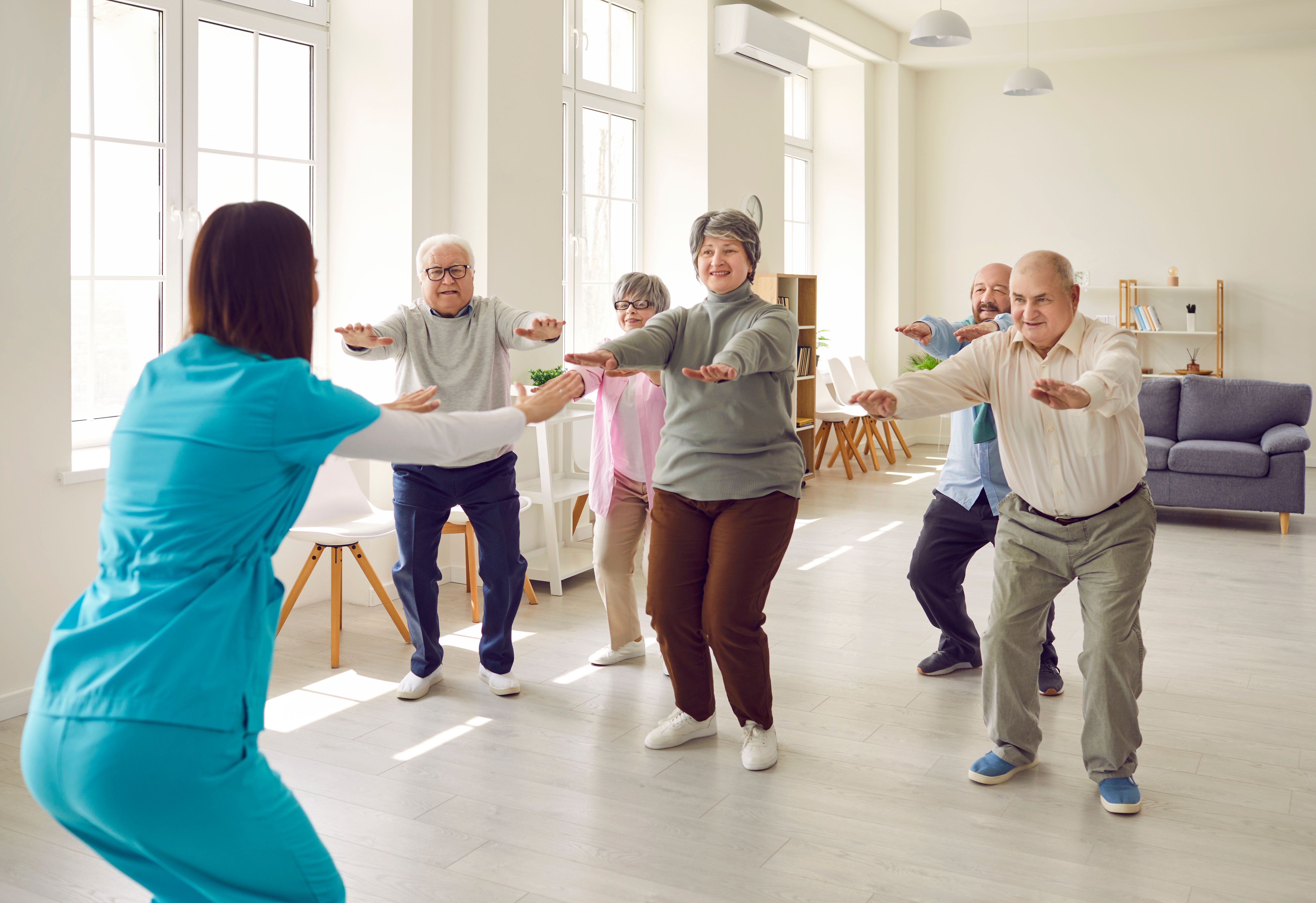
Many elderly people aged 65 years or older might experience hip fractures when they fall down, but getting surgery would not be enough to fully recover, and postoperative rehabilitation is crucial for standing up again, the Taiwanese Osteoporosis Association (TOA) said yesterday.

The TOA yesterday held the Fragility Fracture Network (FFN) Taiwan founding conference in Taipei, where TOA chairperson and Kaohsiung Municipal Da-Tung Hospital orthopedist Chen Chung-hwan (陳崇桓) was named adjunct chairperson of the network.

The FFN addresses the full pathway of care for fragility fracture patients, and has been promoting improvements across four main pillars of care that include perioperative care, surgical treatment, rehabilitation, secondary prevention and policy change, the association said, adding that it has expanded its congress in different regions worldwide with participants from 102 countries.

Photo: Lin Liang-che, Taipei Times

Chen said the FFN Taiwan consists of orthopedists, geriatricians, rehabilitation physicians, nurses, physical therapists, occupational therapists, nutritionists and other healthcare professionals, and it aims to integrate resources for maximizing effectiveness in improving the quality of fragility fracture treatment in Taiwan.

According to the National Health Promotion Administration’s national nutrition survey between 2017 and 2020, the prevalence of osteoporosis among adults aged 50 or older was 8.1 percent, but the prevalence among women was higher — about 10 percent were estimated to have osteoporosis.

Chen said most hip fractures occur in elderly people and are caused by falling, and as women have higher rates of weakened bones by osteoporosis and weaker muscle strength, they have a higher risk of getting a hip fracture from a fall than men.

However, the postoperative healthcare they receive is crucial for recovery, as many who return home only to lay in bed, instead of undergoing physical rehabilitation, would have poor outcomes of rapid muscle loss and the inability to stand up and walk, becoming long-term bedridden patients, he said.

West Garden Hospital rehabilitation physician Hsu Po-cheng (徐伯誠) said about half of hip fracture patients need assistive devices or other people’s help to stand up after surgery, and about 90 percent need assistance to climb stairs, so many might lose their mobility to enjoy a full and active life.

However, if people do not practice moving around after the surgery, they could lose bone mass and muscle strength rapidly within a few months, he said, adding that therefore, they should try to leave their bed as soon as possible and get physical rehabilitation to gradually recover to preoperative conditions.

As people would need to add different exercises into their rehabilitation routine depending on their preoperative exercise habits, health conditions, complications and medications, customized plans and supervision might need a medical team with different health professionals, Hsu said.

Chen said the National Health Insurance policy says that large hospitals are responsible for acute treatment, but patients must be transferred to local hospitals or facilities for post-acute care.

Therefore, the FFN Taiwan would launch a hip fracture registry program with a hospitals this year, he said.

The FFN is to collect data from people to better understand what can be improved in the nation’s fragility fracture healthcare system, and try to obtain more resources to improve it, Chen added.

Additional reporting by CNA

Comments will be moderated. Keep comments relevant to the article. Remarks containing abusive and obscene language, personal attacks of any kind or promotion will be removed and the user banned. Final decision will be at the discretion of the Taipei Times.

Source link

The global medical dynamometer market is experiencing rapid expansion, driven by the rising prevalence of musculoskeletal disorders and the growing emphasis on physical rehabilitation. Advanced technologies such as wireless connectivity and real-time data monitoring are enhancing the efficiency and accuracy of muscle strength assessment, fueling market growth. Additionally, increasing awareness about proactive healthcare management is driving demand for dynamometers in fitness centers, gyms, and home-based rehabilitation programs.

BOSTON, March 27, 2024 /PRNewswire/ -- "According to the latest BCC Research study, the demand for Global Medical Dynamometer Market is estimated to increase from $547.5 million in 2023 to reach $780.1 billion by 2028, at a compound annual growth rate (CAGR) of 7.3% from 2023 through 2028."

This comprehensive report delves into the multifaceted landscape of the medical dynamometer market, spanning healthcare, rehabilitation, and research sectors. It provides an exhaustive analysis covering various types of dynamometers including hand, chest, squeeze, and push-pull variants. Targeting key stakeholders such as hospitals, rehabilitation centers, sports medicine facilities, and research labs, the report offers profound insights into market dynamics, current trends, and future potentials. By addressing drivers, challenges, and opportunities, it equips market players with strategic intelligence to navigate the evolving landscape. Moreover, the report elucidates industry status, recent developments, and emerging trends, enabling informed decision-making and fostering growth. Geographical nuances are meticulously examined, making it an invaluable resource for companies seeking to expand their market footprint. Whether for market incumbents, potential entrants, government agencies, or other interested parties, this report serves as a definitive guide to understanding and capitalizing on the dynamic medical dynamometer market.

The medical dynamometer has emerged as a pivotal tool in addressing clinical needs, particularly in precise ankle strength measurements. With its portability and high reliability, it proves to be indispensable for routine muscle strength monitoring across diverse populations, exhibiting robust agreement with the gold standard handheld dynamometer (HHD). Its versatility extends from children to the elderly and individuals with weak ankle strength, promising accurate measurements in clinical settings pending further validation. Adding to this landscape, Ashva WearTech's Fitmust introduces a handheld muscle strength dynamometer, poised to revolutionize muscle strength assessment. Launched in April, this microprocessor-based device offers precise and medically accurate measurements, aiding in injury assessment and rehabilitation. Fitmust addresses the critical aspect of muscle strength, recognized by physiotherapists and orthopedic surgeons as paramount in treatment protocols. By providing a comprehensive testing solution for various muscle groups, it empowers healthcare professionals with valuable insights for optimizing health and fitness outcomes.

Explore the full report on the burgeoning Global Medical Dynamometer Market for comprehensive insights and forecasts – click here to Learn More

Key Drivers of the Global Medical Dynamometer Market

Increase in Sports Injury: With the surge in the popularity of sports and physical activities, there has been a corresponding increase in sports-related injuries. Medical dynamometers are indispensable in this scenario, as they play a crucial role in assessing muscle strength and monitoring rehabilitation post-injury. These devices are in high demand due to their effectiveness in tracking progress during the recovery process, ensuring athletes and individuals can safely return to their activities.

Rise in Prevalence of Bone Injuries and Osteoporosis: The prevalence of bone injuries, fractures, and conditions such as osteoporosis is on the rise, particularly among the aging population. In addressing these challenges, medical dynamometers prove invaluable. They aid healthcare professionals in evaluating bone health and assessing muscle strength around affected areas. Early detection and continuous monitoring facilitated by these devices significantly contribute to better patient outcomes and improved quality of life.

Rehabilitation and Physiotherapy Trends: There has been a significant emphasis on rehabilitation and physiotherapy in recent years. In this evolving landscape, medical dynamometers serve as essential tools for therapists to design personalized exercise programs tailored to individual patient needs. By facilitating targeted muscle strength assessment and monitoring, these devices help patients regain strength, improve mobility, and enhance overall functional capacity, thereby accelerating the rehabilitation process.

Growing Geriatric Population and Increase in Orthopedic Surgeries: As the global population ages, there is a concurrent increase in age-related conditions and orthopedic surgeries. Joint replacements, fracture repairs, and other orthopedic procedures are becoming more prevalent. Medical dynamometers play a crucial role in this scenario, assisting healthcare providers in preoperative assessments, postoperative rehabilitation, and long-term monitoring of patients. By accurately assessing muscle strength and functional capacity, these devices contribute to optimized outcomes and improved post-surgery recovery trajectories.

Request a Sample Copy of the Global Medical Dynamometer Market Report.

Report Synopsis 

Report Metrics

Details

Base year considered

2022

Forecast Period considered

2023-2028

Base year market size

$516.0 million

Market Size Forecast

$780.1 million

Growth Rate

CAGR of 7.3% for the forecast period of 2023-2028

Segment Covered

Technology, Product Type, Application, End-User, and Geographic Region

Regions covered

North America, Europe, Asia-Pacific, and Rest of the World (RoW)

Countries covered

U.S, Canada, France, Germany, Italy, Spain, UK, Russia, China, India, Japan, and Australia

Key Market Drivers

•  Increase in Sports Injury

•  Rise in Prevalence of Bone Injuries and Osteoporosis

•  Rehabilitation and Physiotherapy Trends

•  Growing Geriatric Population and Increase in Orthopedic Surgeries

Global Medical Dynamometer Market Segmentation Analysis

Technology Segmentation

Within the medical dynamometer market, technology segmentation categorizes devices into two main types: Electronic Dynamometers and Mechanical Dynamometers. Electronic Dynamometers employ electronic sensors to accurately measure muscle strength, offering real-time data for assessment. On the other hand, Mechanical Dynamometers rely on mechanical principles such as springs or levers for muscle strength assessment, providing a portable and cost-effective alternative.

Product Type Segmentation

The market for medical dynamometers further segments products into various types to cater to different assessment needs. Handheld Dynamometers are compact and easy-to-use devices suitable for assessing muscle strength in different body parts. Pinch-gauges are specialized tools designed to measure pinch strength, particularly useful in hand rehabilitation. Push-Pull Dynamometers evaluate upper and lower limb strength during pushing and pulling movements, while Squeeze Dynamometers measure grip strength, crucial for assessing hand function.

Application Segmentation

Medical dynamometers find application across various medical fields, including Orthopedics, Medical Trauma, Cardiology, and Neurology. In Orthopedic settings, these devices assist in preoperative assessments, postoperative rehabilitation, and monitoring orthopedic conditions. Medical Trauma applications involve evaluating muscle strength post-traumatic injuries. Cardiology utilizes dynamometers to assess cardiac muscle strength and endurance, while Neurology employs them in diagnosing and monitoring neurological conditions.

End-User Segmentation

End-user segmentation categorizes users of medical dynamometers into different groups. Hospitals commonly use dynamometers for patient assessment, rehabilitation, and monitoring. Physiotherapy Clinics utilize these devices for designing personalized exercise programs, while Rehabilitation Centers rely on them for patient recovery and functional improvement.

This report on the Global Medical Dynamometer Market provides comprehensive insights and analysis, addressing the following key questions:

1.  What is the projected market size and growth rate of the market?

The Global medical dynamometer market is projected to grow from $516.0 million in 2022 to $780.1 million in 2028, at a compound annual growth rate (CAGR) of 7.3 % during the forecast period.

2.  What are the key factors driving the growth of the market?

  •  Increase in sports injuries.
  •  Rise in prevalence of bones injuries and Osteoporosis
  •  Rehabilitation and Physiotherapy trends
  •  Growing Geriatric Population and increase in orthopaedic surgeries.

3.  What segments are covered in the market?

This report segments the market based on technology, type, application, end-user, and region. Based on the technology, the market is segmented into electronic dynamometer and mechanical dynamometer. Based on product type, the market is segmented into handheld dynamometer, pinch gauges, push-pull dynamometer, squeeze dynamometer and others. Based on application, the market is segmented into orthopedics, medical trauma, cardiology, neurology and others. Based on region, the market is segmented as North America, Europe, Asia-Pacific and Rest of the World.

4.  By Type, which segment will dominate the market by the end of 2028?

Handheld Dynamometer will dominate the market by the end of 2028.

5.  Which region has the highest market share in the market?

North America holds the highest share in the market.

Some of the Key Market Players Are:

  •  3B SCIENTIFIC GMBH
  • BIODEX MEDICAL SYSTEMS INC.
  • BIOMETRICS LTD.
  • BTE TECHNOLOGIES
  • CHARDER ELECTRONICS CO. LTD.
  • FABRICATION ENTERPRISES INC.
  • HERKULES KUNSTSTOFF OBERBURG AG
  • HOGGAN SCIENTIFIC LLC.
  • JLW INSTRUMENTS
  • JTECH MEDICAL INDUSTRIES INC.
  • KERN & SOHN GMBH
  • LAFAYETTE INSTRUMENT CO.
  • MARSDEN GROUP
  • NORTH COAST MEDICAL INC.
  • PERFORMANCE HEALTH
  • VERNIER SCIENCE EDUCATION
  • ZHONGSHAN CAMRY ELECTRONIC CO. LTD.

Browse Related Reports:

Global Market for Pain Management Drugs and Devices: This report refers to a comprehensive analysis of the global market for pain management drugs and devices, focusing on both current trends and future projections up to 2028. It delves into the market drivers, restraints, and challenges, while also providing insights into the competitive landscape and regulatory framework. Segmented by geography, product type (pharmaceuticals and medical devices), and application areas such as post-operative pain, neuropathic pain, and musculoskeletal pain, among others, the report offers a detailed examination of key market players and their strategies. Notably, the report excludes drugs or devices targeting specific conditions like rheumatoid arthritis but includes treatments aimed at alleviating associated pain symptoms. For instance, Enbrel is highlighted for its role in treating moderate-to-severe rheumatoid arthritis, while Celebrex is recognized for its efficacy in managing the signs and symptoms of osteoarthritis and rheumatoid arthritis, alongside the relief of acute pain.

Directly Purchase a copy of the report with BCC Research.

For further information or to make a purchase, please get in touch with [email protected].  

About BCC Research

BCC Research provides objective, unbiased measurement and assessment of market opportunities with detailed market research reports. Our experienced industry analysts' goal is to help you make informed business decisions, free of noise and hype.

Contact Us

Corporate HQ: BCC Research LLC, 49 Walnut Park, Building 2, Wellesley, MA 02481, USA

Email: [email protected],

Phone: +1 781-489-7301

For media inquiries, email [email protected] or visit our media page for access to our market research library.

Data and analysis extracted from this press release must be accompanied by a statement identifying BCC Research LLC as the source and publisher.

Logo: mma.prnewswire.com/media/2183242/BCC_Research_Logo.jpg

SOURCE BCC Research LLC

Source link

Abstract

Introduction

Polyhandicap (PLH) is currently defined as a complex disability condition corresponding to a chronic affliction occurring in an immature brain [1], leading to the combination of severe/profound mental retardation and serious motor impairment, resulting in an extreme restriction of autonomy and communication [2,3].

As comorbidities accumulate in patients with PLH throughout their lives, impairments, activity limitations and health-related quality of life (QoL) worsen [4]. Among comorbidities in patients with PLH, scoliosis represents one of the most prevalent conditions with chronic respiratory insufficiency [57], chronic digestive disorders, or epilepsy [8]. Indeed, Rousseau et al. [9] estimated that the prevalence of severe scoliosis (scoliosis with Cobb angle >50°) prevalence was 60.2%, accounting for a large proportion of deaths induced by respiratory failure. The latter, represents the main cause of death in adults with PLH [9] (63.2%).

Even if the natural history [10], risk factors, treatments [11,12] and complications [13] of scoliosis with Cobb angle >50° are well known in nonambulant cerebral palsy (CP) patients (Gross Motor Function Classification System—GMFCS level 5), few data are available in PLH [3].

Hodgkinson et al. [14] found severe neuro-orthopaedic impairments in adolescents with PLH were associated with poor general condition. They hypothesized that multidisciplinary therapeutic strategies must include surgical scoliosis treatment to improve pain, comfort, and positioning. However, Cassidy et al. [15] found no differences in terms of pain, function, or time for daily care when comparing adolescents with PLH who underwent thoracolumbar spinal fusion with the nonoperated control group. De Lattre et al. [16] also failed to demonstrate improvements in health status between non operated and operated adolescents with PLH. They also reported a high rate of per- and postoperative complications (93,7%). McCarthy et al. [17] reported a rate of perioperative death up to 7% in nonambulant CP patients.

Previous studies estimated that 12% to 32% of subjects with PLH underwent scoliosis surgery [9]. Thus, there is still considerable interest in investigating whether scoliosis surgery truly confers measurable benefits on subsequent health and daily life comfort in adolescents with PLH. If it is hypothesized that surgeries lead to measurable clinical and radiological benefits, valid clinical evaluations are lacking.

The aims of this study were first to evaluate whether the health status variation in adolescents with PLH was improved after scoliosis surgery and second to evaluate the incidence of perioperative complications.

Material and methods

Participants

All adolescents with PLH who underwent definitive spinal fusion surgery for scoliosis with Cobb angle >50° were eligible. Additionally, surgical indication was determined through a multidisciplinary evaluation, considering various parameters including respiratory and abdominal disorders, complications in wheelchair installation, and severe axial hypotonia. PLH was defined as a combination of cerebral lesions onseted under 3 years old and responsible for severe motor deficiency with restricted mobility (GMFCS V), associated with profound intellectual impairment, and daily life dependence (Functional Independence Measure—FIM < 55) [18]. A minimum of 12 months follow-up was required for inclusion and postoperative data collection, except in adolescents presenting lethal complications during procedure or in the immediate follow-up, who were also considered in the analysis. Patients were operated on by one surgeon to ensure population homogeneity. Surgery consisted in all patients in an hybrid instrumentation of the spine: patients in supine position; pelvic fixation to correct the pelvis obliquity; instrumentation of the lumbar area with pedicular screws; thoracic area with sublaminar ligaments and hook-claw at the upper part of the instrumentation; rods were self-bowing by surgeon during the procedure; all implants from Medtronic (Medicrea, Rillieux la Pape 69140, France).

Postoperative care systematically included one night in ICU for monitoring. Pain medication started peroperatively with intrathecal injection of morphine (5 μg/kg). A peridural catheter was maintained during 48h to 5 days with continual perfusion of low dose ropivacaine 0.2% (related to patient weight), and if necessary, paracetamol, ketoprofen, diazepam and oral morphine (related to patient weight and respiratory status) could be used. Esomeprazole for gastric ulcer prevention, lactulose per oral for constipation prevention, and enoxaparin if necessary (related to risk factors) were also administered. Feeding started one day after spinal fusion, through various modalities depending on patients. Lower limbs mobilizations were started two days postoperatively in sitting position, then performed every day. Analysis of targeted medical, paramedical records and X-ray data prior to (in the 6 months preceding) and after surgery (12 months postoperative) in patients operated on between 2009 and 2020 was performed (see Fig 1).

thumbnail

Fig 1. Study design.

M Months; PSS Polyhandicap Severity Score; mCDS modified Clavien-Dindo-Sink classification; FCA Frontal Cobb Angle; FB Frontal Balance; OBL Pelvic Obliquity; SCA Sagittal Cobb Angle; SB Sagittal Balance; PI Pelvic Incidence; SS Sacral Slope; PT Pelvic Tilt.


doi.org/10.1371/journal.pone.0300065.g001

Data

Data were collected by independent evaluators using a specific research algorithm corresponding to the French nomenclature of surgically performed acts. All variables of interest were subsequently extracted from the eligible charts. In addition, we contacted families through phone calls to document parent’s feelings about their adolescent’s QoL after surgery. Two independent evaluators (HB and EO) performed X-ray measurements to reduce ascertainment bias. In cases of disagreement (>6° or >7° differences between two measurements, respectively in frontal and sagittal planes), the referring surgeon (BD) arbitrated the decision. For all outcomes, we only considered the earliest preoperative measurements and the latest postoperative ones.

Data extraction was completed from computerized charts by the first author (HB) who was not involved in the surgical procedure nor medical postoperative follow-up. Method for fulfilling evaluation form was as exhaustive as possible to limit risk of bias. Age at surgery, sex, care facility, preoperative FIM, past surgical history, PLH aetiology, type of physical disability, pain, body mass index (BMI), spontaneous posture, communication level, feeding strategies, and presence of medical devices were extracted. These data were compiled to rate the severity health status of each participant [19] according to the Polyhandicap Severity Scale (PSS). The PSS provides an accurate evaluation of health status regarding abilities, comorbidities and impairments, and assesses the level of global severity of the health status of persons with PLH. This scale ranges scores from 0 to 129, from less to higher levels of severity. A global PSS score was calculated as the sum of the two PSS subscores, respectively “comorbidities and impairments” (coPSS, ranging from 0 to 69) and “abilities” (abPSS, ranging from 0 to 60), according to the International Classification of Functioning, Disability and Health (ICF). Semi structured interviews (see Fig 2) were conducted with families to collect verbatim interviews. We also rated pre- and postoperative PolyQoL scores [20]. This validated short-scale is composed of two domains called health and social, ranging QoL scores from 0 (worst) to 100 (better).

X-ray parameters were extracted from pre- and postoperative X-rays (sagittal and frontal). A specific software program (Surgimap Spine® 2021, Nemaris, Inc.) was used for all spinal and pelvic measurements, as it offers semiautomatic procedures with good inter- and intraobserver reliability, even on instrumented spines. Frontal X-ray data included skeletal maturity (Risser test), frontal Cobb angles (FCA), pelvic obliquity (OBL) and frontal balance (FB). Sagittal parameters included sagittal Cobb angles (SCA) and sagittal balance (SB). Using the same software, we measured pelvic incidence (PI), pelvic tilt (PT) and sacral slope (SS). Frontal and sagittal curves were added separately to obtain a “full curvature degree” for each participant, reflecting the amount of deformity.

Peri- and postoperative complications ranged according to the modified Clavien-Dindo-Sink classification [21] (mCDS). We collected blood loss data (loss of more than 1 blood volume), deep wound infections (DWI) and operative durations. We also calculated hospital lengths of stay, composed of the sum of paediatric surgery, intensive care unit (ICU), and rehabilitation department stays (days of hospitalization).

Results

Descriptive data

The mean age at surgery was 15 (3) years old. The preoperative mean FIM was 22 (10). PLH aetiology was unknown for 8 adolescents and progressive for 6 adolescents. Twenty patients had undergone previous orthopaedic surgery, mainly multilevel tenotomy surgery (n = 9). The mean BMI before surgery was 17.4 (3.9) kg/m². Descriptive data are reported in Table 1.

The mean preoperative scores were 53 (17), 14 (5), and 39 (13) for PSS (/129), coPSS (/69) and abPSS (/60) respectively. For postoperative scores, we found a score of 49 (16) for PSS, a score of 12 (4) for coPSS and a score of 37 (13) for abPSS. Data were missing in 3 cases: 1 adolescent died during surgery and 2 medical records were insufficient for exhaustive PSS grading before and after surgery. Another adolescent died 7 days after surgery, but postoperative PSS scoring was possible. Seven caregivers agreed to answer semistructured interviews. The mean preoperative PolyQoL score was 64 (12). Mean postoperative PolyQoL score was 77 (10). Pre- and postoperative data, with related means of difference, are shown in Table 2. A scatter plot of individual preoperative versus postoperative PSS scores is provided in Fig 4.

thumbnail

Fig 4. Scatter plot of individual preoperative versus postoperative PSS scores.

Mean PSS evolution with standard error is represented by the large red solid line with error bar. PSS evolution i.e improvement (n = 18; dark solid line), stability (n = 8; grey solid line), worsening (n = 1; grey dashed line) are represented for each individual.


doi.org/10.1371/journal.pone.0300065.g004

For complications, all the subjects presented at least 1 per- or postoperative complication. Among them, 9 were rated grade 1, and 10 were rated grade 4 according to the mCDS classification (Fig 5). More accurately, 6 presented with a deep wound infection (DWI), and 13 with a major blood loss. Neither neurologic, nor digestive or pancreatic complications were noted postoperatively in the patients. Finally, we found a mortality rate of 6.7% during follow-up: 1 death occurred during the surgical procedure, which was related to heart failure during anaesthesia, and 1 in the ICU due to multisystemic failure 7 days after surgery.

thumbnail

Fig 5. Proportions (%) of complications as classified by mCDS scoring (minor to severe complications, from grade 1 to 5).

Grade 1 (n = 9): Wound leakage, constipation, nonsevere lung infection; Grade 2 (n = 5): Non severe dura mater breach, delayed wound healing; Grade 3 (n = 2): Deep wound infection, treated with second surgery + IV antibiotics; Grade 4 (n = 10): Any complication requiring ICU admission: Complex deep wound infection, renal failure, urinary sepsis, severe lung infection, delayed awakening after anaesthesia, central venous catheter infection; Grade 5 (n = 2): Per operative death or early postoperative death (within 7 days); NA (n = 2): Missing data.


doi.org/10.1371/journal.pone.0300065.g005

The mean hospital length of stay was 44.7 days (20 to 83 days), respectively comprising 3.6 (2.7) days (1 to 10) in the ICU, 14.3 (8.3) days (1 to 34) in the paediatric surgery department, and 26.9 (12.9) days (0 to 56) in the rehabilitation department.

Discussion

In the present study, we aimed to describe comorbidities, impairments, activity limitations and QoL before and after spinal fusions in adolescents with PLH. We hypothesized that these surgeries lowered the severity health status and conferred clinical benefits. Our hypothesis was confirmed as PSS, coPSS and abPSS scores were significantly improved. We have nevertheless highlighted a 100% rate of complications and 2 deaths, testifying to the complexity of these surgeries.

Comorbidities, impairments, and activity limitations were evaluated, as outlined above, with the PSS. This scale allows us to quantify the severity health status of patients with PLH, providing a new ICF-standardized and exhaustive approach for clinical assessments. We believe in that better preoperative clinical specifications could help in identifying patients who may better tolerate and benefit from spinal fusions. Scoliosis surgery seems to represent a strong contribution to the long-term management of health in persons with PLH [2]. The results of our study demonstrate that spinal fusion surgery in adolescents with PLH leads to a reduction in comorbidities (respiratory, digestive, skin, behavioural, pain, etc.) and seems to allow a slight improvement in neurodevelopmental status.

Moreover in this study, we could interview seven of all families through semistructured questionnaires: witness families reported that spinal fusions conferred a global improvement in QoL. The parents’ statements were mostly positive: during the preoperative period, they had difficulty accepting the vital risk of spinal surgery, but in retrospect, they recognized 1) that the surgery was necessary, and 2) that their child’s health (breathing, posture…) had improved even when peroperative and immediate postoperative complications surrounded. Moreover, we used a validated tool called the PolyQoL questionnaire to objectively document QoL. We did not show any significant improvement regarding PolyQoL scores, but these results must be taken with caution, likely due to our small sample of answers.

Parents and caregivers were particularly attentive to the experience before and during the surgical period and reported having psychologically benefited from preoperative comprehensive explanations. Half of them verbalized the “inevitability” of this surgery. As highlighted by Adams et al. [22], agreements on the goals of surgery between surgeons and caregivers are important. While surgeons and physicians tend to give top priority to sitting considerations, caregivers put head control and physical appearance first. The expected benefits seem to remain superior to the risk of complications, but the therapeutic strategy requires an individualized evaluation rigorously explained to caregivers of PLH adolescents [22,23]. The question of long-term clinical benefits remains important, as follow-up is rarely conducted for more than 1 year [24].

Families are often afraid when scoliosis surgery is considered for their adolescent [25]. The postoperative complication rate (100%) and perioperative mortality rate (6,7%) must be included in the decision and balanced with improvements in severity health status. In our study, the DWI rate was 20%, which is almost twice the rate as previous studies reported in CP [26] (but CP adolescents present a less severe health status than PLH adolescents we analysed). Nutritional status of patients with PLH could be questioned to explain the difference.

Geometric spine readjustment could be one of the reasons for pain alleviation by costo-pelvic impingement prevention and sitting comfort improvement [27]. We observed a reduction in bed sore frequency and gluteal erosions after surgery. We confirmed a significant improvement in BMI after surgery (mean variation of +1,37 kg/m²). As presumed in some studies, straightening the thoracolumbar spine could increase the abdominal space, allowing better peristalsis and less regurgitation [28]. However, severe postoperative pancreatitis cases have been reported after extensive arthrodesis, but we did not report any case in our population.

Regarding medical devices, we observed that IT baclofen pumps did not increase the complication rate, which is consistent with previous studies [29]. Similarly, gastrostomies were not associated with a major complication occurrence in our PLH patients, unlike CP GMFCS V patients in whom an increased risk has been shown previously [30]. Except for DWI, severe complications were mostly respiratory. Lung developmental defects, impaired neurologic command over added lung restrictive syndromes and postoperative intensive care with lung infection risks can partially explain our findings [6]. Preoperative breathing hyper insufflation strategies could be worthwhile as they demonstrated interest in spinal fusions in children with neuromuscular flaccid scoliosis [31]. As preoperative primary care visits led to lower costs and shorter hospitalizations in complex scoliosis surgeries, we support the idea of efficient preoperative rehabilitation programs before spinal fusions. Rehabilitation modalities could be determined according to the domains impacted in the preoperative PSS scoring.

Cobb corrections were 58% for FCA and 30% for SCA. Spinal fusions in adolescents with PLH provide comparable results to those found in GMFCS IV and V adolescents with CP (50 to 68% as reported). A mean PT variation of -7.8° (22.4) was observed, corresponding to a significant reduction in pelvis back-tilting. The mean correction for OBL was 47%. This X-ray parameter was correlated with the postoperative PSS score and PSS variations induced by surgery.

Strengths and limitations

Our study demonstrates for the first time, that spinal fusions reduce the global severity of adolescents with polyhandicap. However, it contains several limitations: our study was monocentric and retrospective data collection may have provided methodological bias. Broadening inclusions to various specialized centers could be of interest in a prospective approach. Longer follow-up (2–5 years) would enable us to assess the long-term effect of spinal fusion surgery on the severity health status of patients with PLH, thus further prospective studies with a longer follow-up are needed (therefore, the present one-year follow-up evaluation should not be considered as the final evaluation). By increasing the duration of follow-up and through the methodical use of standardized tools such as PSS [19] and PolyQoL [20], establishing accurate and repeatable ICF-based descriptions in patients with PLH appears to be easier. An increased sample size could also help in analysing predictive factors of clinical improvement after spinal fusions, keeping in mind the potential confounding factors we highlighted.

One of the main issues when performing spinal fusions in patients with PLH is the assessment of predicted benefits. This study aimed to support the rationale for spinal fusion, acknowledging the already known complication rates and risks in PLH adolescents. Considering the severity of the health status evaluation of these patients and the expected improvement of the global health status and comfort after spine surgery for scoliosis, each case requires careful consideration and evaluation. We support that, under the condition of acceptable preoperative health status, the indication for spinal fusion in that population could be relevant and reasonable. While retrospective evaluation is questionable, the rigorous method used during data collection should limit this bias. The PolyQoL questionnaire has been supported by the verbatim collected in a part of the population, and obviously the good health status of the patients at final follow-up confirms our hypothesis.

References

  1. 1.
    Chabrier S, Pouyfaucon M, Chatelin A, Bleyenheuft Y, Fluss J, Gautheron V, et al. From congenial paralysis to post-early brain injury developmental condition: Where does cerebral palsy actually stand? Ann Phys Rehabil Med. 2019 Aug;S1877065719301137. pmid:31421273
  2. 2.
    Rousseau MC, Baumstarck K, Auquier P, Billette de Villemeur T. Health characteristics and health care trajectory of polyhandicaped person before and after 1990. Rev Neurol (Paris). 2020 Jan-Feb;176(1–2):92–99. Epub 2019 Jun 27. pmid:31255322.
  3. 3.
    Maes B, Nijs S, Vandesande S, Van Keer I, Arthur-Kelly M, Dind J, et al. Looking back, looking forward: Methodological challenges and future directions in research on persons with profound intellectual and multiple disabilities. J Appl Res Intellect Disabil. 2021 Jan;34(1):250–62. pmid:33073444
  4. 4.
    Mensch SM, Echteld MA, Lemmens R, Oppewal A, Evenhuis HM, Rameckers EAA. The relationship between motor abilities and quality of life in children with severe multiple disabilities: Motor abilities and quality of life in children with SMD. J Intellect Disabil Res. 2019 Feb;63(2):100–12.
  5. 5.
    Gautheron V, Mathevon L, Bayle B, Boulard C, Paricio C, Seeman E, et al. Problèmes respiratoires des personnes polyhandicapées: le point de vue du médecin de médecine physique et de réadaptation. Mot Cérébrale Réadapt Neurol Dév. 2015 Jun;36(2):49–53.
  6. 6.
    Proesmans M, Vreys M, Huenaerts E, Haest E, Coremans S, Vermeulen F, et al. Respiratory morbidity in children with profound intellectual and multiple disability: Respiratory Morbidity and Neurocognitive Impairment. Pediatr Pulmonol. 2015 Oct;50(10):1033–8.
  7. 7.
    Boel L, Pernet K, Toussaint M, Ides K, Leemans G, Haan J, et al. Respiratory morbidity in children with cerebral palsy: an overview. Dev Med Child Neurol [Internet]. 2018 Oct 15 [cited 2019 Jan 23]; doi.wiley.com/10.1111/dmcn.14060 pmid:30320434
  8. 8.
    Arvio M, Sillanpää M. Prevalence, aetiology and comorbidity of severe and profound intellectual disability in Finland. J Intellect Disabil Res. 2003 Feb;47(Pt 2):108–12. pmid:12542576
  9. 9.
    Rousseau MC, Mathieu S, Brisse C, Motawaj M, Grimont E, Auquier P, et al. Aetiologies, comorbidities and causes of death in a population of 133 patients with polyhandicaps cared for at specialist rehabilitation centres. Brain Inj. 2015 Jul 3;29(7–8):837–42. pmid:25950262
  10. 10.
    Saito N, Ebara S, Ohotsuka K, Kumeta H, Takaoka K. Natural history of scoliosis in spastic cerebral palsy. Lancet Lond Engl. 1998 Jun 6;351(9117):1687–92. pmid:9734885
  11. 11.
    Tsirikos A. Development and treatment of spinal deformity in patients with cerebral palsy. Indian J Orthop. 2010;44(2):148. pmid:20419001
  12. 12.
    Dohin B. The spastic hip in children and adolescents. Orthop Traumatol Surg Res. 2019 Feb;105(1):S133–41. pmid:30056240
  13. 13.
    Jain A, Sponseller PD, Shah SA, Samdani A, Cahill PJ, Yaszay B, et al. Subclassification of GMFCS Level-5 Cerebral Palsy as a Predictor of Complications and Health-Related Quality of Life After Spinal Arthrodesis: J Bone Jt Surg. 2016 Nov;98(21):1821–8. pmid:27807115
  14. 14.
    Hodgkinson I, Jindrich ML, Metton G, Berard C. Bassin oblique, luxation de hanche et scoliose dans une population de 120 adultes polyhandicapés. Étude descriptive. Ann Réadapt Médecine Phys. 2002 Feb;45(2):57–61.
  15. 15.
    Cassidy C, Craig CL, Perry A, Karlin LI, Goldberg MJ. A reassessment of spinal stabilization in severe cerebral palsy. J Pediatr Orthop. 1994 Nov 1;14(6):731–9. pmid:7814585
  16. 16.
    de Lattre C, Hodgkinson I, Bérard C. Retentissement de la scoliose chez les patients polyhandicapés: étude descriptive de 61 enfants et adultes polyhandicapés avec ou sans arthrodèse vertébrale. Ann Réadapt Médecine Phys. 2007 May;50(4):218–24.
  17. 17.
    McCarthy JJ, DʼAndrea LP, Betz RR, Clements DH. Scoliosis in the Child With Cerebral Palsy: J Am Acad Orthop Surg. 2006 Jun;14(6):367–75. pmid:16757676
  18. 18.
    Rousseau M-C, Winance M, Baumstarck K. Polyhandicap, profound intellectual multiple disabilities: Concept and definition of a highly specific public health issue. Rev Epidemiol Sante Publique 2023;71:102184.
  19. 19.
    Rousseau MC, Baumstarck K, Hamouda I, Valkov M, Felce A, Khaldi-Cherif S, et al. Development and initial validation of the polyhandicap severity scale. Rev Neurol (Paris). 2021 Jun;177(6):683–9. pmid:33069376
  20. 20.
    Hamouda I, Rousseau MC, Aim MA, Anzola AB, Loundou A, De Villemeur TB, et al. Development and initial validation of the quality of life questionnaire for persons with polyhandicap (PolyQoL). Ann Phys Rehabil Med. févr 2023;66(1):101672. pmid:35490967
  21. 21.
    Dodwell ER, Pathy R, Widmann RF, Green DW, Scher DM, Blanco JS, et al. Reliability of the Modified Clavien-Dindo-Sink Complication Classification System in Pediatric Orthopaedic Surgery. JBJS Open Access. 2018 Dec 20;3(4):e0020. pmid:30882054
  22. 22.
    Adams AJ, Refakis CA, Flynn JM, Pahys JM, Betz RR, Bastrom TP, et al. Surgeon and Caregiver Agreement on the Goals and Indications for Scoliosis Surgery in Children With Cerebral Palsy. Spine Deform. 2019 Mar;7(2):304–11. pmid:30660226
  23. 23.
    Tsirikos AI, Chang WN, Dabney KW, Miller F. Comparison of parents’ and caregivers’ satisfaction after spinal fusion in children with cerebral palsy. J Pediatr Orthop. 2004 Jan-Feb;24(1):54–8. pmid:14676534.
  24. 24.
    Miyanji F, Nasto LA, Sponseller PD, Shah SA, Samdani AF, Lonner B, et al. Assessing the Risk-Benefit Ratio of Scoliosis Surgery in Cerebral Palsy: Surgery Is Worth It. J Bone Jt Surg. 2018 Apr;100(7):556–63. pmid:29613924
  25. 25.
    Rousseau MC, Billette de Villemeur T, Khaldi-Cherif S, Brisse C, Felce A, Baumstarck K, et al. Adequacy of care management of patients with polyhandicap in the French health system: A study of 782 patients. Simeoni U, editor. PLOS ONE. 2018 Jul 6;13(7):e0199986. pmid:29979745
  26. 26.
    Sponseller PD, Shah SA, Abel MF, Newton PO, Letko L, Marks M. Infection Rate after Spine Surgery in Cerebral Palsy is High and Impairs Results: Multicenter Analysis of Risk Factors and Treatment. Clin Orthop. 2010 Mar;468(3):711–6. pmid:19543779
  27. 27.
    Roberts SB, Tsirikos AI. Factors influencing the evaluation and management of neuromuscular scoliosis: A review of the literature. J Back Musculoskelet Rehabil. 2016 Nov 21;29(4):613–23. pmid:26966821
  28. 28.
    DeFrancesco CJ, Miller DJ, Cahill PJ, Spiegel DA, Flynn JM, Baldwin KD. Releasing the tether: Weight normalization following corrective spinal fusion in cerebral palsy. J Orthop Surg. 2018 May;26(2):230949901878255. pmid:29938586
  29. 29.
    Buxton K, Difazio R, Morgan A, McCabe M, Forbes PW. Intrathecal Baclofen Therapy Prior to Spinal Fusion for Patients With Gross Motor Function Classification System IV-V Cerebral Palsy: Orthop Nurs. 2018;37(2):136–43. pmid:29570548
  30. 30.
    Nishnianidze T, Bayhan IA, Abousamra O, Sees J, Rogers KJ, Dabney KW, et al. Factors predicting postoperative complications following spinal fusions in children with cerebral palsy scoliosis. Eur Spine J. 2016 Feb;25(2):627–34. pmid:26410446
  31. 31.
    Mills B, Bach JR, Sabharwal S. Posterior Spinal Fusion in Children with Flaccid Neuromuscular Scoliosis: The Role of Noninvasive Positive Pressure Ventilatory Support. J Pediatr Orthop. 2013;33(5):6. pmid:23752144

Source link

Displacement and ongoing fighting continue to impact medical access and mental health in Ukraine.

February 24 marks two years since Russia escalated the war in Ukraine, where hostilities began in 2014. Today, nearly 10 million people are displaced, including 3.7 million internally displaced people and 5.9 refugees abroad. Missile and drone attacks continue and the front line spans more than 600 miles.

Read more about how MSF continues to respond to the needs of people impacted by war in Ukraine.

“About six months ago, everything was shelled—the medical point, the pharmacy, and all the infrastructure destroyed,” said Liudmyla Karatsiuba, a resident near Kupiansk, one of the most volatile areas on the front line in Ukraine, in the northeast of the country. “But it wasn’t the end. We built houses; we strengthened our community.”

Following Ukrainian forces’ partial retaking of the Kharkiv region in September 2022, and the front line shifting further from Kupiansk, an MSF medical team arrived in Liudmyla's village to provide medical treatment. The shelling had left no public buildings for the team to set up a clinic, so Liudmyla agreed to let the team use her home, where they provided medical and psychological consultations to people from the entire community.

“I still follow the advice given by MSF psychologists, and I teach my neighbors the candle breathing exercise for calmness and balance,” said Liudmyla. “It has helped me remain focused on being useful at the age of 75. Currently, I am engaged in farming and raising rabbits.”

The breathing exercise Liudmyla refers to is a simple technique used to ease stress and anxiety. MSF mobile teams in Ukraine have shared breathing exercises that can be easily passed on to people to treat and raise the profile of mental health care. The same teams worked with Liudmyla’s community to rebuild the only local medical point, where Ministry of Health workers have now returned.

Bringing care to communities impacted by fighting

There are many people like Liudmyla living near the front line. Since the dramatic escalation of the war in Ukraine in February 2022, MSF has been conducting mobile clinics in the adjacent regions.

“Most of our patients have been women over the age of 60, many of them suffering from chronic diseases like hypertension and diabetes,” said Maksym Zharikov, MSF deputy medical coordinator in Ukraine. “While some were evacuated, others either couldn’t leave, or chose to remain in their communities. The urgent need remains to provide medical services to patients residing 20–30 kilometers [about 12 to 19 miles] from the front lines.”

This trend has been a constant since the war began in 2014: supplies in markets and medical centers near the frontline dwindle, along with the number of people in the villages. Today, nearly 10 million people are displaced either inside Ukraine or as refugees abroad. Organizations like MSF have been able to support some of these communities with supplies, medical care, and reconstruction. However, more often it is the communities themselves, with the aid of local volunteer organizations, that carry out the work. In the last two years, it has become increasingly difficult to reach areas cut off by fighting or close to the front lines.

MSF runs mobile clinics in 100 different towns and villages near the frontline in the Donetsk, Kharkiv, and Kherson regions. These clinics usually comprise a therapist, a psychologist, a medical doctor, and a social worker.

Psychological support during wartime in Ukraine

“I can see that my younger son, Vania, needs more care and attention now,” said Olena Beda of her nine-year-old son. “He often asks to be hugged and asks how much I love him.”

Olena and her two children have been living in a shelter for displaced people in the Kirovohrad region for over a year after fleeing war in Donetsk. Although they settled in an area relatively far from the frontlines, drones and missiles have become a relentless part of life in the last two years.

Vania began to have trouble sleeping, particularly due to shelling. After a team of MSF psychologists began conducting group sessions for children at the shelter, Olena felt Vania’s anxiety diminish. He was able to go back to school and make new friends.

“However, sudden loud noises and conversations about the war can trigger a sudden change in his condition,” Olena explained.

In the last two years in Ukraine, MSF has provided 26,324 individual psychological consultations. In shelters for internally displaced people, the main group of patients consists of mothers with children.

In the last two years in Ukraine, MSF has provided 26,324 individual psychological consultations. In shelters for internally displaced people, the main group of patients consists of mothers with children.

“At the onset of the escalation, we observed symptoms among children such as anxiety, panic attacks, and fear,” said Alisa Kushnirova, an MSF psychologist. “However, we notice that children have begun to perceive the abnormal situation as normal—they have adapted to the sounds of explosions—though we still observe neurotic reactions.”

Our teams provide psychological support to families, including adults. The mental health of adults is key in maintaining a positive psychological environment within the family, as the parents’ condition often reflects that of their children.

Emergency evacuations and early physical rehabilitation

“On April 18, 2023, I lost my leg,” said Tetiana Doloza. “The market where I worked as a salesperson in the city of Ukrainsk, in Donetsk, was hit by missiles and I was severely injured.”

It’s been 10 months since Tetiana lost her leg. Today, she walks in Kyiv with confidence, relying on a prosthetic limb and crutches for support. Tetiana was evacuated from the market to a hospital and transported by an MSF medical train to the Lviv region, where doctors and physiotherapists have fitted her with a prosthesis.

“When MSF doctors took me to the hospital in the west of the country, I felt lost,” said Tetiana. “I didn't know how I would cope with an amputation. Now, with a prosthetic limb, I live in Kyiv with my son. At 72 years old, I am happy to have survived.”

"At 72 years old, I am happy to have survived.”

TETIANA DOLOZA, MSF PATIENT

“Between March 2022 to December 2023, MSF’s medical evacuation train transported 3,808 patients, 310 of whom were in critical condition,” said Albina Zharkova, MSF project coordinator. “In 2022 and early 2023, the evacuation train was essential for referring people to safer locations and hospitals for treatment. Now the needs have shifted, and our ambulances are the ones doing shorter referrals.”

Today, due to a change in the war’s dynamic, patients are now staying in eastern Ukraine rather than being referred to the west. But our teams continue to operate 15 ambulances that refer patients wounded by shelling and those with chronic illnesses to medical facilities farther away from the front.

The fighting on the frontlines of Ukraine remains as devastating as ever, despite waning international attention on the humanitarian consequences. From 2014 to 2022, more than 14,000 people were killed. Since February 2022 this number has multiplied, with hundreds of thousands wounded physically and psychologically, and almost 10 million people displaced.

Source link

After two years since the escalation of war in Ukraine, the death toll has surged, leaving hundreds of thousands injured and nearly 10 million displaced. We hear from our patients and teams about the mental health and physical rehabilitation needs on the ground.

"About six months ago, everything was shelled -- the medical point, the pharmacy, and all the infrastructure was destroyed... but it wasn't the end. We built houses, we strengthened our community," says Liudmyla Karatsiuba, a resident in a village near Kupiansk, one of the most volatile areas on the frontline in Ukraine, in the northeast of the country.

Following the Ukrainian forces' partial retaking of the Kharkiv region in September 2022 and the frontline shifting further from Kupiansk, a Médecins Sans Frontières (MSF) medical team arrived in Liudmyla's village to offer medical treatment.

The shelling had left no public buildings for the team to set up a clinic, so Liudmyla agreed to let the team use her home, where they provided medical and psychological consultations to people from the entire community.

"I still follow the advice given by MSF psychologists, and I teach my neighbours the candle breathing exercise for calmness and balance," says Liudmyla. "It has helped me remain focused on being useful at the age of 75. Currently, I am engaged in farming and raising rabbits."

The breathing exercise Liudmyla refers to is a simple technique used to ease stress and anxiety. MSF mobile teams in Ukraine have shared breathing exercises that can be easily passed on to people as part of their work to treat and raise the profile of mental health care. The same teams worked with Liudmyla's community to rebuild the only local medical point, where Ministry of Health workers have now returned.

"Our medical centre is now referred to as the 'museum' because it's so new. Now there's somewhere to go when we need treatment or medicine," says Liudmyla.

Liudmyla is typical of the types of patients our teams see near the frontline. Since the dramatic escalation of the war in February 2022, MSF has been conducting mobile clinics in the adjacent regions.

"Most of our patients have been women over the age of 60, many of them suffering from chronic diseases like hypertension and diabetes," says Maksym Zharikov, MSF deputy medical coordinator in Ukraine.

"While some were evacuated, others couldn't leave or chose to remain in their communities. The urgent need remains to provide medical services to patients residing 20-30 kilometres from the frontlines."

This trend has been a constant since the war began in 2014; villages near the frontline dwindle, with fewer supplies in the markets and medical centres, and fewer people. However, following the war's escalation, nearly 10 million people are displaced today, either inside Ukraine or as refugees abroad.

Our teams have been able to support some of these communities with supplies, medical care, and reconstruction. However, more often it is the communities themselves, with the aid of local volunteer organisations, that carry out this work. In the last two years, it has become increasingly difficult to reach areas cut off by fighting or close to the frontlines.

Today, we run mobile clinics in 100 different towns and villages near the frontline in the Donetsk, Kharkiv, and Kherson regions. These clinics usually comprise a therapist, a psychologist, a medical doctor, and a social worker.

Psychological support during wartime in Ukraine

"I can see that my younger son, Vania, needs more care and attention now. He often asks to be hugged and asks how much I love him," says Olena Beda, who has been living in a shelter for displaced people in the Kirovohrad region for over a year with her two children, after fleeing war in the Donetsk region.

Although they settled in an area relatively far from the frontlines, drones and missiles have become a relentless part of life in the past two years. Vania began to have trouble sleeping, particularly after shelling.

After our team of psychologists started conducting group play sessions for the children at the shelter, Olena felt that Vania's anxiety began diminishing; he was able to go back to school and made new friends.

"However, sudden loud noises and conversations about the war can trigger a sudden change in his condition," says Olena.

In the past two years in Ukraine, our teams have provided 26,324 people with psychological consultations. In shelters for people who have been displaced, the main group of patients consists of mothers with children.

"At the onset of the escalation, we observed symptoms in children such as anxiety, panic attacks, and fear," says Alisa Kushnirova, an MSF psychologist. "However, we now notice that children have begun to perceive the abnormal situation as normal - they have adapted to the sounds of explosions, though we still observe neurotic reactions."

Our teams also provide psychological support to families, including adults; the mental health of adults is key in maintaining a positive psychological environment within the family, as parents' condition is often reflected in children.

Emergency evacuations and early physical rehabilitation

"On 18 April 2023, I lost my leg," says Tetiana Doloza, an MSF patient. "The market where I worked as a salesperson in the city of Ukrainsk, in Donetsk region, was hit by missiles, and I was severely injured."

It's been 10 months since Tetiana lost her leg. Today, she walks in Kyiv with confidence, relying on a prosthetic limb and crutches for support. Tetiana was evacuated from the market to a hospital and transported by our medical train to the Lviv region, where doctors and physiotherapists fitted her with a prosthesis.

"When MSF doctors took me to the hospital in the west of the country, I felt lost. I didn't know how I would cope with an amputation," says Tetiana. "Now, with a prosthetic limb, I live in Kyiv with my son, and at 72 years old, I am happy to have survived."

"Between March 2022 to December 2023, our medical evacuation train transported 3,808 patients, 310 of whom were in critical condition," says Albina Zharkova, MSF project coordinator. "In 2022 and early 2023, the evacuation train was essential for referring people to safer locations and hospitals for treatment. Now the needs have shifted, and our ambulances are the ones doing shorter referrals."

Today, due to a change in the war's dynamic, patients are now staying in eastern Ukraine, rather than being referred to the West. But our teams continue to operate 15 ambulances that refer people wounded by the shelling or chronically ill patients to medical facilities farther away from the front.

As international attention on the humanitarian consequences of the war in Ukraine diminishes, the fighting on the frontlines remains as devastating as ever. From 2014 to 2022, more than 14,000 people were killed. Since February 2022 this number has multiplied, with hundreds of thousands wounded physically and psychologically, and almost 10 million people displaced.

Source link

A surge in patients with amputations and complex injuries has created a critical need for physiotherapists to provide post-operative rehabilitation care.

With Russia’s full-scale invasion in February 2022, Ukraine has now become the most heavily mined country in the world—a title that no nation wants.

An estimated 30 percent of Ukraine's territory may be contaminated with landmines and unexploded ordnance, according to the State Emergency Service of Ukraine.

The accompanying surge in patients with amputations and complex injuries has created a critical need for medical specialists—particularly physiotherapists—to provide post-operative rehabilitation care. According to the Ministry of Social Policy, since the escalation of the war in February 2022, the number of Ukrainians with disabilities has increased by 300,000, and the demand for physiotherapists with experience treating acute or chronic injuries has doubled.

Building physiotherapy capacity in Ukraine

In mid-2022, Doctors Without Borders/Médecins Sans Frontières (MSF) initiated a project in response to this need, focusing on the early rehabilitation of war-wounded patients at hospitals in Vinnytsia and Kyiv. In collaboration with the Ministry of Health and the Ministry of Internal Affairs, MSF medical teams started implementing new and evidence-based approaches to rehabilitation to support the recovery of injured patients.

“Physiotherapy is not well developed in Ukraine. There are not enough physiotherapists,” said Viktoriia Vantsarovska, a physiotherapist who joined MSF a year ago. “We are treating amputations, multiple traumas, and nerve injuries. At first, it was very difficult to mentally accept what I was seeing. I have never encountered such war-related traumas before.”

OVERVIEW

To support the training and capacity-building of local medical staff and university students, MSF invited physiotherapists with experience working in international armed conflicts—in places including Afghanistan, Yemen, Palestine, and Sudan—to share their experiences and the latest techniques in physiotherapy. MSF also conducted dozens of training sessions to teach evidence-based and progressive methods of physical rehabilitation.

“The international staff helped us a lot with their teaching and providing us with knowledge,” said Viktoriia. “We have learned how to provide treatment to patients’ stumps in preparation for prosthetics.”

"At first, it was very difficult to mentally accept what I was seeing. I have never encountered such war-related traumas before."

VIKTORIIA VANTSAROVSKA, MSF PHYSIOTHERAPIST

MSF’s physiotherapy team began employing a range of techniques with patients, using stretches and exercises with equipment like parallel bars, fitness balls, and a climbing structure called the “Swedish Wall.” A method of electrical stimulation called transcutaneous electrical nerve stimulation (TENS) was implemented to alleviate the pain of patients experiencing acute and chronic pain as a result of damaged nerve endings in the affected limbs.

“We explain to our patients, ‘If you follow our exercises, you will be able to become independent again,'" said Viktoriia. “When [I] see progress and the patients not giving up [...] I realize that my job is not in vain and I become really happy.”

Since the rehabilitation project commenced, MSF has provided more than 19,000 physiotherapy sessions with 668 patients. One of these patients is Andrii, who has been receiving treatment at the hospital in Vinnytsia.

“I stepped on a landmine in Donetsk in July and since then I’ve had six surgeries,” said Andrii. “My right leg was amputated, and I also have nerve damage in my left arm. The physiotherapist always tries new activities and approaches to help me. I would be immobile without physio.”

The role of mental health in recovery

Mental health care is also an important part of rehabilitation. MSF counseling services help patients process their traumatic experiences, gain motivation to maintain their physiotherapy treatment, and better accept and adapt to their new body and living conditions. MSF teams have provided more than 2,600 psychological sessions to over 500 patients, as well as more than 100 psychosocial group activities.

One particularly innovative component of psychological services for amputee patients involves a "mirroring" exercise, which is performed by putting a mirror between the surviving leg and stump. The patient sees their leg in the reflection of the mirror and moves it, creating the perception that they are moving both limbs.

The exercise helps patients adapt to and accept the prosthesis in the future, with the psychologist playing an essential role in explaining the perception of movements and addressing patients’ fears and worries about their new body. It’s also used to address phantom pains in the missing limb, whereby the patient can address imaginary pain by rubbing or moving the existing limb while looking at the mirror.

The process of treatment and rehabilitation can be quite lengthy, and some patients with serious injuries remain in the hospital for many months. A change of environment is therefore essential for patients’ mental health, which is why MSF psychologists and social workers also organize recreational trips to the zoo, cinema, or fishing. In addition, there are regular excursions to the prosthetic and orthopedic enterprise, where doctors explain the types of prostheses available and how to choose them.

For patients who cannot join these trips due to their health condition, MSF has been bringing activities to them. Art activities are organized on a weekly basis, and special events such as barbecues and visits from trainers with horses and dogs are set up in the hospital gardens.

These activities not only allow patients to have some respite, but also builds their confidence, showing them that they are still capable of doing many of the things they did before they were injured.

Addressing ongoing needs

As the war in Ukraine continues, injuries from landmines, explosions, and shelling are still constant, and the need for rehabilitation services remains high. MSF continues to support Ukraine's Ministry of Health by providing physiotherapy training and technical support to hospitals in the Zhytomyr region, and implementing a new rehabilitation project for war-wounded patients in Cherkasy, where many patients arrive within the first days of being injured due to its proximity to the frontline. Rehabilitation should begin shortly after surgery or limb amputation, which is crucial to the recovery process. If physiotherapy is delayed, joints can become immobilized, which can make the use of prosthetics impossible in the future.

If physiotherapy is delayed, joints can become immobilized, which can make the use of prosthetics impossible in the future.

“Patients with war-related trauma receive early and comprehensive treatment from our multidisciplinary team that provides physiotherapy, mental health support, and nursing care,” said Katerina Serbina, MSF project coordinator for Cherkasy and Zhytomyr. “We work in close partnership with the Ministry of Health [staff], who are very dynamic, but also face a huge challenge because of the war. Our team supports them by building their capacity, so that MSF’s presence has an added value to the whole system of rehabilitative care in the country.”

After setting up the rehabilitation facilities and services in Vinnytsia and Kyiv and providing 18 months of patient care and building staff capacity, MSF handed over these projects to Mehad, a French NGO managing health and development projects in December 2023. All the Ukrainian health specialists, including the physiotherapists and psychologists, are being retained by Mehad, and MSF has donated the medical and office equipment, as well as two vehicles to support the continuation of rehabilitation activities.

Source link

PHYSIOTHERAPY IS treatment to restore, maintain, and make the most of a patient’s mobility, function, and well-being. Physiotherapy helps through physical rehabilitation, injury prevention, and health and fitness. Physiotherapists get you involved in your own recovery.

Physiotherapy, or physical therapy, is a healthcare discipline focused on enhancing and restoring physical function and mobility. It addresses a variety of conditions, including injuries, illnesses, or disabilities that affect the musculoskeletal, neurological, or respiratory system.

According to Dr Dave Foster, president of the Jamaica Physiotherapy Association (JPA), physiotherapists use a combination of exercises, manual therapy, education, and other techniques to reduce pain, improve strength, flexibility, and coordination.

“The goal is to help individuals regain or enhance their ability to perform daily activities, promoting overall health and well-being. Physiotherapy is often an integral part of rehabilitation programmes and can be beneficial in both preventing and managing various health issues,” Dr Foster said.

Physiotherapy is crucial for restoring and maintaining physical function and mobility in individuals with injuries, illnesses, or disabilities. It employs a holistic approach, addressing musculoskeletal, neurological, and respiratory issues.

Dr Foster said physiotherapists use exercises, manual therapy, and other interventions to alleviate pain, improve strength, and enhance flexibility.

“This proactive form of healthcare not only aids in recovery, but also prevents future injuries, promoting overall well-being. By tailoring treatments to individual needs, physiotherapy plays a pivotal role in enhancing quality of life, promoting independence, and optimising functional abilities for a diverse range of patients,” he said.

Physiotherapy, he said, is utilised across a broad spectrum of health conditions and situations. It is commonly employed in the rehabilitation of musculoskeletal injuries, such as fractures, sprains, and strains.

“Individuals recovering from orthopaedic surgeries, like joint replacements, also benefit from physiotherapy to regain strength and mobility,” Dr Foster said.

“Neurological conditions, including strokes, spinal cord injuries, and multiple sclerosis, often necessitate physiotherapy to address mobility challenges and enhance coordination. Patients with respiratory issues, such as chronic obstructive pulmonary disease, can benefit from breathing exercises and chest physiotherapy to improve lung function,” he said.

Chronic pain conditions like arthritis or lower back pain, he said, are frequently managed with physiotherapy interventions aimed at reducing discomfort and improving function. Sports injuries, whether acute or chronic, often involve physiotherapy to facilitate a safe return to physical activity.

Physiotherapy, he said, also extends its reach to cardiovascular health, aiding individuals recovering from heart attacks or cardiac surgeries through tailored exercise programmes. Additionally, it plays a crucial role in geriatric care, promoting balance and preventing falls among the elderly.

“Preventively, physiotherapy educates individuals on posture, ergonomic practices, and exercises to reduce the risk of injuries or musculoskeletal issues. Overall, physiotherapy is a versatile discipline that contributes significantly to improving the quality of life for individuals across various health conditions and stages of life,” Dr Foster said.

Dr Foster is the president of the Physiotherapy Association of Jamaica that represents the interests of Jamaican physiotherapists. “We are committed to promoting and protecting the profession of physical therapy in Jamaica; standardising clinical techniques and practices so as to maintain quality control; and achieving bargaining status,” he said.

The need for the local association for physical therapists was recognised, he said, to promote and protect the profession of physical therapy in Jamaica standardise clinical techniques and practices to help to maintain quality control achieve bargaining status.

The strategic goals of the organisation, he said, are to develop a culture of lifelong learning among the membership, and to make the JPA the advocate group for physiotherapy in Jamaica.

Other goals include to actively encourage and promote membership and participation of physiotherapists in Jamaica within the JPA; to facilitate continued research, with a view to increase evidence-based practice and professional recognition; to provide accessible information on physiotherapy through various media and extend services to the community through outreach programme; and to be actively involved in the development and implementation of health policies which impact on delivery of physiotherapy care to all persons in Jamaica.

Physiotherapists can join the association by visiting the JPA’s website, jpa.org.jm, and click the link ‘Join Us’, or the group may be contacted at (876) 202-0852 for more details.

WHAT YOU CAN EXPECT AT PHYSIOTHERAPY

Your session will be unique, because it is all about you and your particular needs. In general, here is what happens:

• The physiotherapist learns about your medical history;

• The physiotherapist assesses and diagnoses your condition;

• You receive a treatment plan that sets goals for you;

• You are prescribed a course of exercises and any assistive devices needed.

[email protected]

Source link

We can collectively concur that after enduring a strenuous and exhausting day at work, there is nothing quite like the rejuvenation that comes from immersing oneself in a luxurious, warm bath. Indeed, for those fortunate enough to possess a bathtub, this experience can be unparalleled. However, the therapeutic embrace of a soothing, warm shower is no less remarkable. 

Now, envision an alternative to this relaxation ritual, one that swaps the soothing embrace of water for the enveloping resonance of sound. Enter the Gong bath, a practice replete with an array of benefits that extend far beyond the mere solace it offers, serving as a profound method for unwinding and revitalization. 

What is Gong bath meditation?

The Gong Bath is a unique and immersive experience, using the vibrations and harmonious tones produced by gongs to induce a profound sense of relaxation and well-being. The therapeutic resonance of gongs can be likened to the soothing embrace of water, providing a multi-sensory journey that not only eases the strains of a demanding day but also offers a myriad of holistic advantages.

As per Bambi Mathur, Sound and Vibration therapist, Mandala Wellness, "Gong Bath Meditation is not a recent trend but an ancient practice that dates back to various
cultures such as ancient Indian, Tibetan, Egyptian, Greek, and Roman civilizations. The Gong has the
widest range of sound that can sometimes instantly heal the mind body and soul from its core levels.
Sound can be an effective way to reprogram the vibrations in the body to a more optimal
state. For example, at the mental level, the drone-like sounds from a Gong can entrain the
brainwaves to a lower state like the alpha and theta states where the body's natural ability to
heal and restore occurs." 

gong-bath-julio-lopez-lwvjxewgqcg-unsplash-654ca27ee2df8
Pexels

"Stress, anxiety and feelings of being overwhelmed are a result of the
mind going into overdrive. stressed, anxious and overwhelmed. When the mind can enter a
deep state of rest, that also helps relax the entire physical body. On the contrary, When the
body is in an optimal state of physical and mental health, all of the systems in your body are
working in harmony," she adds. 

What are the benefits of Gong bath meditation?

Now that we know what a gong bath actually is, there are many physical, mental and emotional benefits one can derive out of the same. Here are some of them: 

It helps in releasing stress and anxiety

The harmonic resonance of gongs induces a state of deep relaxation, lowering cortisol levels and alleviating stress and anxiety, providing a sanctuary for the mind to find tranquillity.

It helps nurture trauma issues

Gong bath meditation can act as a gentle yet powerful therapy for individuals dealing with emotional trauma, offering a non-invasive method to nurture and heal these deep-seated wounds.

It helps remove emotional blockages

The ethereal vibrations of gongs facilitate the release of pent-up emotions, allowing individuals to confront and resolve emotional blockages, leading to a sense of emotional liberation and inner peace.

It helps improve sleep patterns

Regular practice of Gong bath meditation is linked to improved sleep quality. The soothing sounds help calm a restless mind, promoting deeper and more restful slumber. 

It helps improve breathing and oxygen intake

The practice enhances mindfulness of breath, promoting efficient oxygen intake, which can be invaluable for respiratory health, reducing the likelihood of issues like shallow breathing.

gong bath conscious-design-vsi-74zrzao-unsplash-654ca2bb2d3ca
Pexels

It helps relax the mind 

Gong bath meditation offers a sanctuary of serenity, where the mind can shed its burdens and experience profound relaxation, fostering mental clarity, creativity, and inner calm. 

It helps repair muscles and bones

The therapeutic vibrations can aid in the recovery of injured or fatigued muscles and bones, making it particularly advantageous for athletes or those on the path to physical rehabilitation.

It helps promote weight loss

By addressing stress-induced overeating and fostering mindfulness, gong bath meditation can complement weight loss efforts, making it a valuable addition to a wellness regimen. 

It improves blood pressure

Regular participation in this practice has been associated with improved blood pressure regulation, potentially reducing the risk of hypertension and cardiovascular complications. 

It removes the stagnant energy

The resonant waves of the gongs help dislodge stagnant energy within the body, facilitating a sense of rejuvenation and revitalisation, while also reducing feelings of stagnation or inertia.

gong bath pexels-ekaterina-bolovtsova-7113296-654ca50707894
Pexels

It helps unblock the chakras

The vibrations align with the body's energy centres (chakras), aiding in the removal of blockages and promoting harmonious energy flow, which can lead to greater balance and vitality. 

It improves the emotional and mental health 

The practice is known for reducing negative thought patterns and fostering a positive mindset, contributing to improved mental and emotional well-being. It also helps in bringing in confidence and a better self-image. 

It heals the nervous system

The gentle yet profound impact on the nervous system is particularly beneficial for individuals dealing with stress-related conditions, aiding in their healing journey. 

It helps complement fertility treatments

Gong bath meditation can create an environment of reduced stress and enhanced relaxation, which can be supportive for individuals undergoing fertility treatments, potentially increasing their chances of success.

gong bath pexels-antoni-shkraba-6252094-654ca528eb46c
Pexels

What is the process?

Bambi Mathur explains the entire process of this meditation bath. "A typical gong bath session lasts around 60 minutes. Before the session begins, participants are encouraged to work on their breathing to relax the mind and body. As the gong’s vibrations
wash over them, a profound healing process unfolds, nurturing emotional, physical, mental, and
spiritual levels. Gong baths have come to the forefront of healing. Science and technology have
been able to prove the rapid benefits of this ancient modality. It is safe to say that alongside medical help if required, it compliments one's recovery from their illness. 

Finally, through the harmonising effects of sound and vibrations, gong bath meditation provides
a powerful tool for soothing the mind, body, and soul. As stress and anxiety become more
prevalent in modern life, embracing this transformative technique may hold the secret to
regaining inner serenity and well-being."

So, a gong bath meditation is something one can definitely consider at least once. After all, who does not like to be rejuvenated? 

Source link

Comprehensive Services Offered by ATI Physical Therapy Inc

ATI Physical Therapy Inc. has established itself as a top choice for physical rehabilitation, thanks to its comprehensive range of services. With a commitment to providing the highest quality care, ATI Physical Therapy Inc. offers a wide array of treatments and programs to help patients recover from injuries, manage chronic conditions, and improve their overall physical well-being.

One of the key services offered by ATI Physical Therapy Inc. is orthopedic rehabilitation. This specialized area focuses on the treatment of musculoskeletal injuries, such as fractures, sprains, and strains. ATI Physical Therapy Inc. employs a team of highly skilled physical therapists who are trained in the latest techniques and technologies to help patients regain strength, mobility, and function. Whether it’s post-surgical rehabilitation or non-operative treatment, ATI Physical Therapy Inc. has the expertise to provide effective care.

In addition to orthopedic rehabilitation, ATI Physical Therapy Inc. also offers sports medicine services. Athletes of all levels can benefit from the specialized care provided by ATI Physical Therapy Inc. The team of sports medicine experts at ATI Physical Therapy Inc. understands the unique needs of athletes and works closely with them to develop personalized treatment plans. From injury prevention to performance enhancement, ATI Physical Therapy Inc. is dedicated to helping athletes reach their full potential.

Another area of expertise offered by ATI Physical Therapy Inc. is hand therapy. Hand injuries can be debilitating, affecting a person’s ability to perform everyday tasks. ATI Physical Therapy Inc. has a team of certified hand therapists who are skilled in the treatment of hand and upper extremity conditions. Through a combination of manual therapy, therapeutic exercises, and custom splinting, ATI Physical Therapy Inc. helps patients regain strength, dexterity, and function in their hands.

ATI Physical Therapy Inc. also recognizes the importance of addressing the unique needs of older adults. The company offers geriatric rehabilitation services to help seniors maintain their independence and improve their quality of life. With a focus on fall prevention, balance training, and functional mobility, ATI Physical Therapy Inc. helps older adults stay active and engaged in their daily activities.

Furthermore, ATI Physical Therapy Inc. provides comprehensive women’s health services. Women often face unique physical challenges, such as pelvic pain, incontinence, and pregnancy-related musculoskeletal issues. ATI Physical Therapy Inc. has a team of specialized therapists who are trained in women’s health and can provide effective treatment for these conditions. By addressing these issues, ATI Physical Therapy Inc. helps women improve their overall well-being and quality of life.

In conclusion, ATI Physical Therapy Inc. stands out as a top choice for physical rehabilitation due to its comprehensive range of services. From orthopedic rehabilitation to sports medicine, hand therapy to geriatric rehabilitation, and women’s health services, ATI Physical Therapy Inc. offers specialized care for a wide range of conditions. With a team of highly skilled therapists and a commitment to providing the highest quality care, ATI Physical Therapy Inc. is dedicated to helping patients recover, improve their physical well-being, and achieve their goals.

Source link

Newswise — Scientists have showed that medicine on the base of vitamin B6 can be used for treatment of post-COVID asthenia – condition when patients complain of rapid fatigability, problems with memory and sleep. Taking of this product enabled 35% patients to improve memory, 40% patients began to sleep well, 42% of people began to get tired more slowly. Besides this taking this medicine enabled patients to experience physical activity easier. Results of the research are published in Magazine of Infectology.

Prevalence of COVID -19 is decreasing all over the world, by this most cases proceed easily. However, many people, who had suffered from COVID, noticed that symptoms remained significantly longer than usually. In such cases doctors speak about “post-COVID syndrome” when symptoms remain during more than three weeks after the disease. Among people with mild and middle disease this syndrome occurs in 10-30% cases, and its frequency among patients with severe illness can reach 80%. In this case patients feel tired, have residual short breathing and chest pain, their smelling and sense of taste are worser, they complain of trouble sleeping, anxiety and depression. In general, these symptoms are called asthenic disturbances.

Scientists from Immanuel Kant Baltic Federal University estimated efficiency and safety of usage of combined medicine ethylmethylhydroxypyridine succinate with vitamin B6 (EMHPS / vitamin B6). Such combination is included in such medicine as MexiB6. EMHPS is antioxidant that means that it prevents harmful oxidative stress in brain cells. It also increases resistance of cells to oxygen deficit, protects cell membranes from injuries, that are caused by oxidative stress, and normalizes metabolism in neurons. EMHPS is used in treatment of cerebral blood flow when patients suffer from a stroke, psychosomatic diseases, neurotic disorder, decrease of attention and mental capacity. This medicine is used in Russia and neighboring countries, although there are no authoritative proofs of its efficiency. Vitamin B6, in its turn, is vitally necessary element, that people need for normal functioning of nervous system. It is included in many enzymes, takes part in synthesis and amino acid exchange and neuromediators.

Authors conducted research with participation of 33 people, both male and female, at the age from 22 to 68. During one-month patients took a combined medicine. Researchers visited their patients twice:  before taking medicine and at the end of the month. During their visits doctors questioned patients about their health and made a test using 6-minutes walking.

At the beginning of the experiment most patients complained of decreased memory, disturbance of sleep, easy fatiguability during normal physical and emotional activity. After taking of medicine 35% patients noticed that their memory improved, 40% mentioned that problems with sleep disappeared, 42 % patients made less complaints about easy fatiguability. Besides this after taking a medicine people passed during 6 minutes a distance that was in average by 7% longer than before taking, and that was a statistically reliable result. Thus, a medicine EMHPS/Vitamin B6 improved health of patients and helped to experience physical activity better. It can be recommended as an additional product for physical rehabilitation after CJVID -19.

“We were motivated to conduct this research and study the question of post-covid asthenia by frequent occurrence of “post-covid syndrome”. We estimated efficiency and safety of complex medicine among patients with asthenic and cognitive disorders after covid and discovered that it influenced patients positively. This direction will be developed, patients take part in such researches with great enthusiasm and note the improving of health, and we are glad to help them”, – Yana Belousova, clinical pharmacologist, member of Immanuel Kant Baltic Federal University.


Source link

Nearly 65 million individuals who were infected with COVID-19 have been reported to have post-COVID-19 condition (PCC), with ranging symptoms that include mild impairment to severe systemic disease and multiple symptoms involving various organ systems. However, this number is increasing and is believed to be higher due to undocumented cases of infection. Published in JAMA Network, researchers conducted an analysis of 14 randomized clinical trials that examined different rehabilitation intervention programs for individuals with PCC that found improvements in PPC with functional exercise.

According to The World Health Organization, PCC is defined as a progression or development of new symptoms within 3 months of the initial diagnose of the SARS-CoV-2 infection that last for 2 months without signs of other illness. The press release noted that the most commonly observed symptoms include fatigue, headache, attention disorder, hair loss, and dyspnea.

An increased risk of PCC was found in women who reported a lower income. This is due to the challenge of attaining adequate rest in the early weeks after being infected with COVID-19, according to the researchers.

“There is an urgent need for evidence-based rehabilitation interventions to support people affected by PCCbecause current guidelines are primarily based on expert opinion and observational data,” said the study authors. “The purpose of this meta-analysis is to assess whether rehabilitation interventions are associated with improvements in physical capacity (functional exercise capacity, muscle function, dyspnea, and respiratory function) and quality of life in adults with PCC.”

Data searches were completed from January 2020 to February 2023 from MEDLINE, Scopus, CINAHL, and the Clinical trials Registry to find eligible clinical trials. The researchers used 14 randomized clinical trials that compared rehabilitation interventions with placebo, usual care, waitlist, or control in individuals with PCC. The rehabilitation interventions included respiratory training, aerobic exercises, and resistance exercises.

Image credit: Studio Romantic - stock.adobe.com

The researchers used eligible data from 1244 individuals with a median age of 50 years and 45% of female participants. Six of the trials included individuals who were hospitalized for COVID-19 infection and 5 of the trials included hospitalized individuals who were not hospitalized with COVID-19 infection.

Using a 6-minute walking test, the researchers’ primary outcome was the measure of functional exercise capacity with the closest postintervention time point. The individuals had a 6-week follow up that found an improvement in functional exercise capacity compared to usual care. The secondary outcomes involved fatigue, lower limb muscle function, dyspnea, respiratory function, and quality of life.

“The analysis consistently showed that rehabilitation interventions had a greater probability of being superior to usual care across all outcomes, with probabilities ranging between 85% and 99%. Additionally, rehabilitation interventions were associated with higher probability of reaching the predefined between-group MID threshold for functional aerobic capacity, functional lower limb strength and endurance, dyspnea, and quality of life, with probabilities ranging between 84% and 95%,” said the study authors.

However, due to a lack of data on safety outcomes, individuals should be closely monitored for adverse events and more trials must be conducted to assess results.

Reference

Rehabilitation Interventions for Physical Capacity and Quality of Life in Adults With Post–COVID-19 Condition. JAMA Network. News release. September 19, 2023. Accessed September 25, 2023. jamanetwork.com/journals/jamanetworkopen/fullarticle/2809670.

Source link

Introduction

The fact that small airway disease is a part of the pathology of chronic obstructive pulmonary disease (COPD) was first described in the 19th century,1 and the concept was incorporated into the definition of COPD in the American Thoracic Society statement in 1987.2 More recently, expiratory mechanical constraints due to airflow obstruction in COPD, which is the main cause of exertional dyspnoea, and are caused primarily by small airway narrowing resulting in dynamic airway collapse and dynamic hyperinflation, have become more well-recognised.3–7 Although, to date, strategies centred on bronchodilators acting on peripheral airways and pulmonary rehabilitation including pursed-lip breathing have been tried to reduce airflow obstruction and improve the patients’ health status or endurance capacity, there is limited evidence on improvement in incremental exercise tolerance, that is, aerobic capacity, especially in patients with advanced COPD.8–12

Dynamic airway collapse of not only small airways, but also the glottis that is, the extramediastinal central airway, might play a role in expiratory mechanical constraints, often with dynamic hyperinflation, leading to the exertional dyspnoea and exercise intolerance in COPD. Baz et al13 reported that with progression of COPD, expiratory glottic narrowing occurs from rest to exertion, leading to exertional prolonged expiration and exercise intolerance. Separately, we reported that patients with COPD with exertional prolonged expiration and poor exercise tolerance tend to depend more on expiratory muscle strength for adequate expiration, and we hypothesised that expiratory pressure load training (EPT) might promote glottic widening, which would enable deep strong breathing that is fundamentally different from pursed-lip breathing, potentially improving exercise tolerance in these patients.14 Next, we conducted a pilot study of 11 patients with severe and very severe COPD who received 3-month EPT to test our hypothesis, and we demonstrated significant improvements in endurance time on the constant work rate exercise test (WRET) and in peak oxygen uptake (VO2) on the incremental exercise test.15 Further verification of the hypothesis might expand our understanding of how the underlying disorder in COPD causes pathophysiological abnormalities via airflow obstruction and enable us to identify the therapeutic targets that should be treated to improve exertional airflow obstruction.

This multicentre, prospective, randomised, controlled study was conducted to validate the effect and safety of 3-month EPT: (1) in patients with severe-to-very severe COPD using confirmatory analyses; (2) those with mild-to-moderate COPD as exploratory research; and (3) to show how EPT confers these benefits related to effective ventilation.

Materials and methods

This trial enrolled patients at two sites in Japan between August 2020 and March 2022. The study as shown in the online supplemental protocol was approved by the respective ethics committees at each site (National Hospital Organisation Osaka Toneyama Medical Centre, TNH-R-2020018; and Kitano Hospital, Tazuke Kofukai Medical Research Institute, P201200601), conducted in accordance with the Declaration of Helsinki, and registered with the University Hospital Medical Information Network-Clinical Trials Registry (UMIN000041250). All participating subjects provided written informed consent. Based on the results of our pilot study,15 a total sample size of n=40 patients was planned (for details regarding sample size determination, see the online supplemental methods).

Study subjects

Patients with mild-to-very severe COPD (Global Initiative for Chronic Obstructive Lung Disease (GOLD) grades I–IV) according to airflow limitation severity, as categorised by the GOLD classification,3 were eligible for the study if they were aged between 40 and 80 years, were in stable condition and able to tolerate cardiopulmonary exercise testing (CPET) to ensure adequate evaluation, and provided signed agreement for study participation. Participants were excluded for any of the following reasons: (1) malignant tumours; (2) active infection; (3) severe heart disease; (4) asthma; (5) change in the treatment regimen during the study; (6) receiving pulmonary rehabilitation; (7) receiving oxygen therapy during exercise; (8) a history of pneumothorax or having the risk of developing pneumothorax; and (9) judged by their physician to be ineligible to participate in this study.

Randomisation and blinding

Adaptive random allocation was performed by a person not involved in data collection or recruitment. A computer-generated randomisation list was created by a senior manager independent of this research department. After baseline assessment including pulmonary functions, in each group with mild-to-moderate COPD (GOLD I–II) and severe-to-very severe COPD (GOLD III–IV), patients were randomly allocated at a ratio of 1:1 to receive EPT with standard of care or standard of care alone (control) with equal populations of GOLD grades in each group (figure 1). Owing to the nature of the intervention, it was not possible to mask the participants. Data collection and assessment were performed by a person outside our hospital blinded to the details of the intervention.

Figure 1
Figure 1

Consolidated Standards of Reporting Trials diagram for enrolment and study completion. AS, aortic valve stenosis; EGPA, eosinophilic granulomatosis with polyangiitis; EPT, expiratory pressure load training; GOLD, Global Initiative for Chronic Obstructive Lung Disease.

Intervention

Patients in the EPT group performed EPT of 30 repetitions a set, two sets a day for 3 months at home. The expiratory muscle strength trainer (EMST) 150 or EMST 75 (Aspire Products LLC, Atlanta, Georgia, USA) device was used for EPT. A cylindrical mouthpiece rather than a flat mouthpiece was used in this EPT. The load applied was divided into six levels. The training load was started at 20% of the maximum expiratory pressure (MEP) evaluated at baseline. Thereafter, if possible, the level was increased once every 2 weeks up to 50% of the maximum pressure. Patients received monthly instructions and feedback on how to perform their home training from experienced staff. Further details are presented in the online supplemental methods and online supplemental video. Patients in the control group received standard care alone without EPT.

Measures

The primary outcome was endurance time, evaluated by the constant WRET in GOLD III–IV patients with COPD. The secondary outcomes included: (1) endurance time, evaluated by the constant WRET in GOLD I–II patients with COPD; and (2) in both GOLD III–IV and GOLD I–II patients, (i) V′O2, minute ventilation (V′E), expiratory tidal volume (VTex), mean expiratory flow (VTex/TE), inspiratory tidal volume (VTin), VTin−VTex, ratio of inspiratory time to total respiratory cycle time (TI/Ttot) and dyspnoea (10-point modified Borg Scale) during the incremental exercise test or constant WRET, (2) the glottic dilatation ratio (GDR) using laryngoscopy during the incremental exercise test, (3) health status assessed by the St George’s Respiratory Questionnaire (SGRQ), (4) MEP and (5) respiratory function. All participants were included in the safety analyses.

Evaluation

Exertional variables and glottic movement

During the trial, three types of CPETs15 16 were performed for the evaluation of ventilatory variables at baseline and at the 3-month evaluation with a similar protocol. The incremental exercise test, which consisted of 1 min or 2 min increments of exercise intensity up to 10 W, and the constant WRET at 70% of the peak work load in the incremental exercise test at baseline were performed. The inflection points of VTex, end-tidal carbon dioxide pressure (PETCO2) and dyspnoea (Borg Scale) were investigated by confirming the contributions of VTex, PETCO2 and the Borg Scale to V′E during the incremental exercise test by plotting VTex, PETCO2 and the Borg Scale as a function of V′E.5 17–19 The larynx was examined using a fibreoptic nasendoscope,13 during a submaximal incremental exercise test. This test followed a similar protocol to the baseline evaluation, which was performed until 90% of the peak heart rate (HR) observed during the baseline incremental exercise test (defined as iso-90% HR) was reached. From the video recording, the image stills were captured every 1/3 s at iso-90% HR. Defining the period from the point of first maximum glottic opening to the next maximum opening as one breathing cycle, the glottic areas were measured during each of the stable two breathing cycles at iso-90% HR. The GDR was calculated relative to the first maximum glottic area as 1 (ie, respective glottic area/glottic area at first maximum opening, %). Next, a single breathing cycle was divided from 0 to 1 by intervals of 0.1 cycle, and the GDR in each breathing cycle was calculated by proportional calculation between the two GDRs measured at each 1/3 s, as shown in online supplemental figure S1. When the glottic area was the lowest, the GDR was evaluated as the GDRlowest for that breathing cycle. The averages of the two breathing cycles of the GDR, the GDRlowest and the breathing cycle where the GDRlowest was obtained, were evaluated. The area under the GDR curve (AUC) was evaluated (figure 2). More detailed information is provided in the online supplemental methods.

Figure 2
Figure 2

Exertional change in glottic dilatation during a breathing cycle in the incremental exercise test. (A) In patients with GOLD I–II COPD; (B) in patients with GOLD III–IV COPD. Dotted line: at baseline evaluation; solid line: at 3-month evaluation; shaded area: Δ area under the GDR curve (AUC); square: GDRlowest at maximum glottic collapse during a breathing cycle; Δ AUC and Δ GDRlowest: 3-month change from baseline of the AUC of GDR and GDRlowest, respectively. Δ AUCEPT − Δ AUCControl and Δ GDRlowest EPT − Δ GDRlowest Control: difference in estimated treatment effect between the EPT and control groups. EPT, expiratory pressure load training; GDR, glottic dilatation ratio; GOLD, Global Initiative for Chronic Obstructive Lung Disease.

Health status

The validated Japanese version of the SGRQ was used as previously described.20 21

Static respiratory variables

Maximal inspiratory pressure and MEP were measured,15 and pulmonary function tests were performed,22 as previously described.

Statistical analysis

Continuous data are presented as means (SD) or medians (IQR) depending on the normality of the distribution (Shapiro-Wilk test). Changes within each of the groups were compared using a paired t-test or Wilcoxon signed-rank test, depending on the normality of the distribution in within-group differences (Δ 3 months). Differences in response between groups were assessed as follows. When within-group differences were normally distributed in both groups: (1) if the hypothesis of homogeneity of variance was formed, an unpaired t-test was used, and (2) if it was not formed, Welch’s t-test was used, with the mean difference of Δ 3 months between groups (EPT−control) being reported as the treatment effect for both. For other patterns of within-group differences, the Wilcoxon rank-sum test was used with Hodges-Lehmann estimates as a treatment effect. χ2 tests or Fisher’s exact tests were used for categorical variables. If the data were normally distributed, Pearson’s correlation coefficient was used for quantifying correlation; otherwise, Spearman’s correlation coefficient was used. Bidirectional stepwise variable selection was performed to determine the more influential variables that correlate with the change in exercise tolerance among the exertional variables related to expiratory airflow constraints with variance inflation factors less than 3. These variables were evaluated in the incremental exercise test and constant WRET in the present study. First, age and sex, which are widely known to influence exercise tolerance,23 24 were included as mandatory adjustments in the stepwise variable selection process. Second, GDRlowest, peak incremental exercise test results [VTex, VTex/TE, VTin−VTex and TI/Ttot (peak – rest)] and VTex -V′E inflection during the incremental exercise test were added for the analysis related to peak VO2; and GDRlowest, constant WRET results at the limit of tolerance [VTex, VTex/TE, VTin−VTex and TI/Ttot (peak – rest)] and VTex −V′E inflection during the incremental exercise test were added for the analysis related to endurance time. Furthermore, all possible models by stepwise estimation were also investigated to confirm the most appropriate selection model based on the lowest value of the corrected Akaike Information Criterion (AICc). In the stepwise regression models, the p values were set at 0.20 for variables to both enter and stay. No missing data were observed in the present study except for data on laryngoscopy. Of the 40 patients, four were excluded from the per-protocol set to analyse GDR data. A p<0.05 was considered to indicate significance (JMP software, version 11, SAS Institute, Cary, North Carolina, USA).

Results

Forty-seven patients with COPD who were randomised to the EPT or control groups were subdivided into GOLD grades I–II and III–IV (figure 1). Of them, three discontinued participation before study completion, and four met one exclusion criterion during the study; data from the remaining 40 patients (n=20 each in GOLD I–II and III–IV) were analysed. Baseline characteristics were not significantly different between the study groups (table 1). There were no reported adverse events during this study.

Table 1

Baseline characteristics of the study subjects (n=40)

Primary outcome (endurance time)

In GOLD III–IV patients, (1) compared with controls, EPT increased endurance time on constant WRET by an estimated treatment effect (ETE) of +390 (95% CI: 205 to 574) s, p=0.0006 (table 2); with an ETE of +90% (95% CI: 53% to 126%), from baseline, p=0.0002; and (2) the minimum difference in endurance time from baseline in the EPT group was +93 s.

Table 2

Changes in constant work rate exercise test parameters at the limit of tolerance in GOLD stages III and IV

Secondary outcomes

Anaerobic capacity

Compared with controls, EPT increased peak VO2 on the incremental exercise test from baseline by an ETE of +2.1 (95% CI: 1.1 to 3.1) mL⋅min−1⋅kg−1, p=0.0004 (table 3), +17% (95% CI: 10% to 28%), p=0.0002 in GOLD III–IV patients, and +2.1 (95% CI: 0.6 to 3.6) mL⋅min−1⋅kg−1, p=0.0086 (table 4), +13% (95% CI: 4% to 22%), p=0.0085 in GOLD I–II patients. In GOLD III–IV patients, there were no cases in the EPT group in which peak VO2 was reduced from baseline.

Table 3

Changes in incremental exercise parameters at peak exercise and health status in GOLD stages III and IV

Table 4

Changes in incremental exercise parameters at peak exercise and health status in GOLD stages I and II

Peak incremental exercise test variables

The incremental exercise test results are presented in tables 3 and 4. In GOLD III–IV patients, compared with controls, EPT (1) increased VE (p=0.0017) due to the increased VTex (p=0.0120), while maintaining respiratory frequency; (2) reduced VTin−VTex (p=0.0194), improving exertional prolonged expiration, that is, TI/Ttot (peak – rest) (p=0.0462); and (3) reduced exertional dyspnoea, decreasing Borg Scale scores with an ETE of −1.0 (95% CI: ‒2 to 0), p=0.0341. The reasons for stopping exercise changed after 3-month EPT across the GOLD grades (n=20); the number of patients who stopped primarily because of dyspnoea changed from 13 at baseline to 7 after EPT (p=0.0160).

Inflection point during the incremental exercise test

In both GOLD III–IV and GOLD I–II patients, compared with controls, EPT increased: (1) VTex inf. y-component, that is, the VTex value at the VTex −V′E inflection-point (p=0.0075 in GOLD III–IV; p=0.0015 in GOLD I–II) and VTex inf. x-component, that is, the V′E value at the VTex −V′E inflection point (p=0.0097 in GOLD III–IV; p=0.0151 in GOLD I–II) as shown in figure 3A; (2) PETCO2 inf. x-component, that is, the V′E value at the PETCO2V′E inflection point (p<0.0001 in GOLD III–IV: p=0.0019 in GOLD I–II), which was investigated as a post hoc exploratory analysis as shown in figure 3B, and dyspnoea inf. x-component, that is, the V′E value at the dyspnoea-inflection point (p=0.0156 in GOLD III–IV; p=0.0286 in GOLD I–II) as shown in figure 3C.

Figure 3
Figure 3

Change of 3-month EPT in (A) expiratory tidal volume (VTex), (B) end-tidal carbon dioxide pressure (PETCO2) and (C) dyspnoea intensity from rest to peak exercise shown in response to minute ventilation (V′E) in the incremental exercise test in Global Initiative for Chronic Obstructive Lung Disease (GOLD) I–II and III–IV. Grey symbol: at baseline evaluation; black symbol: at 3-month evaluation; square: inflection point. After 3-month EPT, in GOLD I–II and III–IV patients, (1) each x-component of the VTex, PETCO2, and dyspnoea inflection point is significantly shifted to the positive direction of the x-axis and (2) each y-component of the VTex inflection point is shifted to the positive direction of the y-axis. In GOLD III–IV patients, dyspnoea intensity level is significantly reduced at peak exercise after 3-month EPT. Data are presented as mean±SE at rest, at 10 W, at 20 W (if GOLD I–II), at the inflection point and at peak exercise. *p<0.05, **p<0.01, ****p<0.0001 (x-component); p<0.05, ††p<0.01 (y-component): significant difference between groups at a given time-point. EPT, expiratory pressure load training; GOLD, Global Initiative for Chronic Obstructive Lung Disease.

Constant WRET variables

The constant WRET results are presented in table 2 and online supplemental tables S1–S3. In GOLD I–II patients, compared with controls, EPT increased endurance time from baseline by an ETE of +703 (95% CI: 376 to 1031) s, (p=0.0008), +176% (95% CI: 65% to 287%), (p=0.0058). At isotime, EPT improved dyspnoea (p=0.0018 in GOLD III–IV; p=0.0037 in GOLD I–II) and VTex (p=0.0340 in GOLD III–IV; p=0.0091 in GOLD I–II); VTin−VTex (p=0.0140) was decreased only in GOLD III–IV patients compared with controls.

Health status

In only GOLD III–IV patients, the EPT group experienced benefits compared with controls in SGRQ total with an ETE of −9.7 (95% CI: ‒16.6 to ‒2.8) from baseline (p=0.0088) and SGRQ impact (p=0.0165) (tables 3 and 4).

Dynamic glottic widening

GDR responses to 3-month EPT were investigated (figure 2A,B, n=36). Compared with controls, EPT increased the GDRlowest by an ETE of+28% (95% CI: 17% to 38%), p=0.0001 in GOLD III–IV patients and+19% (95% CI: 6% to 32%), p=0.0062 in GOLD I–II patients, but did not change the breathing cycle where the GDRlowest was detected; and increased the AUC of GDR during the breathing cycle in GOLD III–IV patients (ETE+16% ⋅ cycle (95% CI: 8% to 25% ⋅ cycle), p=0.0006), but not in GOLD I–II patients (ETE+6% ⋅ cycle (95% CI: ‒3% to 15% ⋅ cycle), p=0.1954).

Static respiratory variables

The results of static respiratory variables are shown in online supplemental tables S4 and S5. As a post hoc analysis, across all GOLD grades, among patients who received EPT, the 3-month change in MEP was negatively correlated with 3-month change in GDRlowest (r=−0.49, p=0.0413, n=18).

Post hoc analysis for contribution to exercise tolerance

First, the correlations between baseline GDR and other variables were investigated (n=36). Baseline exertional laryngeal collapse, expressed by GDRlowest, was correlated with exercise intolerance and poor health status across all GOLD grades (online supplemental table S6). Next, whether the 3-month changes in exercise tolerance affected 3-month changes in other variables was investigated. Across all GOLD grades, as shown in online supplemental table S7, the 3-month change in peak VO2 was correlated with 3-month changes in: (1) GDRlowest (r=0.70, p<0.0001, n=36, figure 4A); (2) in the incremental exercise test results at peak exercise, VTin − VTex (r=−0.39, p=0.0124, n=40, especially in GOLD III–IV: r=−0.66, p=0.0015, n=20), and VTex/TE (r=0.51, p=0.0009, n=40); and (3) during exercise in the incremental exercise test results, VTex inf. y-component (r=0.56, p=0.0002, n=40), and PETCO2 inf. x-component (r=0.65, p<0.0001, n=40). Moreover, in GOLD III–IV patients, the 3-month change in GDRlowest was negatively correlated with the 3-month change in VTin − VTex (r=−0.55, p=0.0180, n=18). In the constant WRET, across all GOLD grades, the 3-month change in endurance time was correlated with 3-month changes in: (1) GDRlowest (r=0.67, p<0.0001, n=36) and AUC of GDR (r=0.49, p=0.0027, n=36); VTex inf. y-component (r=0.62, p<0.0001, n=40, figure 4B), PETCO2 inf. x-component (r=0.65, p<0.0001, n=40) and dyspnoea inf. x-component (r=0.64, p<0.0001, n=40) during the incremental exercise test; and (3) SGRQ total (r=–0.77, p<0.0001) in GOLD III–IV patients (n=20). The investigations of more influential variables that correlate with the 3-month change in exercise tolerance across all GOLD grades among all peak, throughout exercise or submaximal exercise variables related to expiratory airflow constraints were performed using stepwise variable selection. The bidirectional stepwise method for the aforementioned purpose yielded the same results with and without age and sex adjustment. With adjustment for age and sex, as more influential variables that correlate with the 3-month change in peak VO2, the analysis selected the following three variables in the most appropriate model (R2=0.73, AICc=102.8): the 3-month changes in VTin − VTex (p=0.1124), VTex/TE (p<0.0001) and GDRlowest (p<0.0001) in the incremental exercise test; and as more influential variables that correlated with the 3-month change in endurance time, the analysis selected the following two variables in the most appropriate model (R2=0.47, AICc=518.7): the 3-month changes in GDRlowest (p=0.0117), and VTex inf. y-component (p=0.0017) in the incremental exercise test. Figure 5 shows an example GOLD III case illustrating the dynamic glottic response following 3-month EPT.

Figure 4
Figure 4

Correlation of (A) 3-month changes in peak oxygen uptake (VO2) (n=36) and (B) 3-month changes in endurance time (n=40) across all Global Initiative for Chronic Obstructive Lung Disease (GOLD) grades. Δ 3-month: 3 month change from baseline; endurance time: time to the limit of tolerance on the constant work rate exercise test; GDRlowest: glottic dilatation ratio at maximum glottic collapse evaluated in the incremental exercise test; peak VO2: obtained in the incremental exercise test; closed circle: the EPT group; open circle: the control group. The shaded area indicates the CI. GDR, glottic dilatation ratio.

Figure 5
Figure 5

Example Global Initiative for Chronic Obstructive Lung Disease (GOLD) III case illustrating dynamic glottic narrowing: (A) before and (B) after 3 months of EPT in the incremental exercise test. Each still image was captured every 1/3 s from a continuous video recording at a HR of 85 beats⋅min−1, which was equal to 90% of the peak HR in the incremental exercise test at baseline evaluation (defined as iso-90% HR). With the maximum glottic area as 1 at the start of the breathing cycle (0), the GDR at each 1/3 s is calculated as the ratio of the respective glottic area to the glottic area at the first maximum opening. A single breathing cycle is divided from 0 to 1 at intervals of 0.1 cycles. After 3-month EPT, GDR at maximum glottal collapse (GDRlowest) changes from 6% at 0.71 breathing cycles to 43% at 0.5 breathing cycles.EPT, expiratory pressure load training; GDR, glottic dilatation ratio; HR, heart rate.

Discussion

Our study showed a key finding that EPT is well tolerated and results in pronounced improvement of aerobic capacity, while leading to greater glottic widening with the release of expiratory-mechanical constraints despite advanced COPD grade, thus reducing end-exercise dyspnoea, as well as in prolonged endurance time and improved health status.

Improved aerobic capacity provides physiological benefits. Peak VO2 is a gold standard index of aerobic capacity and is predictive of mortality in both healthy subjects and those with COPD.25 26 There are, however, only a few reports about the minimal clinically important difference (MCID) in peak VO2 in patients with COPD. However, Ward et al12 reported in a systematic review that exercise training produced small changes in peak VO2 with a standardised mean difference of +0.52 mL⋅min−1⋅kg−1, and that patients with advanced COPD achieved smaller improvements in peak VO2 compared with earlier stage patients. The reason for the small response to exercise training might be directly related to the degree of expiratory airflow constraints, although exercise intolerance in COPD occurs by a variety of mechanisms.7 In this study, the incremental exercise test results showed that across all GOLD grades, the 3-month change in peak VO2 had a significant positive correlation with 3-month change in VTex and VTex/TE at peak exercise and VTex inf. y-component during exercise, and a significant negative correlation with residual air in the lungs after expiration per breath, that is, VTin − VTex at peak exercise, more significantly in GOLD III–IV patients than in patients of all GOLD grades. Although, in the present study, exertional measurements of inspiratory capacity as a measure of dynamic hyperinflation were not performed, the VTin − VTex, which is evaluated unconsciously during exercise, might be a surrogate indicator of dynamic hyperinflation. The above findings might mean that improvement of exercise tolerance was more related to the reduction in dynamic hyperinflation with every breath by EPT in GOLD III–IV patients. Furthermore, given the report that the VTex − V′E inflection point can be detected in most COPD cases, and exertional dyspnoea related to the exertional acidosis is associated with the VTex − V′E inflection point regardless of the presence of dynamic hyperinflation,5 17–19 27 28 the release by EPT of the expiratory-mechanical constraints from the middle exercise phase where the inflection point occurs to peak exercise in both GOLD I–II and III–IV patients (tables 3 and 4, figure 3, and online supplemental table S7) may be helpful for increasing peak VO2, because VO2 is calculated using the product of ventilatory flow and oxygen extraction, that is, the difference between inspired O2 concentration (FiO2) and expired O2 concentration (FeO2) (ΔFO2) of the entire body,29 30 and in the present study, significant changes of EPT in ΔFO2 were not obtained in either GOLD I–II or III–IV patients (tables 3 and 4). Therefore, the effective ventilation obtained from EPT significantly increased peak VO2 in GOLD III–IV patients, with an ETE (2.1 mL⋅min−1⋅kg−1) similar to that in GOLD I–II patients (tables 3 and 4), representing contradictory results to those of Ward et al.12 Admittedly, even in patients with GOLD III–IV in whom ventilatory ability can only be minimally increased, EPT increased VE via an increase in VTex, reducing VTin − VTex and exertional dyspnoea, with an ETE of −1.0 on Borg Scale scores at peak exercise during the incremental exercise test (table 3 and figure 3). This improvement in exertional dyspnoea is noteworthy, because end-exercise dyspnoea is reportedly of similar intensity even after intervention,31 and a modified Borg Scale change of 1 is considered an MCID, but is only used as a reference.31 These findings suggest that EPT increases ventilatory variables from the middle exercise phase where the inflection point occurs to peak exercise and reduces residual air in the lungs and exertional dyspnoea evaluated in the incremental exercise test in GOLD III–IV patients, leading to a drastic increase in aerobic capacity, as with GOLD I–II patients. Given that an endurance time change of 100 s or 33% change is considered the MCID,31 the ETEs of EPT on endurance time in both GOLD III–IV and I–II patients are promising (table 2 and online supplemental table S1), although constant WRET in the present study was performed at 70% of the peak work rate in the incremental exercise test, with 75%–80% of peak work rate being typically selected.31 Interestingly, across all GOLD grades, the 3-month change in endurance time in the constant WRET was significantly correlated with 3-month changes in VT ex inf. y-component (figure 4B), PETCO2 inf. x-component and dyspnoea inf. x-component. These findings mean that, after EPT, an increase in VTex at the middle phase in the incremental exercise test was associated with the delayed dyspnoea-inflection point, coinciding with the plateau level of CO2 clearance, that was followed by a steeply accelerated dyspnoea level (figure 3), the response of which correlated with the prolonged endurance time. That is, after EPT, the inflection point could be shifted to the late exercise phase, allowing exercise with less dyspnoea relating to exertional acidosis if exercise load levels were lower. Therefore, the 3-month increase in endurance time correlated with an improvement in health status in GOLD III–IV patients. This is consistent with the already reported finding that the release of the VTex constraint during exercise is closely related to improvement in exercise tolerance and dyspnoea.32 33 Moreover, EPT was well tolerated without adverse events in the present study. The above findings thus verify the exertional benefit and safety of EPT indicated in our previous pilot study,15 which could not, however, show how EPT reduces expiratory airflow obstruction.

In COPD, the development of expiratory-mechanical constraints entails airway narrowing including the larynx, by which intrinsic positive end-expiratory pressure (PEEPi) is generated as one of the mechanisms.13 34 Although an optimal relationship between effective ventilation and exertional PEEPi for exercise performance could not be investigated in the present study, at least, the excessive dynamic laryngeal narrowing might be in large part responsible for expiratory airflow obstruction, as suggested by the fact that dynamic laryngeal widening by EPT correlated with improvement in exercise tolerance and health status especially in patients with advanced COPD. In the present study, at baseline evaluation, as GDRlowest decreased across all GOLD grades, aerobic capacity decreased, along with worsening of health status, which concurs with the report by Baz et al.13 Moreover, the stepwise method selected the greater increase of GDRlowest and expiratory flow as the more influential variables correlated with the major improvement of anaerobic capacity with EPT. This might be driven by the fact that GDR calculation by the area rather than the width of the glottis more directly evaluated the responses to EPT, because the resistance to flow through tubes is inversely related to the reduction in the radius raised to the fourth power, as indicated by the Hagen-Poiseuille law. Furthermore, the response of EPT on laryngeal widening was larger in GOLD III–IV than in GOLD I–II patients. Therefore, not only the release of expiratory-mechanical constraints but also a greater response in dynamic laryngeal widening by EPT might be more helpful for advanced COPD. Conversely, after EPT, the endurance time increased to a greater degree in GOLD I–II than in GOLD III–IV patients. This might be primarily attributed to the improved ventilation during exercise as indicated by the VTex − V′E inflection point rather than dynamic laryngeal widening, as the stepwise method selected. Unexpectedly, in patients who received EPT across all GOLD grades, the 3-month change in MEP was negatively, not positively, correlated with the 3-month change in GDRlowest. This reminds us that the respiratory or abdominal muscle fatigue might be eliminated secondarily following adequate ventilation with EPT, given that expiratory muscle activity is relatively increased in COPD, but fails to reduce airflow obstruction.18 35 36 However, based on the hypothesis in our previous study14 that more expiratory muscle strength might be needed for adequate expiratory ventilation to improve exercise intolerance, this unexpected post hoc result suggests that measurement of PEEPi may be helpful in clarifying the mechanism of EPT in the future. Since our previous study,14 we have assumed that EPT might train the laryngeal muscle groups to open the central airway like a stent to withstand obstruction for effective ventilation, even though higher levels of PEEPi or maintenance of a certain level of PEEPi are required for exercise. Though we were unable to investigate whether EPT results in an optimal relationship among respiratory system pressure, laryngeal narrowing and expiratory ventilation in the present study, our findings deepen our understanding of how excessive obstruction in the extramediastinal central airway, especially in advanced COPD, leads to stressful breathing by expiratory airflow constraints. Our findings suggest that excessive laryngeal narrowing should be considered a therapeutic target for improving expiratory airflow obstruction, and demonstrate that, even in advanced COPD, a greater response in dynamic laryngeal widening with the release of expiratory mechanical constraints by EPT might have effects on exercise tolerance and health status.

This study has some limitations. First, the number of participants was small. Second, during this trial, people’s movements were curtailed due to the COVID-19 pandemic, and some might have become more immobile, which might have affected the results to a greater degree in the control group than the EPT group. Third, it is possible that patients can still exercise without dyspnoea even if they are hypoxaemic after EPT, because ventilatory limitation rather than hypoxaemia is often the cause of exercise limitation in COPD.27 28 In one patient with GOLD III who received EPT, 3-month differences in peak VO2 and dyspnoea at peak exercise were, respectively, +1.1 mL⋅min−1⋅kg−1 and 0 on the Borg Scale, even though SpO2 at peak exercise decreased from 84% to 79%. Therefore, some patients with advanced COPD might need supplemental oxygen therapy despite an adequate EPT effect. Fourth, exertional expiratory laryngeal narrowing occurs even in healthy people.13 In the present study, although the severity of laryngeal narrowing increased with advancement of COPD grade, the response of GDR to EPT was greater. It is assumed that exertional laryngeal narrowing occurs with increased PEEPi13 or as a compensatory response to tracheal obstruction.37 38 For further clarification of the mechanism, investigation of how EPT affects such narrowing might provide further clues for ensuring adequate ventilation during exercise. Further investigations using measurements of respiratory system pressure or dynamic radiological imaging techniques37–40 might be helpful.

In conclusion, a large effect of EPT on anaerobic capacity, as well as endurance capacity, was found, and it was observed to improve the health status of patients with advanced COPD. On post hoc analysis, the optimal relationship for improvement in exercise intolerance between the release of expiratory mechanical constraints and laryngeal widening after EPT was assumed, which may be considered hypothesis generating and is a potential novel therapeutic target for COPD. While confirmation in a larger population is required, these results might suggest the importance of regulating the larynx as part of the upper airway to reduce expiratory airflow obstruction in lower respiratory tract diseases, such as COPD. We believe these findings are a step towards further large trials to verify the suggestion that EPT could be part of physical rehabilitation of patients with COPD in different clinical scenarios.

Source link

Long Covid patient Shona Jagger at home with her dogs Paddy and Jet. Photo by Maxine Gordon <i>(Image: Maxine Gordon)</i>

Long Covid patient Shona Jagger at home with her dogs Paddy and Jet. Photo by Maxine Gordon (Image: Maxine Gordon)

AN award-winning York teacher told by her GP she was the worst long Covid patient they had ever seen is preparing to return to the classroom and wants more NHS support and treatments for patients.

Shona Jagger, 43, a PE teacher at Fulford School, became a shadow of herself as long Covid ravaged her health.

Breathlessness, brain fog, chronic fatigue, pins and needles, pain, digestive problems, insomnia and tinnitus are just some of the symptoms she has been left battling daily since the end of 2021 - and for which she takes a range of daily medicines.

Her breathing difficulties affected her speech and she has had to learn to talk again.

Miraculously, Shona returned to the job she loved last September and is preparing to go back again this week, although she admits every day is a battle.

She said: "I used to be fit and healthy. I played county hockey, taught PE and played five-a-side football with the men at school."

York Press: Shona Jagger enjoying a day out before long Covid struck

York Press: Shona Jagger enjoying a day out before long Covid struck

Shona Jagger enjoying a day out before long Covid struck

When she was first ill, she struggled to move from room to room in the house. But through determination, rehabilitation workshops, and her medications, she is now able to cycle the short distance to work each day and walk her dogs Jet and Paddy for up to half an hour at a time.

But all the while, she is battling fatigue and faces the risk that her legs may "give up" and she will need to sit or lie down.

She said: "You just have no energy. It is like when your phone runs out of charge. You physically cannot pick up your legs and arms and your brain doesn't work properly."

She added: "The hardest part is: 'why me?' But I can't just sit around asking that."

She said getting back to the job she loved would not have been possible without the support of her husband Joe and the staff and pupils of Fulford School, who she said have been "fantastic".

Shona made the headlines in 2015 along with other Fulford School staff who helped save the life of a pupil who had collapsed. Shona and her colleagues won the Teacher of the Year award in The Press's Community Pride awards afterwards.

York Press: Shona Jagger and fellow teachers winning a Community Pride award in 2015 for saving a pupil's life

York Press: Shona Jagger and fellow teachers winning a Community Pride award in 2015 for saving a pupil's life

Shona Jagger and fellow teachers winning a Community Pride award in 2015 for saving a pupil's life

As she prepares to return to Fulford for the new school year she is determined to keep going.

She said: "Everything is about being back at work.

"I have had to adapt how I do my lessons. I sit down at break times. I have learned to send emails rather than go find someone to talk to them face to face."

The same adaptations apply at home.

"At home, if I wash my hair I have to sit on a stool in the shower. If I am making tea I sit down at the breakfast bar to do it, or take it in stages."

Shona said she has had a lot of support along the way - from her GP, a long Covid clinic in York, as well as through the Nuffield, where she took part in two trials. The first trial was for physical rehabilitation which gave her basic exercises to do at home to improve her mobility and balance. The second was for CBD oil which she said helped with the pain in her legs and improved her sleep.

York Press: Shona with her husband Joe

York Press: Shona with her husband Joe

Shona with her husband Joe

The toll of long Covid affected her mental health too and Shona benefited from the IAPT service in York, which offers psychological treatments for stress, anxiety and depression. People can self refer into this service or access it via their GP.

Shona said: "They did behaviour activation therapy with me - it is brilliant for anyone suffering with their mental health. It works by setting little goals, from mastering easy skills like making a cup of tea, to harder ones like riding a bike or walking the dog."

"I have had very low moments but Joe, my family and school have all been fantastic."

And she has this advice for other people suffering with long Covid. "You can't wait around for the NHS so you need to set yourself little goals.

"Some nights I sit on the sofa and I can't move my legs, but I am not going to let this beat me."

She said long Covid needed to be recognised as an illness in its own right and more needs to be done to diagnose patients correctly and offer treatments.

Source link

Plain Language Summary

Chronic obstructive pulmonary disease, COPD, is a common chronic disease and is treated with medication and exercises. To be beneficial, it is important to adhere to the exercise program prescribed by the physiotherapist or rehabilitation doctor. Research in the field has introduced various tools to measure this adherence. One such tool is the Rehabilitation Adherence Measure for Athletic Training, or RAdMAT for short. The results of these studies demonstrated that the RAdMAT has good psychometric properties in physiotherapy in general. In the Netherlands, a Dutch version of this questionnaire, known as the RAdMAT-NL, is available to rate adherence to prescribed exercises. The quality of these ratings – that is, the reliability and validity of the RAdMAT-NL - is unknown. Our study aimed to explore the structure and accuracy of the RAdMAT-NL in patients with COPD.

Almost 200 patients with COPD who received an exercise program from their physiotherapist in the Netherlands and Belgium participated in our study. For three months, these patients and their physiotherapist provided information about the exercises, including the scores given to patients on the RAdMAT-NL. By analyzing these scores, we found that the RAdMAT-NL consists of two subscales, “Participation” with 13 items and “Communication” with 3 items. The 13 items of Participation may be summed to a single score of adherence to the exercises that may help the doctor or physiotherapist in improving adherence to the exercises if needed.

Introduction

Physical rehabilitation involves self-management, and requires effort from patients themselves in following prescribed exercises at home. Adherence to exercise is therefore essential to enhance rehabilitation outcomes and to improve patients’ health and quality of life.1 Adherence is important in many fields of healthcare and patients who fail to adhere, may experience prolonged treatment and less favorable outcomes.2 To keep healthcare affordable and for improving patient outcomes, attention must be paid to adherence.3

Adherence is of particular importance in chronic obstructive pulmonary disease (COPD). Fewer than half of therapies for COPD are taken as prescribed.4 Medication adherence can be monitored by eg, confirming drug use and dose or pill counts.5 In rehabilitation, the assessment of exercise adherence is less straightforward because prescribed exercises largely take place at home, with patients and their caregivers making decisions as to whether exercises should be started or continued, often without consulting their healthcare provider.6 As a result, it is not always clear to professionals whether patients are adherent or not. Professionals tend to make their own judgements on patients’ adherence by simply asking or by observing treatment progress.7 The accuracy of their judgements however is uncertain. Therefore, standardized instruments are available to rate the extent and reasons for non-adherence.7 On the basis of early signals, the treatment might be adapted to the specific needs of a patient or measures can be taken for improvement.8

Literature shows that instruments are available to measure adherence to exercise in a rehabilitation setting.9 Two appear to be valid and reliable: the Sport Injury Rehabilitation Adherence Scale (SIRAS)10 and the Rehabilitation Adherence Measure for Athletic Training (RAdMAT).11 The SIRAS, with only three items, has been criticized for its limited ability to adequately capture the various behavioral aspects important for adherence.11 The RAdMAT allows for a more detailed and comprehensive assessment of adherence and includes 16 items on patients’ attitudes, communication and clinic behaviors. The RAdMAT has been examined within a broader physiotherapy setting in patients with shoulder problems and showed good psychometric properties.12 Previous validation (dimensionality and construct validity) of the RAdMAT recommended the use of three subscales, but suggested that a single score for overall adherence also might be calculated.10,11 Based on the acceptability of the RAdMAT, a Dutch version (RAdMAT-NL) was designed to be used for the Dutch-speaking population. This version was prepared by a native speaker based on the guidelines of translating questionnaires.13 The RAdMAT-NL showed a high inter-rater reliability in patients from a primary physiotherapy setting.14 Therefore, the RAdMAT-NL seems to be appropriate for quantifying adherence in patients with COPD. However, the validity of the RAdMAT-NL, especially its dimensionality and construct validity, is unknown.

The aim of this study was to evaluate the dimensionality and construct validity of the RAdMAT-NL in patients with chronic obstructive pulmonary disease undertaking pulmonary rehabilitation in a primary physiotherapy practice. Secondly, we examined if the items of the RAdMAT-NL could be summed to a single score of adherence.

Methods

The reporting of this study complies with the Strengthening The Reporting of Observational Studies in Epidemiology (STROBE) checklist for cohort studies.15

Participants

Participants of this prospective study were Dutch/Flemish-speaking patients aged ≥18 years who were recruited between January 2021 and December 2021 from 53 primary physiotherapy practices and from the COPD patient organizations from the Netherlands and Belgium. Included were patients with COPD, with airflow limitation stage Global Initiative for Chronic Obstructive Lung Disease (GOLD) II–IV16 and having rehabilitation sessions for at least once a month, for at least one month. The exclusion criteria were home-based rehabilitation and insufficient mastery of the Dutch/Flemish language to complete the questionnaires.

Physiotherapy practices were approached by email and social media for participation and patients were recruited by their attending physiotherapist. Patients who were willing to participate were contacted by the researcher per email for further information on their participation, and to obtain informed consent. For patients without an email address, this was done by their physiotherapist at their visit to the physiotherapy practice. Patients were also recruited via the COPD patient organization in the Netherlands and Belgium who, in turn, invited their physiotherapist for participation.

The study complies with the Declaration of Helsinki and was approved by the Ethical Committee Psychology of the University of Groningen (PSY-1920-S-0504).

Procedures

Before the start of the study, all participating physiotherapists received an email about the implementation of the study, including a manual on how to use and administer the RAdMAT-NL, the SIRAS and the Five Times Sit to Stand Test (FTSST).

After signing informed consent and inclusion, patients completed an online (Qualtrics; online survey software)17 or paper form on their demographic characteristics; age (years), gender (male/female), level of education (lower/middle/high), smoking status (never/stopped/still smoking). If applicable, a paper form was sent by the researcher to the participating physiotherapy practice for completion.

At the same time, the physiotherapist provided information on the characteristics of the disease; GOLD classification as determined by the pulmonologist (II/III/IV), Medical Research Council (MRC) dyspnea score18 as determined by the patient (0/1/2/3/4/5), time since onset of COPD (years), duration of physiotherapeutic rehabilitation (0–3 months/3-6 months/6-12 months/1-5 years/>5 years), and administered the FTSST to the patient. The patients provided information about their homework exercises and their health at one, two and three months after inclusion. After three months, the physiotherapist provided information and measurements on the patient’s rehabilitation.

Assessment Instruments

RAdMAT-NL

The RAdMAT-NL evaluates clinic-based adherence with 16-items using a four-point rating scale (never = 1, always = 4).14 The original RAdMAT consists of three subscales: Attendance/participation “follows the prescribed rehabilitation plan” (items 1–5, range 5–20 points), Communication “asks questions about his/her rehabilitation” (items 6–8, range 3–12 points), and Attitude/effort “gives 100% effort in rehabilitation sessions” (items 9–16, range 8–32 points). The total scale range is 16–64 points (maximal adherence). The RAdMAT-NL was completed by the physiotherapist after three months of rehabilitation, independent of the patient and not in their presence.

Sport Injury Rehabilitation Adherence Scale (SIRAS)

The SIRAS rates, on a five-point scale, adherence to clinic-based rehabilitation, using three items on amount of effort taken, on extent of following instructions and advice, and on receptiveness to change in the rehabilitation program.12 Scores may range between 3 and 15 points (maximal adherence).10 Like the RAdMAT-NL, the SIRAS was completed by the physiotherapist after three months of rehabilitation independent of the patient, and not in their presence.

Five Times Sit to Stand Test (FTSST)

Measuring exercise skills, eg, lower extremity strength, may be a way to assess adherence.19 If patients adhere to their rehabilitation program, their muscle strength will increase (or is at least stable) over time.19 The FTSST is a test of lower limb function to assess the fastest time taken to stand five times from a chair with arms folded.20 The FTSST was assessed by the physiotherapist during consultation two times, first after signing informed consent, and second after three months of rehabilitation.

Home-Based Adherence Form

Patients completed a form with four items, to rate the extent they had performed their prescribed exercises the past month21 using a five-point rating scale (as often as I had to = 1, not performed = 5).

The home-based adherence form was filled out by the patient after one, two and three months of rehabilitation. Patients could also indicate whether or not they had experienced a COVID-19 infection or a lung attack22 in that month. If so, those data were excluded from the psychometric analysis of the RAdMAT-NL.

Perceived Adherence and Percentage of Attendance

Perceived adherence and percentage of attendance were rated by patients after three months of rehabilitation. Perceived adherence (home-based and clinic-based) included the extent to which they succeeded to attend appointments, performed prescribed exercises, and the extent to which they asked questions and gave feedback about their rehabilitation. Adherence was rated on a five-point scale (did not succeed at all = 1, totally succeeded = 5).19

The percentage of attendance was calculated for each patient by the number of appointments attended divided by the number of appointments scheduled, multiplied by 100.12

Statistical Analysis

Data were analyzed using R version 4.0.3.23 using the {psych} and {eRm} package.

Only complete datasets were included in the analysis. Descriptive statistics were used to summarize the baseline demographic and clinical characteristics of the patients. Variables were expressed in percentages or as the median with interquartile range (IQR).

The dimensionality of the RAdMAT-NL ie, the number of dimensions present in the RAdMAT-NL itemset, was evaluated with Horn’s parallel analysis.24 Parallel analysis is a technique that works by creating a random dataset with the same numbers of observations and variables as the original data. The associated scree plots based on the actual and random data allow to distinguish true factors from factors that are obtained by chance.24 Based on the results of the parallel analysis, a forced exploratory factor analysis (EFA, principal axis factoring) was performed. Specificity of the extracted factors was evaluated by the absence of cross-loading items (>0.45) using the cut-offs according to sample size outlined by Hair.25 Fit of the factor model was assessed with the root mean square residuals (RMSR, ≤0.05), the Fit based upon off diagonal values (>0.95), and the Tucker Lewis Index (TLI, >0.90).26 Prior to performing the EFA, the appropriateness of the data for factor analysis was assessed with the Kaiser–Meyer–Olkin measure of sampling adequacy (KMO, >0.70) and Bartlett’s test of sphericity (p < 0.05).24

Subsequently, essential unidimensionality of the extracted RAdMAT-NL subscales was tested with the one parameter logistic Rasch model for binary item responses. The Rasch model is a formula describing the probability of an item response (0 or 1) as a function of trait level.27 The basic assumption is that with increasing trait levels an increasingly higher probability of a positive score is expected; 1 instead of 0 for a binary scored item.27 Rasch analysis estimates item measures (difficulty to endorse) and person trait measures which are both expressed on a common logit unit scale. To facilitate the Rasch model analysis, the item scores were dichotomized according to their mean (item score < mean = 0; item score > mean = 1). Item fit to the Rasch model was examined by mean square infit and outfit effect sizes (ranges 0.5 to 1.5 were considered acceptable).28

Construct validity of the RAdMAT-NL subscales was examined by calculating Spearman correlations with; SIRAS, percentage change on FTSST, self-reported home-based adherence, overall adherence and percentage attendance at scheduled rehabilitation appointments. The strength of correlations was interpreted based on the guidelines described by Cohen: small = 0.10–0.29, medium = 0.30–0.49 and large is 0.50–1.0.29

Results

Patients from 53 different physiotherapy practices participated in the study. Out of 199 patients who gave informed consent, data from 193 patients were analyzed (Additional File 1). The most common reasons for exclusion were; the physiotherapists did not provide any information, or the patient left the study (died or diagnosed with cancer).

Table 1 summarizes the demographic and disease characteristics of the patients. Characteristics of the participating physiotherapists are summarized in Additional File 2.

Table 1 Demographic and Disease Characteristics Patients (n = 193)

Dimensionality of the RAdMAT-NL

The KMO was 0.90, and all values for individual items were >0.76, exceeding the recommended minimum value of 0.7. Bartlett’s test of sphericity was, χ2 (120) = 1759.6, p < 0.001, below the needed p < 0.0523, indicating the appropriateness of conducting a factor analysis.

The parallel analysis indicated the presence of two dimensions emerging over chance (Figure 1). The blue line with the crosses (the factor eigenvalues) represents the original data scree plot. The red dashed line represents a scree plot based on the random data. The figure shows that eigen value of the second large factor is slightly above that obtained by chance. Table 2 shows the factor loadings of the subsequent forced two-factor model. The factors were labelled as Participation (13 items) and Communication (3 items) and explained 44% and 7% of the variance respectively. There were no cross-loadings, items loading >0.45 on both factors. Model fit statistics were within acceptable ranges (RMSR of 0.06, a Fit based upon off diagonal of 0.98, a TLI of 0.803).

Table 2 Item Frequency Distribution, Factor Loadings, Eigenvalues and Percentage of Explained Variance for the Two Factors of the RAdMAT-NL (n = 193)

Figure 1 Parallel analysis.

The RAdMAT-NL total scale and subscales (“Participation” and “Communication”) were subjected to the unidimensional Rasch model (Table 3). Both subscales showed adequate fit to the unidimensional model, except for item 14 from the Participation subscale where infit and outfit statistics fell slightly above the 1.5 cutoff. For the RAdMAT-NL total scale, the Rasch model was rejected. In line with the extracted subscales, Communication items 6 to 8 interfered with unidimensionality.

Table 3 Item Fit Statistics for Participation and Communication Scales and Total RAdMAT-NL

Construct Validity

The Spearman correlation coefficients between the subscales of the RAdMAT-NL and the other indicators or measures of adherence, SIRAS, percentage change on FTSST, self-reported home-based adherence, overall adherence and percentage attendance at scheduled rehabilitation appointments are presented in Table 4. As expected, the RAdMAT-NL subscale participation was positively associated with all other measures indicative of adherence. Self-reported home-based adherence had a medium positive correlation, all other measures had a large positive correlation with the RAdMAT-NL subscale Participation, demonstrating evidence for construct (convergent) validity.

Table 4 Construct Validity; Correlations with FTSST Change and Adherence Measures

Discussion

This study assessed the dimensionality and the construct validity of the Dutch version of the Rehabilitation Adherence Measure for Athletic Training (RAdMAT-NL) in patients with COPD undertaking pulmonary rehabilitation in a primary physiotherapy practice in the Netherlands and Belgium. The study also examined whether the 16 items of the RAdMAT-NL could be summed to a single score of adherence. The RAdMAT-NL exhibited two subscales (Participation and Communication) that fitted the unidimensional Rasch model for objective measurement. Construct validity was supported by convergence with other established measures of adherence.

Dimensionality of the RAdMAT-NL

The parallel analysis results of this study show a different number of dimensions present in the RAdMAT-NL compared to the original research;11 two factors instead of three factors. Factor 2, Communication, is the same factor as in the original research, however Factor 1 and 3 of the study of Granquist et al11 are reflected in our study as one factor, Participation. Fit statistics indicated a good fit for the two-factor solution. Rasch model fit statistics indicated unidimensionality for both dimensions. Only Participation item 14 had infit and outfit statistics slightly above the 1.5 cut-off, interfering with unidimensionality but not degrading for measurement.28

Granquist et al suggested the RAdMAT could be used as a single score but did not statistically show this.10 Since it may be convenient for research, in practice and in evaluation research to use a single sum score for adherence, it was tested whether this is allowed. The unidimensional Rasch model was rejected for a summed score of all 16 RAdMAT-NL items because of the Communication items. Granquist et al11 also made their own observation regarding the Communication items. They described that two previous studies30,31 addressed communication as an important factor for rehabilitation adherence, but that literature does not identify communication as a rehabilitation-adherence behavior.32 Granquist et al11 concluded that athletic trainers likely use other cues/communication and that this should be kept in mind when interpreting the results of adherence. Generally, we observed more score variation on the Communication items than on the Participation items (with the exception of item 16). The presence of more variation could indicate a wider range of opinions or perspectives among the physiotherapists. Although communication is an important aspect of rehabilitation, the Communication items involve the patient’s communication only, which is less appropriate for assessment of adherence in a medical rehabilitation setting. Items evaluating the quality of patient-therapist communication are probably more appropriate in a medical rehabilitation setting, where shared decision-making is central.

Construct Validity

In line with the results of Granquist et al11 and Clark et al,12 our results demonstrated large correlations between the RAdMAT-NL subscale Participation and SIRAS and between the RAdMAT-NL subscale Participation and percentage attendance. Furthermore, this study supported convergent validity by a medium correlation between the subscale Participation and self-reported home-based adherence, and large correlations with perceived overall adherence and the change on the FTSST. The overall observed positive correlations confirm that the RAdMAT-NL subscale Participation is associated with attendance to appointments, exercise skills and self-rated adherence which suggests the potential clinical relevance of the RAdMAT-NL.

Strengths and Limitations

The strengths of this study are inclusion of patients from 53 physiotherapy practices and from patient-organizations in The Netherlands and Belgium, which enhances the generalizability of the study results. Another strength is the use of parallel analysis; parallel analysis is considered one of the best options for determining the factors to retain in factor analysis because it provides a statistically rigorous approach. It addresses the issue of overestimating the number of factors to retain, which is a common challenge in exploratory factor analysis.24 Finally, a strength of this study is that prospective data were collected in a real-life setting of primary physiotherapy practices, increasing ecological validity.33

This study also has some limitations. This study was conducted during the COVID-19 pandemic and may have affected motivation of patients for physiotherapy and the way physiotherapists work. Counselling may not always have been able to take place physically, but also online and may have influenced the Communication items.

Follow-up research should further explore the validity of the RAdMAT-NL, preferably with a larger sample size, to assess the robustness of the results and especially to further investigate content validity with regard to the formulation of the Communication items and whether or not to retain Communication items to assess adherence in medical rehabilitation.

Implications

No Dutch language instrument for exercise adherence was available. The RAdMAT-NL in its current form could be used with some caution as an interim assessment instrument of exercise adherence for patients who are not progressing as expected. Low scores on the subscales of the RAdMAT-NL may indicate the need for interventions to enhance patient’s adherence. According to the American College of Sports Medicine (ACSM) guidelines, a score of at least 85% must be achieved to be adherent to the rehabilitation program.34 The RAdMAT-NL may also be used or studied in relation to the patient psychological determinants of adherence, such as motivation and perceived control. That is, while the RAdMAT-NL provides the professional’s perspective on the patient’s adherence, the psychological determinants may provide the patient perspective, and together they may predict adherence even better. To investigate whether this is indeed possible, we used the RAdMAT-NL in combination with the Theory of Planned Behavior, among others, to develop a prediction model for adherence.35

Conclusions

The RAdMAT-NL, with two subscales (Participation and Communication) and good construct validity, can be used to quantify exercise adherence in patients with COPD undertaking pulmonary rehabilitation in a primary physiotherapy practice. With only 16 items in a simple format, the RAdMAT-NL is also relatively easy to use, whether by healthcare providers in clinical practice or for research purposes.

Abbreviations

COPD, Chronic Obstructive Pulmonary Disease; EFA, Exploratory Factor Analysis; FTSST, Five Times Sit to Stand Test; GOLD, Global Initiative for Chronic Obstructive Lung Disease; IQR, Interquartile Range; KMO, Kaiser–Meyer–Olkin measure; MRC, Medical Research Council; MSQ, Mean Square; PR, Pulmonary Rehabilitation; RAdMAT-NL, Dutch version of the Rehabilitation Adherence Measure for Athletic Training; RMSR, Root mean square of the residuals; SIRAS, Sport Injury Rehabilitation Adherence Scale; TLI, Tucker Lewis Index.

Data Sharing Statement

Data are available on reasonable request. Data are available on reasonable request through the corresponding author Ellen Ricke, [email protected].

Ethics Approval and Informed Consent

This study is registered with the number METc 2020/392. The METc UMCG has concluded that the study is not clinical research with human subjects as meant in the Medical Research Involving Human Subjects Act (WMO). Also, the study was approved by the Ethical Committee Psychology of the University of Groningen (PSY-1920-S-0504).

Acknowledgments

The authors wish to acknowledge the contributions of the patients and physiotherapists involved in this study.

Author Contributions

All authors made a significant contribution to the work reported, whether that is in the conception, study design, execution, acquisition of data, analysis and interpretation, or in all these areas; took part in drafting, revising or critically reviewing the article; gave final approval of the version to be published; have agreed on the journal to which the article has been submitted; and agree to be accountable for all aspects of the work.

Funding

This research received no specific grant from any funding agency in the public, commercial or not-for-profit sectors.

Disclosure

The authors declare that they have no competing interests.

References

1. Sabaté E. Adherence to Long-Term Therapies. Evidence for Action. Geneva: World Health Organization; 2003.

2. Pisters MF, Veenhof C, Schellevis FG, Twisk JW, Dekker J, De Bakker DH. Exercise adherence improving long-term patient outcome in patients with osteoarthritis of the Hip and/or knee. Arthritis Care Res. 2010;62(8):1087–1094. doi:10.1002/acr.20182

3. Mold J. Goal-directed health care: redefining health and health care in the era of value-based care. Cureus. 2017;9(2):e1043. doi:10.7759/cureus.1043

4. Bender BG. Nonadherence in chronic obstructive pulmonary disease patients: what do we know and what should we do next? Curr Opin Pulm Med. 2014;20(2):132–137. doi:10.1097/MCP.0000000000000027

5. Lam WY, Fresco P. Medication adherence measures: an overview. Biomed Res Int. 2015;2015:217047. doi:10.1155/2015/217047

6. George M. Adherence in asthma and COPD: new strategies for an old problem. Respir Care. 2018;63(6):818–831. doi:10.4187/respcare.05905

7. Bassett S. Measuring patient adherence to physiotherapy. J Nov Physiother. 2012;2(7):e124. doi:10.4172/2165-7025.1000e124

8. Abbott J, Dodd M, Bilton D, Webb AK. Treatment compliance in adults with cystic fibrosis. Thorax. 1994;49(2):115–120. doi:10.1136/thx.49.2.115

9. McLean SM, Holden MA, Potia T, et al. Quality and acceptability of measures of exercise adherence in musculoskeletal settings: a systematic review. Rheumatology. 2017;56(3):426–438. doi:10.1093/rheumatology/kew422

10. Brewer BW, van Raalte JL, Peptitpas AJ, et al. Preliminary psychometric evaluation of a measure of adherence to clinic-based sport injury rehabilitation. Phys Ther Sport t. 2000;1(3):68–74.

11. Granquist M, Gill D, Appaneal R. Development of a measure of rehabilitation adherence for athletic training. J Sport Rehabil. 2010;19(3):249–267. doi:10.1123/jsr.19.3.249

12. Clark H, Bassett S, Siegert R. Validation of a comprehensive measure of clinic-based adherence for physiotherapy patients. Physiotherapy. 2018;104(1):136–141. doi:10.1016/j.physio.2017.07.003

13. Beaton D, Bombardier C, Guillemin F, Ferraz MB. Guidelines for the process of cross-cultural adaptation of self-report measures. Spine. 2000;25(24):3186–3191. doi:10.1097/00007632-200012150-00014

14. Ricke E, Bakker E. Measuring adherence in clinic-based physiotherapy; A study of the inter-rater reliability of a Dutch measurement. Int J Physiother Rehabil. 2019;5(1):1–8.

15. Elm von E, Altman DG, Egger M, Pocock SJ, Gøtzsche CP, Vandenbroucke JP. The Strengthening the Reporting of Observational Studies in Epidemiology (STROBE) statement: guidelines for reporting observational studies. Ann Intern Med. 2007;147(8):573–577.

16. GOLD. Global strategy for the diagnosis, management, and prevention of chronic obstructive pulmonary disease; 2023. Available from: goldcopd.org/2023-gold-report-2/. Accessed August 9, 2023.

17. Qualtrics. Qualtrics XM; 2023. Available from: www.qualtrics.com/nl/?rid=langMatch&prevsite=en&newsite=nl&geo=NL&geomatch=. Accessed August 9, 2023.

18. Bestall JC, Paul EA, Garrod R, Garnham R, Jones PW, Wedzicha JA. Usefulness of the Medical Research Council (MRC) dyspnoea scale as a measure of disability in patients with chronic obstructive pulmonary disease. Thorax. 1999;54(7):581–586. doi:10.1136/thx.54.7.581

19. Babatunde FO, MacDermid JC, MacIntyre N. A therapist-focused knowledge translation intervention for improving patient adherence in musculoskeletal physiotherapy practice. Arch Physiother. 2017;7(1). doi:10.1186/s40945-016-0029-x

20. Jones SE, Kon SS, Canavan JL, et al. The five-repetition sit-to-stand test as a functional outcome measure in COPD. Thorax. 2013;68(11):1015–1020. doi:10.1136/thoraxjnl-2013-203576

21. Bassett S. The assessment of patient adherence to physiotherapy rehabilitation. N Z J Physiother. 2003;31(2):60–66.

22. Holverda S, Rutgers MR, Kerstjens HAM. Time to rename COPD exacerbations: implementing the term lung attack. Lancet Respir Med. 2020;8(4):E25. doi:10.1016/S2213-2600(20)30123-5

23. R Core Team. R: A Language and Environment for Statistical Computing. Vienna, Austria: R Foundation for Statistical Computing; 2020.

24. Field A. Discovering Statistics Using R. London: SAGE Publications Ltd; 2012.

25. Hair JF, Tatham RL, Anderson RE, Black W. Multivariate Data Analysis. Fifth ed. New Jersey: Prentice-Hall; 1998.

26. Xia Y, Yang Y. RMSEA CFI, and TLI in structural equation modeling with ordered categorical data: the story they tell depends on the estimation methods. Behav Res Methods. 2019;51(1):409–428. doi:10.3758/s13428-018-1055-2

27. DeVellis RF, Thorpe CT. Scale Development: Theory and Applications. London: Sage Publications; 2021.

28. Linacre JM What do infit and outfit, mean-square and standardized mean? 2022. Available from: www.rasch.org/rmt/rmt162f.htm. Accessed August 23, 2023.

29. Cohen J. Statistical Power Analysis for the Behavioral Sciences. New York: Taylor & Francis Inc; 1988.

30. Fisher AC, Hoisington LL. Injured athletes’ attitudes and judgments toward rehabilitation adherence. J Athl Train. 1993;28(1):48–50.

31. Fisher AC, Mullins SA, Frye PA. Athletic trainers’ attitudes and judgments of injured athletes’ rehabilitation adherence. J Athl Train. 1993;28(1):43–47.

32. Meichenbaum D, Turk D. Facilitating Treatment Adherence. New York: Plenum; 1987.

33. Stellmann JP, Neuhaus A, Götze N, et al. Ecological validity of walking capacity tests in multiple sclerosis. PLoS One. 2015;10(4):e0123822. doi:10.1371/journal.pone.0123822

34. American College of Medicine. ACSM’s Guidelines for Exercise Testing and Prescription. Lippincott Williams & Wilki; 2021.

35. Ricke E, Bakker EW. Development and validation of a multivariable exercise adherence prediction model for patients with COPD: a prospective cohort study. Int J Chron Obstruct Pulmon Dis. 2023;18:385–398. doi:10.2147/COPD.S401023

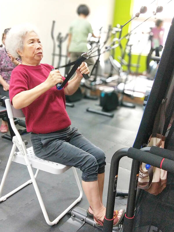
Source link

  • Dewan MC, Rattani A, Gupta S, Baticulon RE, Hung YC, Punchak M, Agrawal A, Adeleye AO, Shrime MG, Rubiano AM, Rosenfeld JV, Park KB. Estimating the global incidence of traumatic brain injury. J Neurosurg. 2018;1:1–18. doi.org/10.3171/2017.10.JNS17352.

    Article 

    Google Scholar
     

  • Wongchareon K, Thompson HJ, Mitchell PH, Barber J, Temkin N. IMPACT and CRASH prognostic models for traumatic brain injury: external validation in a South-American cohort. Inj Prev. 2020;26(6):546–54. doi.org/10.1136/injuryprev-2019-043466.

    Article 
    PubMed 

    Google Scholar
     

  • Global Burden Diseases 2016 Traumatic Brain Injury and Spinal Cord Injury Collaborators. Global, regional, and national burden of traumatic brain injury and spinal cord injury, 1990–2016: a systematic analysis for the Global Burden of Disease Study 2016. Lancet Neurol. 2019;18(1):56–87.

    Article 

    Google Scholar
     

  • Ndoumbe A, Edu PBN, Simeu C, Takongmo S. Epidemiological Analysis of 135 Cases of Severe Traumatic Brain Injury Managed at a Surgical Intensive Care Unit. Open J Mod Neurosurg. 2018;8:119–31.

    Article 

    Google Scholar
     

  • Oberholzer M, Müri RM. Neurorehabilitation of traumatic brain injury (TBI): a clinical review. Med Sci (Basel). 2019;7(3):47.

    PubMed 

    Google Scholar
     

  • Iaccarino MA, Bhatnagar S, Zafonte R. Rehabilitation after traumatic brain injury. Handb Clin Neurol. 2015;127:411–22. doi.org/10.1016/B978-0-444-52892-6.00026-X.

    Article 
    PubMed 

    Google Scholar
     

  • National Academies of Sciences, Engineering, and Medicine 2022. Traumatic brain injury: a roadmap for accelerating progress. Washington, DC: The National Academies Press. doi.org/10.17226/25394.

  • Dash HH, Chavali S. Management of traumatic brain injury patients. Korean J Anesthesiol. 2018;71(1):12–21. doi.org/10.4097/kjae.2018.71.1.12.

    Article 
    PubMed 
    PubMed Central 

    Google Scholar
     

  • Dixon J, Comstock G, Whitfield J, Richards D, Burkholder TW, Leifer N, Mould-Millman NK, Calvello Hynes EJ. Emergency department management of traumatic brain injuries: A resource tiered review. Afr J Emerg Med. 2020;10(3):159–66. doi.org/10.1016/j.afjem.2020.05.006.

    Article 
    PubMed 
    PubMed Central 

    Google Scholar
     

  • World Health Organisation. The WHO Emergency Care System Framework and Assessment Tool, 2007. Department for Management of NCDs, Disability, Violence and Injury Prevention. www.humanitarianresponse.info/sites/www.humanitarianresponse.info/files/documents/files/ems.pdf.

  • Pujari R, Hutchinson PJ, Kolias AG. Surgical management of traumatic brain injury. J Neurosurg Sci. 2018;62(5):584–92. doi.org/10.23736/S0390-5616.18.04533-2.

    Article 
    PubMed 

    Google Scholar
     

  • Meara JG, Leather AJ, Hagander L, Alkire BC, Alonso N, Ameh EA, Bickler SW, Conteh L, Dare AJ, Davies J, Mérisier ED, El-Halabi S, Farmer PE, Gawande A, Gillies R, Greenberg SL, Grimes CE, Gruen RL, Ismail EA, Kamara TB, Lavy C, Lundeg G, Mkandawire NC, Raykar NP, Riesel JN, Rodas E, Rose J, Roy N, Shrime MG, Sullivan R, Verguet S, Watters D, Weiser TG, Wilson IH, Yamey G, Yip W. Global Surgery 2030: evidence and solutions for achieving health, welfare, and economic development. Lancet. 2015;386(9993):569–624. doi.org/10.1016/S0140-6736(15)60160-X.

    Article 
    PubMed 

    Google Scholar
     

  • Alayande B, Chu KM, Jumbam DT, Kimto OE, Musa Danladi G, Niyukuri A, Anderson GA, El-Gabri D, Miranda E, Taye M, Tertong N, Yempabe T, Ntirenganya F, Byiringiro JC, Sule AZ, Kobusingye OC, Bekele A, Riviello RR. Disparities in access to trauma care in sub-Saharan Africa: a narrative review. Curr Trauma Rep. 2022;8(3):66–94. doi.org/10.1007/s40719-022-00229-1.

    Article 
    PubMed 
    PubMed Central 

    Google Scholar
     

  • Buh FC, Taiwe GS, Maas AIR, Motah M, Youm E, Wanyu BY, Wang KW, Hutchinson PJA, Sumbele IUN. Demographics, causes, and outcome of traumatic brain injury among trauma cases in Cameroon: a multi-center five year’s retrospective study. Neurotrauma Rep. 2022;3(1):569–83. doi.org/10.1089/neur.2022.0053.

    Article 
    PubMed 
    PubMed Central 

    Google Scholar
     

  • Aderinto N, Opanike J, Oladipo E, Olakanmi D, Adepoju O. Accessing neurosurgical services in Africa: efforts, challenges, and recommendation. Int J Surg. 2022;5(6):e90. doi.org/10.1097/GH9.0000000000000090.

    Article 

    Google Scholar
     

  • Dada OE, Ooi SZY, Bukenya GW, Kenfack YJ, Le C, Ohonba E, Adeyemo E, Narain K, Awad AK, Barrie U, Sichimba D, Ogunfolaji O, Kitonga LM, Oriaku AJ, Bamimore MA, Okor DE, Rominiyi O. Evaluating the impact of neurosurgical rotation experience in Africa on the interest and perception of medical students towards a career in neurosurgery: a continental, multi-centre, cross-sectional study. Front Surg. 2022;9:766325. doi.org/10.3389/fsurg.2022.766325.

    Article 
    PubMed 
    PubMed Central 

    Google Scholar
     

  • Kanmounye US, Ammar A, Rolle M, El Ouahabi A, Park KB. Global neurosurgical workforce density-you cannot improve what you do not measure. Chin Neurosurg J. 2021;7(1):33. doi.org/10.1186/s41016-021-00252-2.

    Article 
    PubMed 
    PubMed Central 

    Google Scholar
     

  • Ukachukwu AK, Still MEH, Seas A, von Isenburg M, Fieggen G, Malomo AO, Shokunbi MT, Egger JR, Haglund MM, Fuller AT. Fulfilling the specialist neurosurgical workforce needs in Africa: a systematic review and projection toward 2030. J Neurosurg. 2022;138(4):1102–13. doi.org/10.3171/2022.2.JNS211984.

    Article 
    PubMed 

    Google Scholar
     

  • World Health Organisation. Rehabilitation 2021. Available from www.who.int/news-room/fact-sheets/detail/rehabilitation. Last accessed 13.03.2023.

  • Königs M, Beurskens EA, Snoep L, Scherder EJ, Oosterlaan J. Effects of timing and intensity of neurorehabilitation on functional outcome after traumatic brain injury: a systematic review and meta-analysis. Arch Phys Med Rehabil. 2018;99(6):1149-1159.e1.

    Article 
    PubMed 

    Google Scholar
     

  • Turner-Stokes L, Paul S, Williams H. Efficiency of specialist rehabilitation in reducing dependency and costs of continuing care for adults with complex acquired brain injuries. J Neurol Neurosurg Psychiatry. 2006;77(5):634–9.

    Article 
    CAS 
    PubMed 
    PubMed Central 

    Google Scholar
     

  • Turner-Stokes L. Cost-efficiency of longer-stay rehabilitation programmes: can they provide value for money? Brain Inj. 2007;21(10):1015–21.

    Article 
    PubMed 

    Google Scholar
     

  • Eghbali M, Khankeh H, Ebadi A. The importance of early rehabilitation in traumatic brain injury. Nurs Pract Today. 2020;7(2):84–6.


    Google Scholar
     

  • Kreitzer N, Rath K, Kurowski BG, Bakas T, Hart K, Lindsell CJ, Adeoye O. Rehabilitation practices in patients with moderate and severe traumatic brain injury. J Head Trauma Rehabil. 2019;34(5):E66–72.

    Article 
    PubMed 
    PubMed Central 

    Google Scholar
     

  • Mendez-Tellez PA, Nusr R, Feldman D, Needham DM. Early physical rehabilitation in the ICU: a review for the neurohospitalist. Neurohospitalist. 2012;2(3):96–105.

    Article 
    PubMed 
    PubMed Central 

    Google Scholar
     

  • Swaminathan N, Praveen R, Surendran J. The role of physiotherapy in intensive care units: a critical review. Physiother Q. 2019;27(4):1–5.

    Article 

    Google Scholar
     

  • Turner-Stokes L, Pick A, Nair A, Disler PB, Wade DT. Multi-disciplinary rehabilitation for acquired brain injury in adults of working age. Cochrane Database Syst Rev. 2015;2015(12):CD004170.

    PubMed 
    PubMed Central 

    Google Scholar
     

  • Corten L, van As AB, Rahim S, Kleinsmith J, Kleyn A, Kwinana T, Ndaba N, Sillito E, Smith JT, Williams N, Figaji A. Physiotherapy in children hospitalized with traumatic brain injury in a South African tertiary paediatric hospital. Physiother Res Int. 2020;25(4):e1860. doi.org/10.1002/pri.1860.

    Article 
    PubMed 

    Google Scholar
     

  • Lalwani SS, Dadgal R, Harjpal P, Saifee SS, Lakkadsha TM. Perks of early physical therapy rehabilitation for a patient with diffuse axonal injury. Cureus. 2022;14(10):e30886. doi.org/10.7759/cureus.30886.

    Article 
    PubMed 
    PubMed Central 

    Google Scholar
     

  • Hassett L. Physiotherapy for the acute care management of traumatic brain injury, 2005. Liverpool Health Service. BC NSW 1871. Available from www.swslhd.health.nsw.gov.au/Liverpool/BIRU%5Ccontent/pdf/Physioth_for_TBI.pdf. Accessed 8 Feb 2023.

  • Lee SY, Amatya B, Judson R, Truesdale M, Reinhardt JD, Uddin T, Xiong XH, Khan F. Clinical practice guidelines for rehabilitation in traumatic brain injury: a critical appraisal. Brain Inj. 2019;33(10):1263–71.

    Article 
    PubMed 

    Google Scholar
     

  • Dean PJ, Sterr A. Long-term effects of mild traumatic brain injury on cognitive performance. Front Hum Neurosci. 2013;12(7):30. doi.org/10.3389/fnhum.2013.00030.

    Article 

    Google Scholar
     

  • McInnes K, Friesen CL, MacKenzie DE, Westwood DA, Boe SG. Mild Traumatic Brain Injury (mTBI) and chronic cognitive impairment: a scoping review. PLoS One. 2017;12(4):e0174847. doi.org/10.1371/journal.pone.0174847.

    Article 
    CAS 
    PubMed 
    PubMed Central 

    Google Scholar
     

  • Wu X, Li Z, Cao J, Jiao J, Wang Y, Liu G, Liu Y, Li F, Song B, Jin J, Liu Y, Wen X, Cheng S, Wan X. The association between major complications of immobility during hospitalization and quality of life among bedridden patients: a 3 month prospective multi-center study. PLoS One. 2018;13(10):e0205729. doi.org/10.1371/journal.pone.0205729.

    Article 
    CAS 
    PubMed 
    PubMed Central 

    Google Scholar
     

  • Dementovych N. Hospitalisation can be particularly stressful for elderly patients: It is important to be aware of the risk and how to help avoid them. Jefferson Health, Home of Sidney Medical College 2022. Available from www.jeffersonhealth.org/your-health/living-well/the-mental-and-physical-effects-of-a-hospital-stay-on-seniors. Last accessed 08.09.2022.

  • Rodrigues-Gomes RM, Martí JD, Rolán RM, Gelabert-González M. Rapid chest compression effects on intracranial pressure in patients with acute cerebral injury. Trials. 2022;23(1):312. doi.org/10.1186/s13063-022-06189-w.

    Article 
    PubMed 
    PubMed Central 

    Google Scholar
     

  • Powell J, Heslin J, Greenwood R. Community based rehabilitation after severe traumatic brain injury: a randomised controlled trial. J Neurol Neurosurg Psychiatry. 2002;72(2):193–202. doi.org/10.1136/jnnp.72.2.193.

    Article 
    CAS 
    PubMed 
    PubMed Central 

    Google Scholar
     

  • Iemmi V, Kumar KS, Blanchet K, Gibson L, Hartley S, Murthy GVS, Patel V, Weber J, Kuper H. Community-based rehabilitation for people with physical and mental disabilities in low- and middle-income countries. Cochrane Database Syst Rev. 2017;2017(3):CD010617. doi.org/10.1002/14651858.CD010617.pub2.

    Article 
    PubMed Central 

    Google Scholar
     

  • Anwar F, Razaq S, Yasmeen R, Rathore FA. Interdisciplinary inpatient rehabilitation of acquired brain injury — Part II — a proposed model of care for Pakistan. J Pak Med Assoc. 2022;72(12):2573–5.

    PubMed 

    Google Scholar
     

  • National Health Service. Neurorehabilitation Team 2023. Available from gmnisdn.org.uk/patients-and-carers/the-neuro-rehabilitation-team/. Last assessed 30-March-2023.

  • Ramaswamy S, Seshadri S, Bunders-Aelen J. Shifting landscapes of global child mental health: imperatives for transdisciplinary approaches. Asian J Psychiatr. 2022;69:103002. doi.org/10.1016/j.ajp.2021.103002.

    Article 
    PubMed 

    Google Scholar
     

  • Karol RL. Team models in neurorehabilitation: structure, function, and culture change. NeuroRehabilitation. 2014;34(4):655–69. doi.org/10.3233/NRE-141080.

    Article 
    PubMed 

    Google Scholar
     

  • Source link

    4.1. Summary of Evidence

    This systematic review reveals a growing interest in telemedicine-driven pulmonary rehabilitation following the acute phase of COVID-19, although, as the pandemic reaches an end, it will be interesting to observe this trend in the upcoming years. The studies included in this review were conducted across diverse geographical settings, further supporting the potential global applicability of telemedicine interventions in post-acute COVID-19 management. It seems that regardless of the study design, the integration of telerehabilitation in post-acute care significantly improved several health outcomes among COVID-19 patients. These improvements were observed in physical and mental health, quality of life, and pulmonary function, albeit with variations across different studies.

    In terms of rehabilitation protocols, a significant amount of heterogeneity was observed among the studies. This could be reflective of the individualized approach necessary for addressing the distinct needs of post-acute COVID-19 patients, as well as the absence of a unified protocol for telerehabilitation in this setting. The protocols were designed to incorporate a balance of aerobic and resistance training, breathing exercises, and functional activities, often accompanied by counseling or motivational measures to promote adherence. The intensity, duration, and frequency of the sessions varied widely, suggesting the need for further studies to identify optimal parameters for telerehabilitation programs in this population.

    Different rehabilitation protocols emphasize various exercises, including deep breathing, inspiratory muscle training, and breathing control techniques, although the majority were performed in a hospital or institutional setting, contrary to our study [28,29,30,31]. These protocols often also incorporate physical exercises for strength and endurance, which help in functional improvement and disability reduction [32,33]. However, the question that arises is what degree of change and disability reduction is dependent on the PR setting. A holistic approach, encouraged by the American Thoracic Society (ATS) and the European Respiratory Society (ERS), expands beyond mere physical exercises, embracing comprehensive patient evaluation and lifestyle modifications [34]. Some studies have combined their rehabilitation protocols with educational sessions to address such issues as dyspnea, cough, fatigue, anxiety, memory, and daily activity management [35,36]. These integrative approaches have demonstrated significant improvements in functional abilities, patient’s quality of life, and reintroduction into professional life.
    Around 90% of COVID-19 patients in hospitals deal with debilitating lung effects, indicating the importance of physical and respiratory rehabilitation [37,38]. The most common symptoms include dyspnea, fatigue, and exercise intolerance. Thus, telerehabilitation, offering physiotherapy remotely, is a suitable option to address these issues, particularly during social distancing, while its convenience also encourages patient adherence [39]. Improvements in physical health outcomes, such as fatigue, pain, and exercise capacity, suggest that telerehabilitation can be an effective modality to enhance recovery and function after acute COVID-19. The improvements in mental health outcomes further support the potential of telerehabilitation in addressing the psychological impact of the disease, an aspect that is often overlooked in physical rehabilitation programs. The positive effect on quality-of-life measures is particularly encouraging, given the significant impact COVID-19 can have on overall well-being.
    The pandemic added challenges for research and trial participation due to strict control measures and economic struggles. Randomized trials showcased a variety of participant details, disease stage, telerehabilitation methods, and the varying telemonitoring options that could have impacted outcomes, such as smartphones, video conferences, and messaging applications [40,41]. The rush to introduce remote interventions often came with inadequate implementation guidance and professional training, which was evident in some of our included studies and other trials for acute COVID-19 [25,42]. Despite the variability in the measures used to assess pulmonary function, some significant improvements were reported. It is important to highlight that the effects on pulmonary function seem to depend on the severity of the disease, with the less severe cases showing more significant improvements. Thus, one hypothesis that this study suggests is that telerehabilitation might be more effective when initiated in the early post-acute phase before severe pulmonary sequelae develop.
    Moreover, further research is needed to examine the aspects of physical abilities and lung function that better represent changes during PR post-COVID-19. Notably, these parameters encompass Forced Vital Capacity (FVC), Forced Expiratory Volume in 1 s (FEV1), and Diffusing Capacity for Carbon Monoxide (DLCO), as used in other PR programs for COPD and ILD [43,44]. Furthermore, not only do these traditional lung function tests show improvement following a physical rehabilitation program, but the severity of dyspnea, or shortness of breath, also significantly decreased among adult survivors of COVID-19. This provides another practical measurement of improved respiratory function, as reducing the feeling of breathlessness is a critical component of the recovery process. In addition to the aforementioned tests, it may also be beneficial to consider other tests, such as the Total Lung Capacity (TLC) and the Peak Expiratory Flow (PEF), that may provide a more nuanced understanding of the impact of physical rehabilitation programs on lung function among COVID-19 survivors.

    The quality of the studies included in this review was generally good or excellent, suggesting reliable findings. However, it is worth noting that the higher-quality studies were all randomized trials conducted in 2022, indicating that the quality of research in this area is improving. Additionally, the higher-quality studies tended to have larger sample sizes, suggesting that they may provide more reliable evidence for the effectiveness of telerehabilitation in this setting.

    4.2. Limitations

    While the findings of this review are encouraging, it is important to acknowledge its limitations. The diversity of the rehabilitation programs, their duration, intensity, and the measures used to assess outcomes across studies make it challenging to draw definitive conclusions. There was also a wide age range among the participants, which might influence the outcomes of the rehabilitation programs. The gender distribution was not balanced across studies, and the severity of COVID-19 varied, factors that might also affect the response to rehabilitation. Furthermore, many studies did not provide detailed data about the length of hospitalization, making it difficult to explore its potential impact on outcomes. Moreover, the number of included studies was relatively small, indicating a need for more high-quality research in this field.

    By limiting the search to studies published in English, there may have been inadvertently introduced language bias, potentially excluding relevant studies published in other languages. In addition, this review did not include the gray literature, such as conference papers or technical reports, possibly leading to publication bias. Despite the robust measures used to assess this, the possibility of missing some relevant information cannot be entirely negated. Finally, our quality assessment was based on the National Heart, Lung, and Blood Institute (NHLBI) tool. Despite its wide acceptance and use, it possesses inherent limitations and subjectivity, which could potentially influence the conclusions drawn from this review. Future studies should strive for uniformity in their protocols and measures to allow for more direct comparisons and, potentially, meta-analyses.

    Source link

    A traumatic event can cause a person to experience post-traumatic stress disorder (PTSD), a mental health condition that is characterised by signs like anxiety, intrusive thoughts, nightmares and flashbacks. While therapy and medication are frequently used as traditional PTSD treatments, health experts insist that complementary modalities like Yoga and physiotherapy have shown promise in helping people manage their symptoms.

    Post traumatic stress disorder: How Yoga and physiotherapy can help in PTSD (Shutterstock)
    Post traumatic stress disorder: How Yoga and physiotherapy can help in PTSD (Shutterstock)

    In an interview with HT Lifestyle, Dr Amit Deshpande, Founder and Director of Activist, shared, “Post-traumatic stress disorder (PTSD) is a debilitating mental health condition that can occur after experiencing or witnessing a traumatic event. Fortunately, Yoga and physiotherapy offer promising avenues for healing and managing PTSD symptoms. Yoga provides a holistic approach by combining physical postures, breathing exercises and meditation, helping to regulate the nervous system and reduce anxiety and stress.”

    He gushed, “The mindful movements and deep breathing foster a sense of calmness and self-awareness, enhancing emotional resilience. Physiotherapy, on the other hand, focuses on restoring physical function and mobility through targeted exercises and manual therapy. It can address physical symptoms associated with trauma, such as muscle tension and pain. Additionally, both yoga and physiotherapy offer a safe and supportive environment, fostering a sense of community and connection, which can aid in the recovery process.”

    Dr Rajeev Rajesh, Chief Yoga Officer at Jindal Naturecure Institute in Banglore, explained, “Yoga is a mind-body discipline with its roots in the ancient Indian subcontinent. It combines physical postures, breathing techniques and meditation. It has been discovered to be successful in lowering anxiety, enhancing sleep, and fostering general wellbeing. Yoga can give people with PTSD a safe place to re-connect with their body and process their trauma in a nurturing setting. Yoga's physical asanas and deliberate breathing techniques work to control the autonomic nervous system, lowering hyperarousal and enhancing relaxation. Additionally, yoga's emphasis on mindfulness encourages people to be in the present moment, which helps to lessen intrusive thoughts and improve self-awareness.”

    He added, “On the other hand, physiotherapy focuses on enhancing functional movement and physical rehabilitation. Physiotherapy can help people with PTSD deal with the somatic symptoms, chronic pain, and other physical effects of trauma. Physiotherapy can assist people in releasing tension, restoring proper movement patterns, and improving body awareness through a variety of techniques like manual therapy, exercise, and body awareness training. Physiotherapy can indirectly reduce psychological distress and improve general wellbeing by addressing physical symptoms. Yoga and physiotherapy both provide holistic approaches to healing and can support existing PTSD treatments. It is crucial to remember that these methods might not be appropriate for everyone and should only be used under the supervision of trained experts. However, incorporating yoga and physical therapy into PTSD treatment plans can give patients more coping skills and tools to manage their symptoms and enhance their quality of life.”

    Bringing his expertise to the same, Dr Sumit Gupta PT, HOD- Department of Physiotherapy and Rehabilitation at Regency Health, highlighted that PTSD can also increase stress and anxiety leading to tension in the body but revealed how Yoga and physiotherapy is very helpful to prevent this condition:

    1. So many time patient complaint pain. Physiotherapist set goals. Strengthening exercise, stability exercise, joint mobilisation, needling and motion exercise is help to reduce pain.

    2. Breathing exercise - Physiotherapist can give their patient different breathing exercise (deep breathing, chest mobilisation, diaphragmatic breathing) which help to reduce stress or anxiety.

    3.Anxiety-regular exercise is an effective method in management anxiety and stress follow a prescribe exercise plan will allow the body to release chemical that can help in reducing stress and anxiety.

    He listed the benefits of regular exercise on PTSD symptoms as:

    • A few positive results physiotherapy can have on your physical health including generate mobility and flexibility increase strength weight loss and improvement in cardiovascular health.
    • Regular exercise can also have a positive effect on your mental health through the reduce the of depression and anxiety feelings.

    Source link

  • Nalbandian A, Sehgal K, Gupta A, Madhavan MVM, McGroder C, Stevens JS, Cook JR, et al. Post-acute COVID-19 syndrome. Nat Med. 2021;27(4):601–15.

    Article 
    CAS 
    PubMed 
    PubMed Central 

    Google Scholar
     

  • NICE. COVID-19 rapid guideline: Managing the long-term effects of COVID-19. 2020.

  • Soriano JB, Murthy S, Marshall JC, Relan P, Diaz JV. A clinical case definition of post-COVID-19 condition by a Delphi consensus. Lancet Infect Dis. 2021;22(4):102–7.

    Article 

    Google Scholar
     

  • Centers for Disease Control and Prevention. Long COVID or Post-COVID Conditions 2022. Available from: www.cdc.gov/coronavirus/2019-ncov/long-term-effects/index.html. Accessed 27 September 2022.

  • O’Mahoney LL, Routen A, Gillies C, Ekezie W, Welford A, Zhang A, et al. The prevalence and long-term health effects of Long Covid among hospitalised and non-hospitalised populations: a systematic review and meta-analysis. eClinicalMedicine. 2023;55:101762.

  • Maltezou HC, Pavli A, Tsakris A. Post-COVID syndrome: an insight on its pathogenesis. Vaccines (Basel). 2021;9(5):497.

    Article 
    CAS 
    PubMed 

    Google Scholar
     

  • International Primary Care Respiratory Group. What are the risk factors for long-COVID-19 disease/post-COVID syndrome (PCS)? 2021. Available from: www.ipcrg.org/resources/search-resources/what-are-the-risk-factors-for-long-covid-19-diseasepost-covid-syndrome. Accessed 10 February 2022.

  • Menges D, Ballouz TA, Anagnostopoulos A, Aschmann HED, Domenghino A, Fehr JS, Puhan M. Burden of post-COVID-19 syndrome and implications for healthcare service planning: a population-based cohort study. PLoS One. 2021;16(7):e0254523.

    Article 
    CAS 
    PubMed 
    PubMed Central 

    Google Scholar
     

  • Evans RA, McAuley H, Harrison EM, Shikotra A, Singapuri A, Sereno M, et al. Physical, cognitive, and mental health impacts of COVID-19 after hospitalisation (PHOSP-COVID): a UK multicentre, prospective cohort study. Lancet Respir Med. 2021;9(11):13.

    Article 

    Google Scholar
     

  • Aiyegbusi OL, Hughes SE, Turner G, Rivera SC, McMullan C, Chandan JS, et al. Symptoms, complications and management of long COVID: a review. J R Soc Med. 2021;114(9):428–42.

    Article 
    PubMed 
    PubMed Central 

    Google Scholar
     

  • World Health Organisation. Clinical management of COVID-19: Living guideline, 15 September 2022 2022. Available from: www.who.int/publications/i/item/WHO-2019-nCoV-Clinical-2022.2. Accessed 21 December 2022.

  • The Cochrane Collaboration. Cochrane Training 2022. Available from: training.cochrane.org/. Accessed 2 May 2022.

  • PRISMA. PRISMA - transparent reporting of systematic reviews and meta-analyses 2021. Available from: www.prisma-statement.org/. Accessed 2 May 2022.

  • Research Aid Networks Long Covid Library. Available from: www.zotero.org/groups/4411227/research_aid_networks_long_covid_library/items/9ITDFM6W. Accessed 9 May 2022.

  • Resources LongCovid. Available from: docs.google.com/spreadsheets/d/1jy354stmCE30zYoE5Ou3lz0O1hZSbvuLfvxcUGoBroQ/htmlview?fbclid=IwAR1RFFF7YnmeNzGQIH9uf3RcgWzmHkRARzj_oIfu4F00qGiHLe4yH0bP3L4. Accessed 9 May 2022.

  • Cochrane. Available from: covid-19.cochrane.org/. Accessed 9 May 2022.

  • Epistemonikos. Available from: app.iloveevidence.com/loves/5e6fdb9669c00e4ac072701d/advanced-search. Accessed 9 May 2022.

  • World Health Organisation. Available from: search.bvsalud.org/global-literature-on-novel-coronavirus-2019-ncov/. Accessed 9 May 2022.

  • Falconer J. Removing duplicates from an EndNote library 2018. Available from: blogs.lshtm.ac.uk/library/2018/12/07/removing-duplicates-from-an-endnote-library/. Accessed 9 May 2022.

  • Rayyan. Available from: www.rayyan.ai/. Accessed 9 May 2022.

  • Covidence. Available from: www.covidence.org/. Accessed 9 May 2022.

  • CASP UK. CASP CHECKLISTS 2021. Available from: casp-uk.net/casp-tools-checklists/. Accessed 11 January 2022.

  • Sterne J, Savović J, Page M, Elbers R, Blencowe N, Boutron I, et al. RoB 2: a revised tool for assessing risk of bias in randomised trials. BMJ. 2019;366:4898.

    Article 

    Google Scholar
     

  • Guyatt GH, Oxman AD, Vist GE, Kunz R, Falck-Ytter Y, Alonso-Coello P, et al. GRADE: an emerging consensus on rating quality of evidence and strength of recommendations. BMJ. 2008;336(7650):924–6.

    Article 
    PubMed 
    PubMed Central 

    Google Scholar
     

  • GRADE working group. Handbook for grading the quality of evidence and the strength of recommendations using the GRADE approach. 2013. Available from: gdt.gradepro.org/app/handbook/handbook.html. Accessed 12 October 2021.

  • Barbara C, Clavario P, De Marzo V, Lotti R, Guglielmi G, Porcile A, et al. Effects of exercise rehabilitation in patients with long COVID-19. Eur J Prev Cardiol. 2022;29(7):258–60.

    Article 

    Google Scholar
     

  • Betschart M, Rezek S, Unger I, Beyer S, Gisi D, Shannon H, et al. Feasibility of an outpatient training program after COVID-19. Int J Environ Res Public Health. 2021;18(8):3978.

    Article 
    PubMed 
    PubMed Central 

    Google Scholar
     

  • Bouteleux B, Henrot P, Ernst R, Grassion L, Raherison-Semjen C, Beaufils F, et al. Respiratory rehabilitation for Covid-19 related persistent dyspnoea: a one-year experience. Respir Med. 2021;189: 106648.

    Article 
    PubMed 
    PubMed Central 

    Google Scholar
     

  • Daynes E, Gerlis C, Chaplin E, Gardiner N, Singh SJ. Early experiences of rehabilitation for individuals post-COVID to improve fatigue, breathlessness exercise capacity and cognition – a cohort study. Chron Respir Dis. 2021;18:14799731211015692.

  • Hameed F, Palatulan E, Jaywant A, Said R, Lau C, Sood V, et al. Outcomes of a COVID-19 recovery program for patients hospitalized with SARS-CoV-2 infection in New York City: a prospective cohort study. PM&R. 2021;13(6):609–17.

    Article 

    Google Scholar
     

  • Kireyev IV, Zhabotynska NV, Bakumenko MG, Khyzhnyak VM, Knizhenko IB. Rehabilitation in Post COVID-19 neurological syndrome. Acta Balneologica. 2022;64(1):11–5.

    Article 

    Google Scholar
     

  • Martin I, Braem F, Baudet L, Poncin W, Fizaine S, Aboubakar F, et al. Follow-up of functional exercise capacity in patients with COVID-19: It is improved by telerehabilitation. Respir Med. 2021;183: 106438.

    Article 
    PubMed 
    PubMed Central 

    Google Scholar
     

  • Nambi G, Abdelbasset WK, Alrawaili SM, Elsayed SH, Verma A, Vellaiyan A, et al. Comparative effectiveness study of low versus high-intensity aerobic training with resistance training in community-dwelling older men with post-COVID 19 sarcopenia: a randomized controlled trial. Clin Rehabil. 2022;36(1):59–68.

    Article 
    PubMed 

    Google Scholar
     

  • Cahalan RM, Meade C, Mockler S. SingStrong-A singing and breathing retraining intervention for respiratory and other common symptoms of long COVID: a pilot study. Canadian Journal of Respiratory Therapy. 2022;58:20–7.

    Article 
    PubMed 
    PubMed Central 

    Google Scholar
     

  • Liu K, Zhang W, Yang Y, Zhang J, Li Y, Chen Y. Respiratory rehabilitation in elderly patients with COVID-19: a randomized controlled study. Complement Ther Clin Pract. 2020;39: 101166.

    Article 
    PubMed 
    PubMed Central 

    Google Scholar
     

  • Philip KEJ, Owles H, McVey S, Pagnuco T, Bruce K, Brunjes H, et al. An online breathing and wellbeing programme (ENO Breathe) for people with persistent symptoms following COVID-19: a parallel-group, single-blind, randomised controlled trial. Lancet Respiratory Medicine. 2022;10(9):851–62.

    Article 
    CAS 
    PubMed 

    Google Scholar
     

  • Srinivasan V, Kandakurti PK, Alagesan J, Suganthirababu P, Jenifer Augustina S, Anitha A, et al. Efficacy of pursed lip breathing with bhastrika pranayama vs incentive spirometry in rehabilitating post Covid 19 follow up-a randomized control study. Turkish Journal of Physiotherapy and Rehabilitation. 2021;32(3):402–7.


    Google Scholar
     

  • Ahmed I, Inam AB, Belli S, Ahmad J, Khalil W, Jafar MM. Effectiveness of aerobic exercise training program on cardio-respiratory fitness and quality of life in patients recovered from COVID-19. European Journal of Physiotherapy. 2021;24(6):358–63.

    Article 

    Google Scholar
     

  • Dalbosco-Salas M, Torres-Castro R, Leyton AR, Zapata FM, Salazar EH, Bastías GE, et al. Effectiveness of a primary care telerehabilitation program for post-covid-19 patients: a feasibility study. J Clin Med. 2021;10(19):4428.

    Article 
    CAS 
    PubMed 
    PubMed Central 

    Google Scholar
     

  • Kokhan S, Romanova E, Nadeina L, Baatar B, Shagdarsuren O, Purevdorj D. Effect of physical rehabilitation on the functional state of post COVID-19 patients. Laplage em Revista. 2021;7(3A):675–81.

  • Li J, Xia W, Zhan C, Liu S, Yin Z, Wang J, et al. A telerehabilitation programme in post-discharge COVID-19 patients (TERECO): a randomised controlled trial. Thorax. 2021;77(7):697–706.

    Article 
    PubMed 

    Google Scholar
     

  • Scaturro D, Vitagliani F, Di Bella VE, Falco V, Tomasello S, Lauricella L, et al. The role of acetyl-carnitine and rehabilitation in the management of patients with post-COVID syndrome: case-control study. Appl Sci. 2022;12(8):4084.

    Article 
    CAS 

    Google Scholar
     

  • Stavrou VT, Tourlakopoulos KN, Vavougios GD, Papayianni E, Kiribesi K, Maggoutas S, et al. Eight weeks unsupervised pulmonary rehabilitation in previously hospitalized of SARS-CoV-2 infection. Journal of Personalized Medicine. 2021;11(8):806.

    Article 
    PubMed 
    PubMed Central 

    Google Scholar
     

  • D’Ascanio L, Vitelli F, Cingolani C, Maranzano M, Brenner MJ, Di Stadio A. Randomized clinical trial “olfactory dysfunction after COVID-19: olfactory rehabilitation therapy vs. intervention treatment with Palmitoylethanolamide and Luteolin”: preliminary results. Eur Rev Med Pharmacol Sci. 2021;25(11):4156–62.

  • Di Stadio A, D’Ascanio L, Vaira LA, Cantone E, De Luca P, Cingolani C, et al. Ultramicronized palmitoylethanolamide and luteolin supplement combined with olfactory training to treat post-COVID-19 olfactory impairment: a multi-center double-blinded randomized placebo-controlled clinical trial. Curr Neuropharmacol. 2022;20(10):2001–12.

  • Rathi A, Jadhav SB, Shah N. A randomized controlled trial of the efficacy of systemic enzymes and probiotics in the resolution of post-COVID fatigue. Medicines. 2021;8(9):47.

    Article 
    CAS 
    PubMed 
    PubMed Central 

    Google Scholar
     

  • Abdelalim AA, Mohamady AA, Elsayed RA, Elawady MA, Ghallab AF. Corticosteroid nasal spray for recovery of smell sensation in COVID-19 patients: a randomized controlled trial. Am J Otolaryngol. 2021;42(2): 102884.

    Article 
    CAS 
    PubMed 
    PubMed Central 

    Google Scholar
     

  • Denis F, Septans A-L, Periers L, Maillard J-M, Legoff F, Gurden H, et al. Olfactory training and visual stimulation assisted by a web application for patients with persistent olfactory dysfunction after SARS-CoV-2 infection: observational study. J Med Internet Res. 2021;23(5): e29583.

    Article 
    PubMed 
    PubMed Central 

    Google Scholar
     

  • Le Bon SD, Konopnicki D, Pisarski N, Prunier L, Lechien JR, Horoi M. Efficacy and safety of oral corticosteroids and olfactory training in the management of COVID-19-related loss of smell. Eur Arch Otorhinolaryngol. 2021;278(8):3113–7.

    Article 
    PubMed 
    PubMed Central 

    Google Scholar
     

  • Pires ÍAT, Steffens ST, Mocelin AG, Shibukawa DE, Leahy L, Saito FL, et al. Intensive olfactory training in post-COVID-19 patients: a multicenter randomized clinical trial. Am J Rhinol Allergy. 2022;36(6):780–7.

    Article 
    PubMed 

    Google Scholar
     

  • Vandersteen C, Payne M, Dumas LÉ, Cancian É, Plonka A, D’Andrea G, et al. Olfactory training efficiency in Post-COVID-19 persistent olfactory disorders: value normalization for threshold but not identification. J Clin Med. 2022;11(12):3275.

    Article 
    CAS 
    PubMed 
    PubMed Central 

    Google Scholar
     

  • Albu S, Rivas Zozaya N, Murillo N, García-Molina A, Figueroa Chacón CA, Kumru H. Multidisciplinary outpatient rehabilitation of physical and neurological sequelae and persistent symptoms of covid-19: a prospective, observational cohort study. Disabil Rehabil. 2021;44(22):6833–40.

    Article 
    PubMed 

    Google Scholar
     

  • García-Molina A, Espiña-Bou M, Rodríguez-Rajo P, Sánchez-Carrión R, Enseñat-Cantallops A. Programa de rehabilitación neuropsicológica en pacientes con síndrome post-COVID-19: una experiencia clínica. Neurología. 2021;36(7):565–6.

    Article 
    PubMed 
    PubMed Central 

    Google Scholar
     

  • Everaerts S, Heyns A, Langer D, Beyens H, Hermans G, Troosters T, et al. COVID-19 recovery: benefits of multidisciplinary respiratory rehabilitation. BMJ Open Respir Res. 2021;8(1): e000837.

    Article 
    PubMed 

    Google Scholar
     

  • Gloeckl R, Leitl D, Jarosch I, Schneeberger T, Nell C, Stenzel N, et al. Benefits of pulmonary rehabilitation in COVID-19: a prospective observational cohort study. ERJ Open Research. 2021;7(2):00108–2021.

    Article 
    PubMed 
    PubMed Central 

    Google Scholar
     

  • Hayden MC, Limbach M, Schuler M, Merkl S, Schwarzl G, Jakab K, et al. Effectiveness of a three-week inpatient pulmonary rehabilitation program for patients after COVID-19: a prospective observational study. Int J Environ Res Public Health. 2021;18(17):9001.

    Article 
    CAS 
    PubMed 
    PubMed Central 

    Google Scholar
     

  • Nopp S, Moik F, Klok FA, Gattinger D, Petrovic M, Vonbank K, et al. Outpatient pulmonary rehabilitation in patients with long COVID improves exercise capacity, functional status, dyspnea, fatigue, and quality of life. Respiration. 2022;101(6):593–601.

    Article 
    CAS 
    PubMed 

    Google Scholar
     

  • Babliuk L, Fediaeva S, Babova I, Mesoedova V, Tamazlykar S. Rehabilitation of post-COVID patients with chronic fatigue and cognitive disorders syndromes. Balneo and PRM Research Journal. 2022;13(1):497.

    Article 

    Google Scholar
     

  • Fan Y, Shi Y, Zhang J, Sun D, Wang X, Fu G, et al. The effects of narrative exposure therapy on COVID-19 patients with post-traumatic stress symptoms: a randomized controlled trial. J Affect Disord. 2021;293:141–7.

    Article 
    CAS 
    PubMed 
    PubMed Central 

    Google Scholar
     

  • Hawkins J, Hires C, Keenan L, Dunne E. Aromatherapy blend of thyme, orange, clove bud, and frankincense boosts energy levels in post-COVID-19 female patients: a randomized, double-blinded, placebo controlled clinical trial. Complement Ther Med. 2022;67: 102823.

    Article 
    PubMed 
    PubMed Central 

    Google Scholar
     

  • Heald AH, Perrin R, Walther A, Stedman M, Hann M, Mukherjee A, et al. Reducing fatigue-related symptoms in Long COVID-19: a preliminary report of a lymphatic drainage intervention. Cardiovascular Endocrinology & Metabolism. 2022;11(2): e0261.

    Article 
    CAS 

    Google Scholar
     

  • Robbins T, Gonevski M, Clark C, Baitule S, Sharma K, Magar A, et al. Hyperbaric oxygen therapy for the treatment of long COVID: early evaluation of a highly promising intervention. Clin Med. 2021;21(6):629–32.

    Article 

    Google Scholar
     

  • Sathyamoorthy M, Verduzco-Gutierrez M, Varanasi S, Ward R, Spertus J, Shah S. Enhanced external counterpulsation for management of symptoms associated with long COVID. American Heart Journal Plus: Cardiology Research and Practice. 2022;13(9): e18398.


    Google Scholar
     

  • Shogenova LV, Tuet TT, Kryukova NO, Yusupkhodzhaeva KA, Pozdnyakova DD, Kim TG, et al. KalHydrogen inhalation in rehabilitation program of the medical staff recovered from COVID-19. Cardiovascular Therapy and Prevention. 2021;20(6):24–32.

    Article 

    Google Scholar
     

  • Chen H, Shi H, Liu X, Sun T, Wu J, Liu Z. Effect of pulmonary rehabilitation for patients with post-COVID-19: a systematic review and meta-analysis. Front Med. 2022;9: 837420.

    Article 

    Google Scholar
     

  • Fugazzaro S, Contri A, Esseroukh O, Kaleci S, Croci S, Massari M, et al. Rehabilitation Interventions for post-acute COVID-19 syndrome: a systematic review. Int J Environ Res Public Health. 2022;19(9):5185.

    Article 
    CAS 
    PubMed 
    PubMed Central 

    Google Scholar
     

  • Debeuf R, Swinnen E, Plattiau T, De Smedt A, De Waele E, Roggeman S, et al. The effect of physical therapy on impairments in COVID-19 patients from intensive care to home rehabilitation: a rapid review. J Rehabil Med. 2022;54:jrm00242.

    PubMed 

    Google Scholar
     

  • Goodwin VA, Allan L, Bethel A, Cowley A, Cross JL, Day J, et al. Rehabilitation to enable recovery from COVID-19: a rapid systematic review. Physiotherapy. 2021;111:4–22.

    Article 
    PubMed 
    PubMed Central 

    Google Scholar
     

  • Negrini F, de Sire A, Andrenelli E, Lazzarini SG, Patrini M, Ceravolo MG. Rehabilitation and COVID-19: update of the rapid living systematic review by Cochrane Rehabilitation Field as of December 31st, 2021. Eur J Phys Rehabil Med. 2022;58(2):328–31.

    Article 
    PubMed 
    PubMed Central 

    Google Scholar
     

  • Twomey R, DeMars J, Franklin K, Culos-Reed SN, Weatherald J, Wrightson JG. Chronic fatigue and postexertional malaise in people living with long COVID: an observational study. Phys Ther. 2022;102(4):pzac005.

  • Davis HE, Assaf GS, McCorkell L, Wei H, Low RJ, Re’em Y, et al. Characterizing long COVID in an international cohort: 7 months of symptoms and their impact. EClinicalMedicine. 2021;38: 101019.

  • Parker M, Sawant HB, Flannery T, Tarrant R, Shardha J, Bannister R, et al. Effect of using a structured pacing protocol on post-exertional symptom exacerbation and health status in a longitudinal cohort with the post-COVID-19 syndrome. J Med Virol. 2022;95(1): e28373.

    PubMed Central 

    Google Scholar
     

  • Hersche R, Weise A. Occupational therapy-based energy management education in people with post-COVID-19 condition-related fatigue: results from a focus group discussion. Occup Ther Int. 2022;2022:4590154.

    Article 
    PubMed 
    PubMed Central 

    Google Scholar
     

  • Décary S, De Groote W, Arienti C, Kiekens C, Boldrini P, Lazzarini SG, et al. Scoping review of rehabilitation care models for post COVID-19 condition. Bull World Health Organ. 2022;100(11):13.

    Article 

    Google Scholar
     

  • World Health Organisation. Support for rehabilitation: self-management after COVID-19-related illness, second edition 2021. Available from: www.who.int/europe/publications/i/item/WHO-EURO-2021-855-40590-59892. Accessed 21 December 2022.

  • Jimeno-Almazán A, Franco-López F, Buendía-Romero Á, Martínez-Cava A, Sánchez-Agar JA, Sánchez-Alcaraz Martínez BJ, Courel-Ibáñez JP, Pallarés JG. Rehabilitation for post-COVID-19 condition through a supervised exercise intervention: a randomized controlled trial. Scand J Med Sci Sports. 2022;32(12):1791–801.

    Article 
    PubMed 

    Google Scholar
     

  • Del Corral T, Fabero-Garrido R, Plaza-Manzano G, Fernández-de-Las-Peñas C, Navarro-Santana M, López-de-Uralde-Villanueva I. Home-based respiratory muscle training on quality of life and exercise tolerance in long-term post-COVID-19: Randomized controlled trial. Ann Phys Rehabil Med. 2022;66(1): 101709.

    Article 
    PubMed 
    PubMed Central 

    Google Scholar
     

  • Deutsche Gesellschaft für Neurorehabilitation e.V. (DGNR). SARS-CoV-2, COVID-19 und (Früh-) Rehabilitation. 2021.

  • Federatie Medisch Specialisten. Langdurige klachten en revalidatie na COVID-19. 2022.

  • Dillen H, Bekkering G, Bastiaens A, Li A, Van den Broeck A, Spiette A, et al. Richtlijn ‘Opvolging en revalidatie van patiënten met aanhoudende klachten na COVID-19 in de eerste lijn’. 2022.

  • Boscolo-Rizzo PH, Hopkins C, Menini A, Dibattista M, Cancellieri E, Gardenal N, Tofanelli M, et al. Parosmia assessment with structured questions and its functional impact in patients with long-term COVID-19-related olfactory dysfunction. Int Forum Allergy Rhinol. 2022;12(12):1570–4.

  • Source link

    “These investments highlight what the new markets tax credit investment sets out to do in communities all across the country,” said Greg Clements, Novogradac partner and conference chair. “They represent a wide swath of investments that help in rural and urban low-income communities.”

    Thirteen community development entities (CDEs) that made qualified low-income community investments (QLICIs) in five businesses have been named winners of the Novogradac Journal of Tax Credits QLICIs of the Year awards for 2023.

    The awards go to new markets tax credit (NMTC) stakeholders who strive for excellence in community development. This year’s winning CDEs earned the awards for the following investments:


    • DC Central Kitchen’s Klein Center for Jobs and Justice in Washington, D.C., as Metro QLICI of the Year.
    • Lauderdale Community Hospital in Ripley, Tennessee, as Nonmetro QLICI of the Year.
    • The Freelon at Sugar Hill in Detroit as Real Estate QLICI of the Year.
    • Detroit Food Commons in Detroit as Small Business QLICI of the Year.
    • Santa Cruz Community Health Centers–Live Oak Health Clinic in Santa Cruz, California, as Operating Business QLICI of the Year.

    The winners will be honored at the Novogradac 2023 Spring New Markets Tax Credit Conference June 8-9 at The Fairmont in Washington, D.C.

    “These investments highlight what the new markets tax credit investment sets out to do in communities all across the country,” said Greg Clements, Novogradac partner and conference chair. “They represent a wide swath of investments that help in rural and urban low-income communities.”

    DC Central Kitchen’s Klein Center for Jobs and Justice in Washington, D.C., is a new, 36,000-square-foot facility that serves as a 15-hour-a-day alternative to the traditional soup kitchen and includes a culinary training kitchen and production kitchen capable of producing 25,000 meals per day. CDEs CAHEC New Markets, Reinvestment Fund and Chase New Markets Corporation allocated a combined $18.5 million in QLICIs.

    Lauderdale Community Hospital replaces an aging hospital in rural Ripley, Tennessee, giving a multimillion-dollar upgrade to improve and expand its services for patients and staff due to NMTCs allocated by three CDEs. Upgrades include emergency cardiac and pulmonary rehabilitation, surgery services, radiology, laboratory, physical rehabilitation, acute care and respiratory care. DV Community Investment, Hope Enterprise Corporation and CCG Community Partners combined on $22 million in QLICIs.

    The Freelon at Sugar Hill, a mixed-use development in Midtown Detroit’s Sugar Hill Arts District, will provide high-quality modern housing options, including units set aside for veterans, as well as commercial space for properties that are either woman- or immigrant-led. Building America CDE, Michigan Community Capital, Cinnaire New Markets and PNC Community Partners combined for $29.5 million in QLICIs for the endeavor.

    The Detroit Food Commons a 31,000-square-foot, two-story building in Detroit’s North End neighborhood, will house the Detroit People’s Food Co-op–a community-owned, full-service grocery store including a deli and a café–on the first floor. On the second floor, there will be four teaching and shared-use commercial kitchens, a banquet hall/community meeting space and office spaces for the Detroit Black Community Food Security Network. New Markets Support Company, Michigan Community Capital and U.S. Bancorp Impact Finance combined to allocate nearly $20 million in QLICIs.

    Santa Cruz Community Health Centers–Live Oak Health Clinic in Santa Cruz, California, will help an additional 3,000 patients and accommodate 20,000 visits annually at the new health clinic. SCCHC is a federally qualified health center and a Health Resources & Services Administration-designed Healthcare for the Homeless provider. Primary Care Development Corporation and HEDC New Markets combined for $14.4 million in QLICIs for the development.

    Additional details about the award winners and information on how to nominate a development for the 2024 round of awards can be found at www.novoco.com/events/awards.

    About Novogradac

    Novogradac began operations in 1989 and has grown to more than 700 employees and partners with offices in more than 25 cities. Tax, audit and consulting specialty practice areas for Novogradac include affordable housing, community development, historic rehabilitation and renewable energy.

    ###

    Share article on social media or email:

    Source link

    In their quest to relieve stress, many people turn to various tools and techniques. One such popular tool is the stress ball—a small, squeezable toy that promises to alleviate tension and promote relaxation.

    You’ve probably seen some of those semi-soft foam balls adorning virtually every corporate office, enticing people to release tension by squeezing them between their fingers. But do these ridiculous balls actually help people decompress? Should you regularly use them or is it a better idea to blow off some steam by throwing stress balls out the window?

    From work to school to romantic relationships, there’s no shortage of challenges in our lives. Often, this creates an internal conflict that manifests itself as stress. When left unchecked, stress will not only take its toll on our emotional and mental health but also on our physical well-being.

    Enter stress balls — a low-key and effective solution to managing temporary stress.

    How Do Stress Balls Work?

    It may sound implausible, but the answer to the question, “Do stress balls work?” is yes – at least from a physiological standpoint.

    Have you ever noticed how a comforting touch can instantly soothe your frayed nerves? Researchers believe that the power of tactile engagement lies at the heart of stress balls’ effectiveness. When you squeeze a stress ball, the muscles in your hand and forearm contract, triggering a series of physiological responses in your body. As you release the grip, these muscles relax, promoting a sense of relief and relaxation. 

    Stress ball
    Credit: Pixabay.

    In 2006, researchers found that stress balls can improve the focus and attention spans of sixth graders. Another study found that fidgeting with objects — squeezing a stress ball or twirling a pen, for instance — can help boost productivity by giving the mind a break, making it easier to pay attention to the task upon returning to it.

    According to MIT researchers, fidgeting objects designed to soothe or calm have to be smooth or squeezable, whereas fidgets meant to make people alert are generally clickable, sharp, or pokey. Yet another study found that stress balls helped relieve patients’ anxiety during surgery.

    However, the only study that specifically researched the effectiveness of stress balls in reducing the physiological symptoms of stress found that they don’t do much. The researchers at the University of Wisconsin-Madison found that stress balls are not effective in reducing heart rate, blood pressure, or skin conductance following an episode of induced acute stress in college-aged individuals. The sample size was rather small, though, and involved only 30 students.

    Do stress balls also work for anxiety? Studies suggest they do. Stress balls work to reduce stress because the act of squeezing improves the nervous system as well as decreases hormones, which can minimize stress levels. 

    Stress balls can squeeze out stress, but they’re no cure for chronic stress

    In light of its productivity-enhancing and anxiety-relieving properties, stress balls may be worth your time.

    Bear in mind that if you’re chronically stressed, no amount of squishy foam balls or teddy bears will help you in the long run. To release the physical and emotional tension in the body from ongoing stress, doctors recommend exercising, dancing, venting with friends, and — why not — letting it all out by crying or shouting.

    While stress balls offer a range of benefits, it is essential to acknowledge their limitations. While they may provide immediate relief, their effects are often temporary and may not address the underlying causes of stress.

    Additionally, stress balls may not be suitable for everyone. Individuals with certain hand conditions, such as arthritis or carpal tunnel syndrome, should consult with their healthcare provider before incorporating stress balls into their routine.

    What Are The Benefits of Using Stress Balls?

    A stress ball
    Credit: Pixabay.

    America’s favorite squeezable knick-knack has come a long way since it was invented in the 1980s by Alex Carswell, a 29-year-old TV writer who came up with the idea after an angry phone call with his boss compelled him to throw a magic marker at a framed photo of his mother.

    “It made me feel very good at the moment,” Carswell said later that year, “but I also had a broken picture of my mother and her dog I had to get reframed, and a mess to clean up.”

    Today, hundreds of millions of foam balls are being manufactured all over the world. But do they actually do anything?

    It might be tempting to laugh off the potential benefits of these handheld wonders, but in truth, we stand to gain a lot from them.  Stress balls have the capacity to enhance creativity, improve focus, reduce blood pressure, and even help you get a better night’s sleep. 

    And while they’re not exactly meant for bodybuilding, a stress ball can certainly strengthen your grip muscles as well as help alleviate arthritis pain.  So, if you’re wondering how stress balls work – they work on various different levels. Ultimately, a stress ball can be used as a distraction device, or a different focal point, essentially allowing you to manage stress levels in a healthy way.

    Speaking to the Huffington Post, David Posen, a stress expert and author of Is Work Killing You?: A Doctor’s Prescription for Treating Workplace Stress,” says that at least some of that stress energy can be channeled towards a physical object. Stress balls can work really well, Posen says, because they prompt you to squeeze and release, leaving you less tense.

    What Are The Different Types of Stress Balls?

    stress balls
    Credit: Pixabay.

    You might be surprised to learn there are over a dozen different types of stress balls.  In fact, there is a ball for pretty much every type of personality wanting to squeeze their way into a stress-free lifestyle. 

    In the US, the most popular choice is a bean bag type of stress reliever, but there are a variety of types all around the world.  Construction of these anxiety-reducing balls can range from gel-filled, water-filled, solid foam, rubber, or a filling mixed with gel and baking soda

    Since their inception, stress balls have evolved into all sorts of varieties such as porcupine balls, squeezy balls, kooshy balls, splat balls, puffer balls, and the list goes on. 

    The common denominator for all of these different types of stress balls is that, on some level, they are intended to release tension, therefore, reduce stress.    

    How Long Do Stress Balls Last?

    The answer to this question depends upon the type of ball you have and how much you’re using it.  In general, most stress balls last a few months.  If you have a high-quality ball made with durable materials, it could last up to a year or more.  Cheaper balls made with flimsy materials will begin to deteriorate quickly. 

    Of course, if your dog gets hold of your stress ball, it’s not going to last very long at all. In all seriousness, your ball should last a good, long time if it is made of sturdy materials, well-cared for, and used in moderation. 

    Are Stress Balls Good For Your Hands?

    Stress balls can be good for your hands because they build strength.  They can also be beneficial for building strength in your forearms and wrists. Using stress balls can increase flexibility and stimulate blood circulation, which leads to decreased stiffness and swelling. 

    When used in moderation, yes, stress balls can be therapeutic for your hands, fingers, wrists, and forearms. While stress balls are used for certain types of physical therapy, they are not always the best for your hands.  Overuse of stress balls can cause strains in tendons in the wrists or might cause pain.

    What Are the Symptoms of Stress?

    The curious thing about stress is that it manifests differently in each person. For instance, some people may exhibit stress by sweating profusely or hyperventilating.  Other people might show symptoms of stress emotionally by becoming irascible or aggressive, while others might express stress by withdrawing, becoming sullen, or succumbing to depression.  

    Moreover, stress can manifest itself in ways we normally wouldn’t, such as acting out, exhibiting nervous ticks such as nail-biting or using substances. While these are manifestations of stress, they are also behaviors that are symptoms of stress as many of us attempt to compensate or escape the unfavorable experience of stress. All in all, there are innumerable ways symptoms of stress might manifest in different people under various conditions.   

    Chronic stress can lead to headaches, an upset stomach, sleep problems, and fatigue. If left unchecked, stress can contribute to far more serious health problems, such as high blood pressure, diabetes, and obesity.

    One of the symptoms of stress is muscle tension. We literally clench our body’s muscles when feeling psychologically stressed, prompted by a flood of hormones like adrenaline, noradrenaline, and cortisol. Essentially, these chemicals prime the body for “fight or flight”.

    However, it’s not always an option to fight your boss or run away from work — this is where the ubiquitous stress ball might come in handy.

    Can Stress Be Measured? 

    Yes, stress can be measured. Observation of heart rate, pulse, breathing, and emotional responses and recording this data is the most common way to measure stress.  But how can stress be measured? 

    In many instances, we can measure our own stress levels by paying attention to our responses and reactions to stressful situations.  Through self-observation and recording our responses, we are better able to understand our stress levels and adjust accordingly.  

    Are there objective tests for stress? Yes. You can do a stress level test which can provide you with valuable information about your condition and guide you to solutions to reduce stress. You may also opt to get a medical stress test from your doctor or mental health care provider. The types of stress tests you choose will depend upon your desired outcome.  

    For instance, if your stress manifests on a physical level, you might consider getting a cardiac stress test to assess how stress is affecting your heart.  If you respond to stress more emotionally, then a mental health stress test administered by a mental professional will reveal insights that can help you manage your emotions more effectively while encountering stress.

    While stress balls may not hold the key to complete stress eradication, their simple yet effective design offers a tangible means of managing stress and promoting relaxation.

    The act of squeezing a stress ball engages both the body and the mind, providing a momentary respite from the pressures of everyday life.

    Scientific research supports their effectiveness, revealing their potential to reduce stress, enhance cognitive performance, alleviate anxiety, and aid in physical rehabilitation.

    Thanks for your feedback!

    Source link